. 2018 Jan 19;9(2):63.
doi: 10.1038/s41419-017-0094-4.

U-box ubiquitin ligase PPIL2 suppresses breast cancer invasion and metastasis by altering cell morphology and promoting SNAI1 ubiquitination and degradation

Affiliations

U-box ubiquitin ligase PPIL2 suppresses breast cancer invasion and metastasis by altering cell morphology and promoting SNAI1 ubiquitination and degradation

Zhaojun Jia et al. Cell Death Dis. .

Abstract

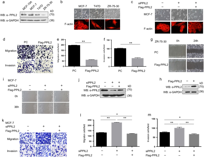
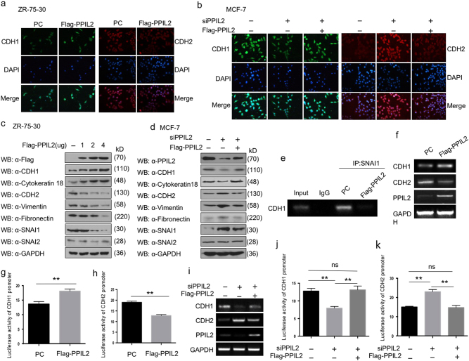
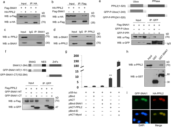
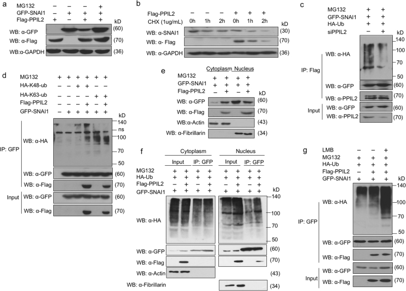
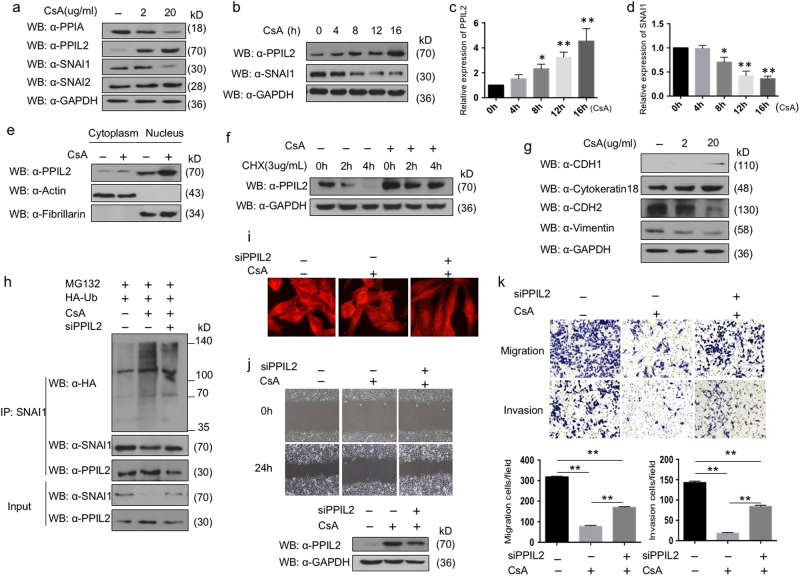
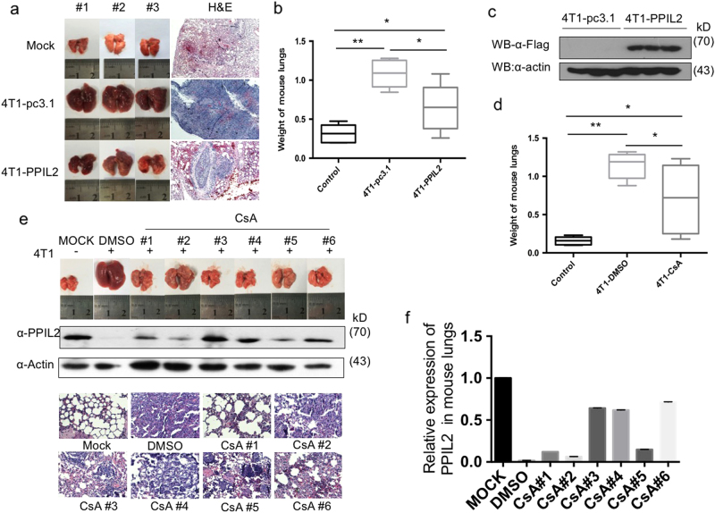
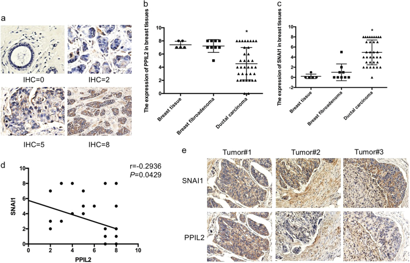
Metastasis is the leading cause of breast cancer fatalities. To develop new therapeutic strategies, the mechanisms underlying breast cancer invasion and metastasis need to be further investigated. Peptidylprolyl isomerase (cyclophilin)-like 2 (PPIL2) is a U-box-type E3 ubiquitin ligase belonging to the cyclophilin family. Proteins within this family are the major cytosolic binding proteins of the immunosuppressant drug cyclosporine A (CsA). Although PPIL2 has been reported to potentially be involved in cell migration, its role in breast cancer is still unclear. Herein, we demonstrate that PPIL2 suppressed metastasis in a breast cancer model by altering cell morphology and suppressing the epithelial-mesenchymal transition (EMT) process. Moreover, elevated PPIL2 inhibited EMT and breast cancer invasion by interacting with the classical EMT transcription factor, SNAI1, to enhance its ubiquitin-dependent degradation. Furthermore, PPIL2 protein level and stability was upregulated after CsA treatment, indicating that PPIL2 might be involved in CsA-mediated repression of EMT in breast cancer. Analysis of tissue samples taken from breast cancer patients showed a significant correlation between the expression of PPIL2 and the degree of cancer invasion and metastasis. In summary, these results would shed light on a potential clinical use of CsA in breast cancer patients.


Conflict of interest statement

The authors declare that they have no competing interests.

Figures

Fig. 1
Fig. 1. PPIL2 repressed metastasis in breast cancer cells
a Western blot analysis of the expression of PPIL2 in MCF10A and three different breast cancer cells. b The morphology of MCF-7, T47D, and ZR-75-30 was checked by phase-contrast microscopic images (top, 20×), and F-actin of MCF-7, T47D, and ZR-75-30 was checked by immunofluorescence (bottom, 40×). c The changes of cell-to-cell junction were checked by phase-contrast microscopic images (top, 20×), and the F-actin deposition in MCF-7 cells with PPIL2 knockdown restored was checked by immunofluorescence (bottom, 40×) df, Transwell assay was used to assess the function of PPIL2 on migration and invasion in ZR-75-30 cells. Cell migration and invasion assays were performed in transwell chambers without or with Matrigel. Representative images from triplicate experiments are shown. The bar graphs show the number of migrating and invading cells for each category of cells. Each bar represents the mean ± S.D. from five independent experiments. ** means P < 0.01. g Scratch wound-healing assay assessing the effects of PPIL2 on the motility of ZR-75-30 cells (20×). h Western blot analysis was used to ensure the overexpression of PPIL2 in ZR-75-30 cells used for the Transwell assay and the scratch wound-healing assay in dg. i Scratch wound-healing assay assessing the effects of PPIL2 knockdown on the motility of MCF-7 cells (10×). j Western blot analysis was used to confirm the level of PPIL2 in MCF-7 cells treated with siPPIL2 and Flag-PPIL2. Those cells were used in c, I, km. The siPPIL2 was designed to target the 3′UTR sequence of human PPIL2 gene, Flag-PPIL2 that contained the exon of PPIL2 only. Thus, Flag-PPIL2 could be used as a siPPIL2-resistant isoform. km Transwell assay was used to evaluate the function of PPIL2 on migration and invasion in MCF-7 cells transfected with siPPIL2 and Flag-PPIL2. The bar graphs show the number of migrating and invading cells for each category of cells (right). **P < 0.01. *P < 0.05
Fig. 2
Fig. 2. PPIL2 modulated the expression of EMT markers in breast cancer
a The expression of CDH1 (E-cadherin) and CDH2 (N-cadherin) in ZR-75-30 cells that overexpressed Flag-PPIL2 was detected using immunofluorescence, and the level of PPIL2 was confirmed in Fig. 1h. b The expression of CDH1 and CDH2 in MCF-7 cell transfected with siPPIL2 and Flag-PPIL2 was detected using immunofluorescence, and the level of PPIL2 was confirmed in Fig. 1j. c Western blot was used to detect the level of EMT markers in ZR-75-30 cells overexpressed with PPIL2. CDH1 and cytokeratin 18 are epithelial EMT markers. CDH2, vimentin, and fibronectin are mesenchymal markers. d The protein level of EMT markers in MCF-7 cells was examined using western blot when PPIL2 was silenced or restored. e Chromatin immunoprecipitation assay showed the influence PPIL2 has on the binding of SNAI1 to the E-box of the CDH1 promoter in ZR-75-30 cells. f RT-PCR analysis showed the mRNA levels of CDH1 and CDH2 with PPIL2 overexpression in ZR-75-30 cells. g, h Reporter-gene assay showed the regulation of CDH1 and CDH2 luciferase reporter activity by PPIL2 overexpression in ZR-75-30 cells. Each bar represents the mean ± S.D. from five independent experiments. **P < 0.01. i RT-PCR analysis showed the mRNA levels of CDH1 and CDH2 with PPIL2 knockdown and restoration in MCF-7 cells. j, k Reporter-gene assay showed the regulation of CDH1 and CDH2 luciferase reporter activity by PPIL2 silencing and restoration in MCF-7 cells. Each bar represents the mean ± S.D. from five independent experiments. **P < 0.01. ns no significant difference
Fig. 3
Fig. 3. PPIL2 interacted with SNAI1
a, b Co-IP assay showed the interaction between exogenous PPIL2 and SNAI1 in T47D cells. c,d Co-IP assay showed the interaction between endogenous PPIL2 and SNAI1 in T47D cells. e Co-IP assay showed that PPIL2 interacted with SNAI1 via its U-box domain in T47D cells. f Co-IP assay showed that SNAI1 interacted with PPIL2 via its N terminal in T47D cells. g The direct interaction between PPIL2 and SNAI1 was observed using mammalian two-hybrid system. h GST pull-down assay showed the interaction between GST-SNAI1 and endogenous PPIL2 in MCF cells. i Immunofluorescence showed that PPIL2 (red) and SNAI1 (green) colocalized in the nucleus of MCF-7 cells (20×)
Fig. 4
Fig. 4. PPIL2 modulated SNAI1 stability and ubiquitination
a Western blot assay showed that the decreased expression of SNAI1 in PPIL2-transfected ZR-75-30 cells was reversed 4 h after treatment with 10 μM MG132. b Western blot assay that showed overexpression of PPIL2 resulted in a shorter half-life of SNAI1 in ZR-75-30 cells treated with CHX. c Co-IP assay showed that the ubiquitin levels associated with SNAI1 were reduced when PPIL2 was silenced in MCF-7 cells. d Immunoblot analysis of lysates in 293T cells transfected for 24 h with GFP-SNAI1, along with HA-K63-Ub, HA-K48-Ub, and Flag-PPIL2, followed by immunoprecipitation with anti-GFP magnetic beads. e The cells were collected 24 h after transfection of Flag-PPIL2 and GFP-SNAI1, and the NE-PER™ Nuclear and Cytoplasmic Extraction Reagents (Themofisher, Beijing, China) were used to extract nuclear protein and cytoplasmic protein. Western blot assay showed that elevated PPIL2 reduced nucleus location of SNAI1 in ZR-75-30 cells. f Cytoplasmic protein and nuclear protein were extracted for separate immunoprecipitation. The results showed that PPIL2 increased the ubiquitination of SNAI in the nucleus but had no effect on the ubiquitination of SNAI1 in the cytoplasm. Western blot assay showed that elevated PPIL2 increased the enrichment of SNAI1 ubiquitination in the nuclear but not the cytoplasmic fractions. g Co-IP assay showed that PPIL2 prompted the ubiquintination of SNAI1 in the nucleus in ZR-75-30 cells after incubation with 5 ng/ml Leptomycin B for 3 h
Fig. 5
Fig. 5. The effect of CsA on PPIL2 and EMT in breast cancer cells
a Concentrations of 2 and 20 μg/ml CsA were added into the medium 16 h after the ZR-75-30 cells were plated. DMSO was taken as negative control. The cells were collected 12 h later and prepared for western blot. The expression of PPIL2, SNAI1, and SNAI2 was detected. bd A concentration of 20 μg/ml CsA was added 16 h after the ZR-75-30 cells were plated. The cell lysate was collected at 4, 8, 12, and 16 h, respectively. The result of western blot showed that PPIL2 level increased in a time-dependent manner with CsA treatment within 16 h. The bar graphs showed the relative level of PPIL2 and SNAI1 in three independent experiments. *P < 0.05, **P < 0.01 e CsA raised PPIL2 abundance both in the cytoplasm and the nucleus in ZR-75-30 cells. f CsA prolonged the half-period of PPIL2 in ZR-75-30 cells. g The expression of EMT markers was examined in ZR-75-30 cells treated with 2 and 20 μg/ml CsA. h The level of SNAI1 ubiquitination was upregulated with CsA treatment, but PPIL2 silence attenuated the effect of CsA. i CsA induced F-actin arrangement in ZR-75-30 cells. Segments of F-actin fiber evenly distributed throughout the cell. The fibrous construction was reconstructed and prolonged after PPIL2 was silenced (40×). j, k the migratory and invasive ability of ZR-75-30 cells treated with 20 μg/ml was dampened compared to cells treated with DMSO (20×). The effect of CsA on cell motility was negated with siPPIL2 transfection. The bar graphs show the number of migrating and invading cells for each category of cells. Each bar represents the mean ± S.D. from three independent experiments. **P < 0.01
Fig. 6
Fig. 6. PPIL2 inhibited metastasis in vivo
a Mice injected with PPIL2-overexpressing 4T1/Luc cells showed a significant reduction of lung metastases compared to injection with 4T1-pc3.1 cells. n = 5 mice per group. b The weight of lungs in the 4T1-PPIL2-injected group was also significantly decreased compared to the 4T1-pc3.1-injected group. Each bar represents the mean ± S.D. from five independent experiments. *P < 0.05. **P < 0.01. c The level of Flag-PPIL2 in lung tissue was examined to ensure that 4T1/Luc cells had migrated to the lung. d Mice injected with CsA (25 mg/kg/day, n = 8) showed a significant reduction of lung metastases compared to injection with equal volume of 10% DMSO (n = 5). Each bar represents the mean ± S.D. from five independent experiments. *P < 0.05. **P < 0.01. e The lung metastases and endogenous PPIL2 expression in the mice injected with CsA in mouse lung tissue. The lungs tissue slices were observed after hematoxylin–eosin staining (4×). f The bar graphs show the quantitative expression of PPIL2 in e
Fig. 7
Fig. 7. The expression of PPIL2 in clinical cases
a A IHC score was assigned to the expression level of PPIL2 and SNAI1 in normal/pericarcinomatous breast tissues (n = 5), breast fibroadenoma tissues (n = 9), and ductal breast cancer tissues (n = 34, 20×). b, c The figures showed that the expression of PPIL2 was significantly less in ductal breast cancer tissues. The expression of SNAI1 was higher in ductal breast cancer tissues than the others. *P < 0.05. d There was a negative correlation between the expression of PPIL2 and SNAI1 level in different breast tissues (r = −0.2936 P = 0.0429). e Lower PPIL2 levels were observed in areas where cancer cells appeared to be traversing the basement membrane to the peripheral tissue (10×)

Similar articles

Cited by

References

    1. DeSantis C, Ma J, Bryan L, Jemal A. Breast cancer statistics, 2013. CA Cancer J. Clin. 2014;64:52–62. doi: 10.3322/caac.21203. - DOI - PubMed
    1. Chen W, et al. Cancer statistics in China, 2015. CA Cancer J. Clin. 2016;66:115–132. doi: 10.3322/caac.21338. - DOI - PubMed
    1. DeSantis CE, et al. Breast cancer statistics, 2015: convergence of incidence rates between black and white women. CA Cancer J. Clin. 2016;66:31–42. doi: 10.3322/caac.21320. - DOI - PubMed
    1. Hanahan D, Weinberg RA. Hallmarks of cancer: the next generation. Cell. 2011;144:646–674. doi: 10.1016/j.cell.2011.02.013. - DOI - PubMed
    1. Wheelock MJ, Johnson KR. Cadherins as modulators of cellular phenotype. Ann. Rev. Cell Dev. Biol. 2003;19:207–235. doi: 10.1146/annurev.cellbio.19.011102.111135. - DOI - PubMed

Publication types