

Offline stimulation of human parietal cortex differently affects resting EEG microstates
- PMID: 29352255
- PMCID: PMC5775423
- DOI: 10.1038/s41598-018-19698-z
Offline stimulation of human parietal cortex differently affects resting EEG microstates
Abstract
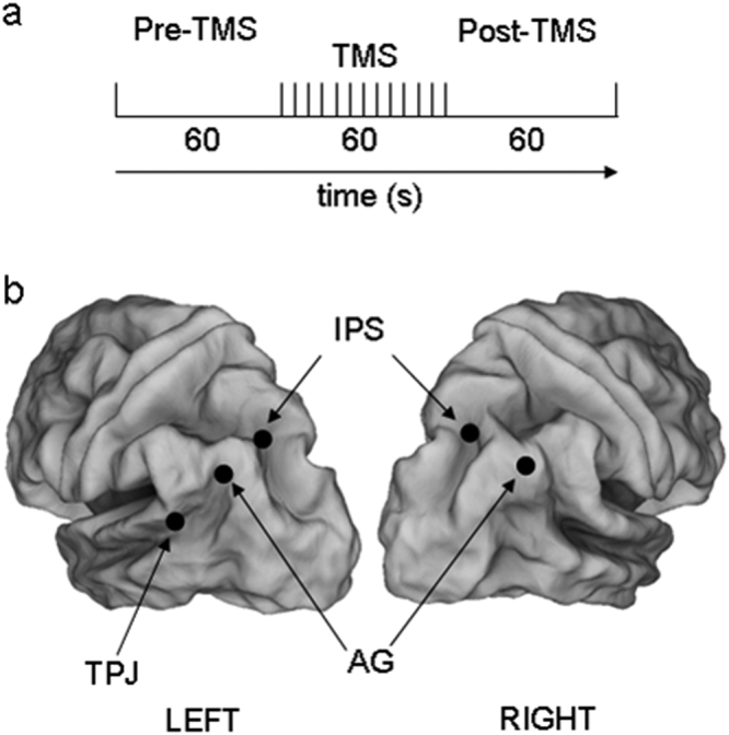
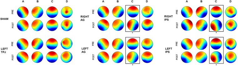
The interference effects of transcranial magnetic stimulation (TMS) on several electroencephalographic (EEG) measures in both temporal and frequency domains have been reported. We tested the hypothesis whether the offline external inhibitory interference, although focal, could result in a global reorganization of the functional brain state, as assessed by EEG microstates. In 16 healthy subjects, we inhibited five parietal areas and used a pseudo stimulation (Sham) at rest. The EEG microstates were extracted before and after each stimulation. The canonical A, B, C and D templates were found before and after all stimulation conditions. The Sham, as well as the stimulation of a ventral site did not modify any resting EEG microstates' topography. On the contrary, interfering with parietal key-nodes of both dorsal attention (DAN) and default mode networks (DMN), we observed that the microstate C clearly changes, whereas the other three topographies are not affected. These results provide the first causal evidence of a microstates modification following magnetic interference. Since the microstate C has been associated to the activity in regions belonging to the cingulo-opercular network (CON), the regional specificity of such inhibition seems to support the theory of a link between CON and both DAN and DMN at rest.
Conflict of interest statement
The authors declare that they have no competing interests.
Figures
Similar articles
-
Magnetic stimulation selectively affects pre-stimulus EEG microstates.Neuroimage. 2018 Aug 1;176:239-245. doi: 10.1016/j.neuroimage.2018.04.061. Epub 2018 Apr 30. Neuroimage. 2018. PMID: 29723638
-
rTMS affects EEG microstates dynamic during evoked activity.Cortex. 2021 May;138:302-310. doi: 10.1016/j.cortex.2021.02.014. Epub 2021 Mar 4. Cortex. 2021. PMID: 33774580
-
Resting-state connectivity in the prodromal phase of schizophrenia: insights from EEG microstates.Schizophr Res. 2014 Feb;152(2-3):513-20. doi: 10.1016/j.schres.2013.12.008. Epub 2014 Jan 2. Schizophr Res. 2014. PMID: 24389056
-
Microstates in resting-state EEG: current status and future directions.Neurosci Biobehav Rev. 2015 Feb;49:105-13. doi: 10.1016/j.neubiorev.2014.12.010. Epub 2014 Dec 17. Neurosci Biobehav Rev. 2015. PMID: 25526823 Free PMC article. Review.
-
Machine learning detects EEG microstate alterations in patients living with temporal lobe epilepsy.Seizure. 2018 Oct;61:8-13. doi: 10.1016/j.seizure.2018.07.007. Epub 2018 Jul 10. Seizure. 2018. PMID: 30044996 Review.
Cited by
-
Causal link between prefrontal cortex and EEG microstates: evidence from patients with prefrontal lesion.Front Neurosci. 2023 Dec 14;17:1306120. doi: 10.3389/fnins.2023.1306120. eCollection 2023. Front Neurosci. 2023. PMID: 38161794 Free PMC article.
-
A Quantum-Classical Model of Brain Dynamics.Entropy (Basel). 2023 Mar 30;25(4):592. doi: 10.3390/e25040592. Entropy (Basel). 2023. PMID: 37190380 Free PMC article.
-
Altering Temporal Dynamics of Sleepiness and Mood During Sleep Deprivation: Evidence from Resting-State EEG Microstates.Brain Sci. 2025 Apr 21;15(4):423. doi: 10.3390/brainsci15040423. Brain Sci. 2025. PMID: 40309897 Free PMC article.
-
Pre-stimulus EEG Microstates Correlate With Anticipatory Alpha Desynchronization.Front Hum Neurosci. 2020 May 27;14:182. doi: 10.3389/fnhum.2020.00182. eCollection 2020. Front Hum Neurosci. 2020. PMID: 32536858 Free PMC article.
-
Low-Frequency TMS Results in Condition-Related Dynamic Activation Changes of Stimulated and Contralateral Inferior Parietal Lobule.Front Hum Neurosci. 2021 Jul 23;15:684367. doi: 10.3389/fnhum.2021.684367. eCollection 2021. Front Hum Neurosci. 2021. PMID: 34366812 Free PMC article.
References
-
- Taylor PCJ, Nobre AC, Rushworth MFS. Subsecond changes in top down control exerted by human medial frontal cortex during conflict and action selection: a combined transcranial magnetic stimulation electroencephalography study. J Neurosci. 2007;27:11343–11353. doi: 10.1523/JNEUROSCI.2877-07.2007. - DOI - PMC - PubMed
MeSH terms
LinkOut - more resources
Full Text Sources
Other Literature Sources
Miscellaneous
