. 2018 Jan 19;13(1):e0191497.
doi: 10.1371/journal.pone.0191497. eCollection 2018.

Perfusion decellularization of a human limb: A novel platform for composite tissue engineering and reconstructive surgery

Affiliations

Perfusion decellularization of a human limb: A novel platform for composite tissue engineering and reconstructive surgery

Mattia Francesco Maria Gerli et al. PLoS One. .

Abstract

Muscle and fasciocutaneous flaps taken from autologous donor sites are currently the most utilized approach for trauma repair, accounting annually for 4.5 million procedures in the US alone. However, the donor tissue size is limited and the complications related to these surgical techniques lead to morbidities, often involving the donor sites. Alternatively, recent reports indicated that extracellular matrix (ECM) scaffolds boost the regenerative potential of the injured site, as shown in a small cohort of volumetric muscle loss patients. Perfusion decellularization is a bioengineering technology that allows the generation of clinical-scale ECM scaffolds with preserved complex architecture and with an intact vascular template, from a variety of donor organs and tissues. We recently reported that this technology is amenable to generate full composite tissue scaffolds from rat and non-human primate limbs. Translating this platform to human extremities could substantially benefit soft tissue and volumetric muscle loss patients providing tissue- and species-specific grafts. In this proof-of-concept study, we show the successful generation a large-scale, acellular composite tissue scaffold from a full cadaveric human upper extremity. This construct retained its morphological architecture and perfusable vascular conduits. Histological and biochemical validation confirmed the successful removal of nuclear and cellular components, and highlighted the preservation of the native extracellular matrix components. Our results indicate that perfusion decellularization can be applied to produce human composite tissue acellular scaffolds. With its preserved structure and vascular template, these biocompatible constructs, could have significant advantages over the currently implanted matrices by means of nutrient distribution, size-scalability and immunological response.


Conflict of interest statement

Competing Interests: The authors declare no conflicts of interest. HCO is founder and stockholder of IVIVA Medical Inc.; this relationship did not affect the content or conclusions reported in this manuscript. This does not alter our adherence to PLOS ONE policies on sharing data and materials.

Figures

Fig 1
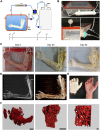
Fig 1. Perfusion decellularization of a human upper extremity.
(A) The scheme depicts the bioreactor designed to allocate and decellularize a full human limb and the detergent perfusion timeline utilized for the experiments presented in the article. (B) The panel presents highlights on the main components of the bioreactor: the perfusion chamber (top), HEPA filters, pressure sensor and UV sterilizer (bottom). (C) Images of the human arm at the beginning of the procedure, after 30 days in perfusion with SDS and at the end of the decellularization procedure. (D) Computer tomography images showing the perfusion of contrast fluid in the brachial, ulnar and radial artery and palmar arch (left), as well as in the medium size vessels of the proximal arm. (E) Macroscopic comparison of the decellularized hand with a native control. The orange 15 gauge stubs have been subsequently utilized to perform Microfil silicon casting. The red rectangle indicates the region of the Abductor Pollicis Brevis muscle explanted and utilized for the μCT scan experiments. (F) 3D reconstructions of μCT scans obtained from the Abductor Pollicis Brevis muscle depicting the preserved microvascular architecture at different magnifications (scale bars: 1 mm, 1 mm, 100 μm).
Fig 2
Fig 2. Histological characterization of the tissue composing the decellularized limb.
(A) The panel shows bright field images of the Haematoxylin and eosin staining performed on muscle, nerve, skin and vessels before and after the decellularization procedure, highlighting absence of nuclei and reduction in the eosin positive structures across the tissues (L indicates the vascular lumen; scale bar: 100 μm) (B) The panel shows bright field images of a Masson’s trichrome staining performed on muscle, nerve, skin, vessels bone and tendon, highlighting the removal of the nuclei and cellular components such as sarcomeres, axons, epidermis, smooth muscle and bone marrow (black and red) and preservation of the collagenous matrix proteins (blue; L indicates the vascular lumen; scale bar: 100 μm). (C) Phase contrast images of a Verhoeff’s elastin stain indicating the preservation of the elastic fibres (black) in section of native and decellularized skin and vessels (L indicates the vascular lumen; scale bar: 100 μm). (D) Bright field microscopy images of a Safranin O / Fast green stain performed on native and decellularized cartilage and bone tissue highlighting removal of the proteoglycans (orange), bone marrow (blue) and nuclei (black; scale bar: 100 μm).
Fig 3
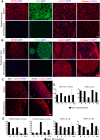
Fig 3. Molecular characterization of the different compartments of the acellular scaffold.
(A) The immunofluorescence images depict native and acellular muscle sections stained for the sarcomeric proteins Myosin (MyHC) and Alpha-Actinin (SAA) and for the extracellular matrix proteins Laminin and Collagen IV. Sections were counterstained with DAPI to confirm the removal of the cell nuclei upon decellularization (scale bar: 100 μm). (B) Immunofluorescent imaging of native and acellular peripheral nerve sections stained for the axon protein Neurofilaments (NF200), the Schwann’s cell marker S100β and for the extracellular matrix proteins Laminin and Collagen IV. Sections were counterstained with DAPI to confirm the removal of the cell nuclei upon decellularization (scale bar: 100 μm). (C) Immunofluorescent staining for Laminin on skin and vessel sections (L indicated the vessel lumen; scale bar: 100 μm) (D) Quantification of the number of DAPI-positive nuclei and residual double strand DNA concentration across native and decellularized nerve (N), muscle (M), skin (S) and vessel (V) biopsies (left). Quantification was performed using a PicoGreen fluorescent assay (measures are presented as: ng of dsDNA / mg of dry tissue). (E) The bar graphs indicate: the concentration of soluble and insoluble collagen measured via Sircol assay; the Elastin concentration measured via Fastin fluorescent assay; the GAGs concentration quantified via Blyscan fluorescent assay across three independent muscle (M), nerve (N), skin (S) and vessel (V) biopsies (measures are presented as: μg of protein / mg of dry tissue).

Similar articles

Cited by

References

    1. Guyette JP, Gilpin SE, Charest JM, Tapias LF, Ren X, Ott HC. Perfusion decellularization of whole organs. Nature protocols. 2014;9(6):1451–68. doi: 10.1038/nprot.2014.097 . - DOI - PubMed
    1. Ott HC, Matthiesen TS, Goh SK, Black LD, Kren SM, Netoff TI, et al. Perfusion-decellularized matrix: using nature's platform to engineer a bioartificial heart. Nature medicine. 2008;14(2):213–21. doi: 10.1038/nm1684 . - DOI - PubMed
    1. Ott HC, Clippinger B, Conrad C, Schuetz C, Pomerantseva I, Ikonomou L, et al. Regeneration and orthotopic transplantation of a bioartificial lung. Nature medicine. 2010;16(8):927–33. doi: 10.1038/nm.2193 . - DOI - PubMed
    1. Gilpin SE, Guyette JP, Gonzalez G, Ren X, Asara JM, Mathisen DJ, et al. Perfusion decellularization of human and porcine lungs: bringing the matrix to clinical scale. The Journal of heart and lung transplantation: the official publication of the International Society for Heart Transplantation. 2014;33(3):298–308. doi: 10.1016/j.healun.2013.10.030 . - DOI - PubMed
    1. Guyette JP, Charest JM, Mills RW, Jank BJ, Moser PT, Gilpin SE, et al. Bioengineering Human Myocardium on Native Extracellular Matrix. Circulation research. 2016;118(1):56–72. doi: 10.1161/CIRCRESAHA.115.306874 . - DOI - PMC - PubMed

Publication types