. 2018 Jan 19;14(1):e1006820.
doi: 10.1371/journal.ppat.1006820. eCollection 2018 Jan.

Activation of PI3K, Akt, and ERK during early rotavirus infection leads to V-ATPase-dependent endosomal acidification required for uncoating

Affiliations

Activation of PI3K, Akt, and ERK during early rotavirus infection leads to V-ATPase-dependent endosomal acidification required for uncoating

Mahmoud Soliman et al. PLoS Pathog. .

Abstract

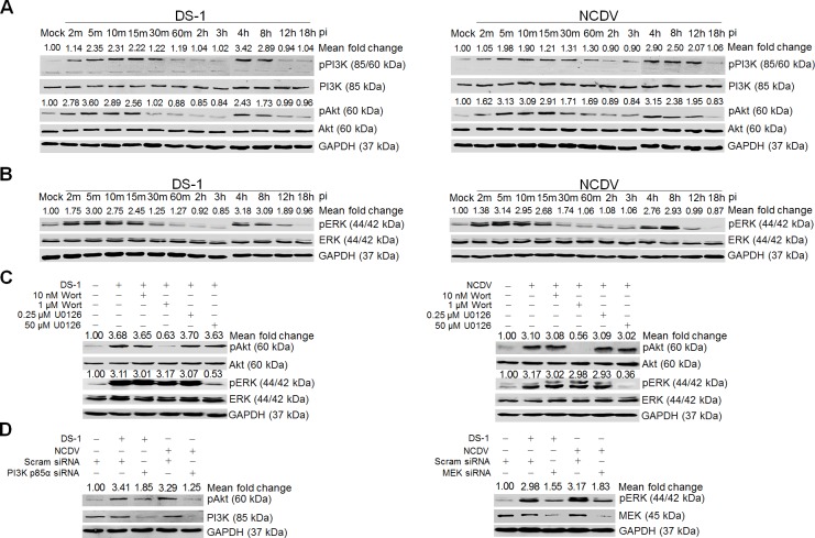
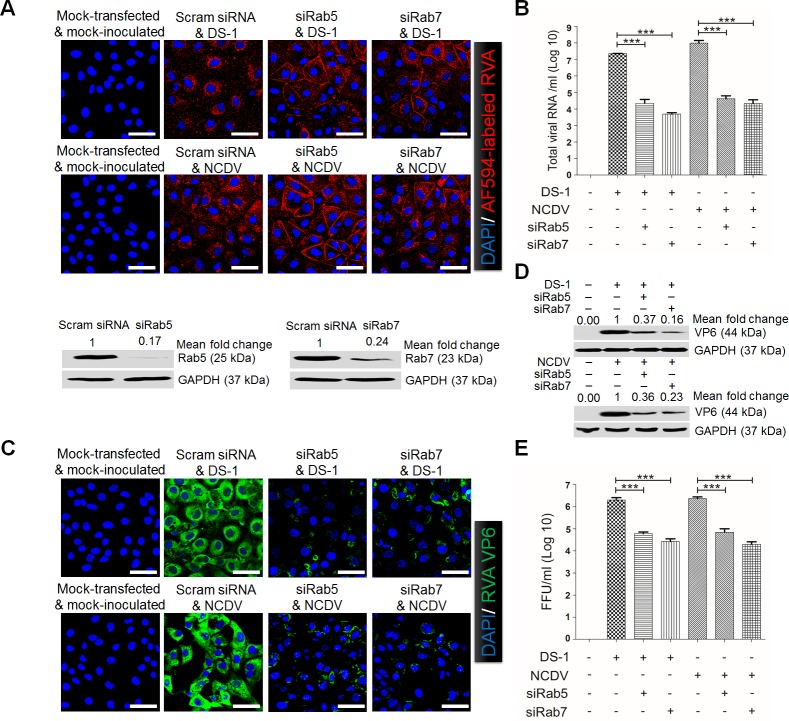
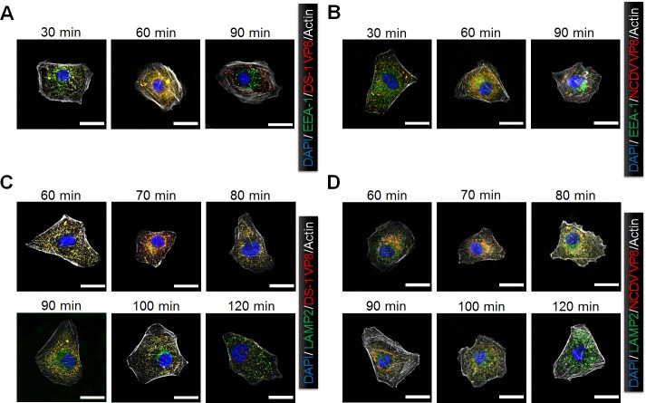
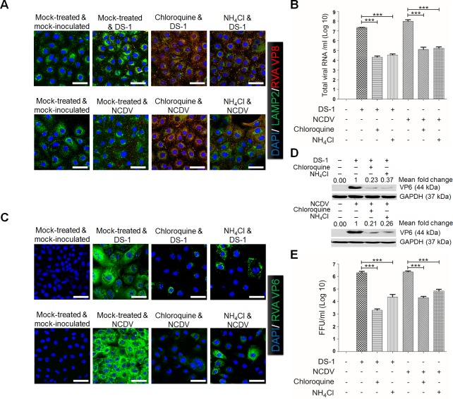
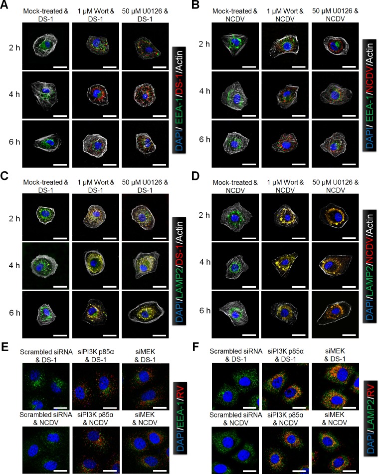
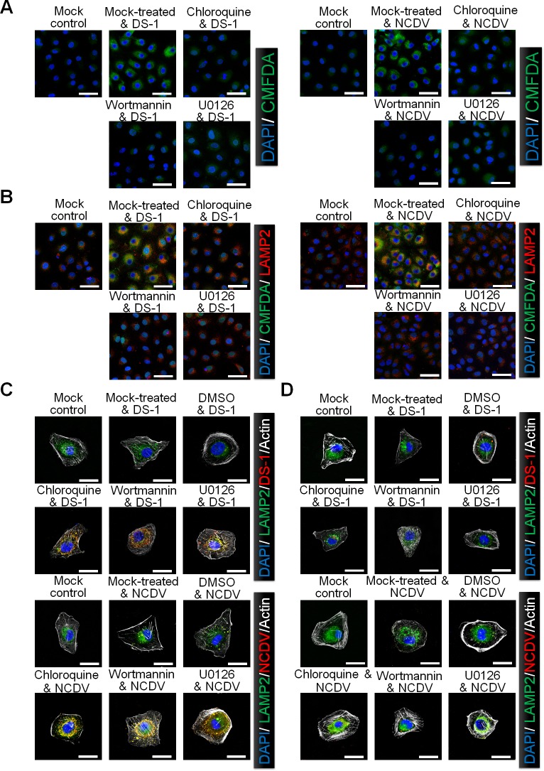
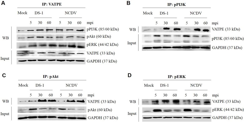
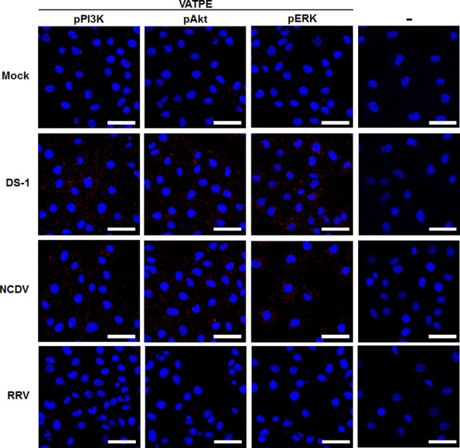
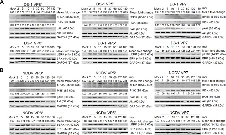
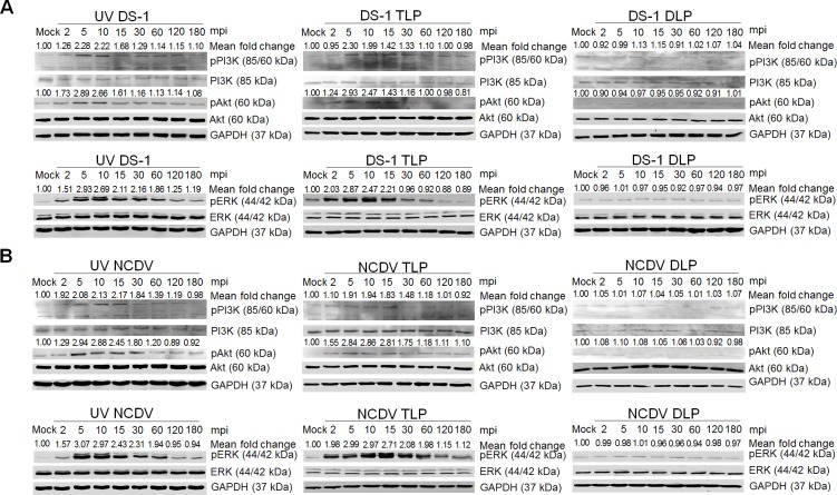
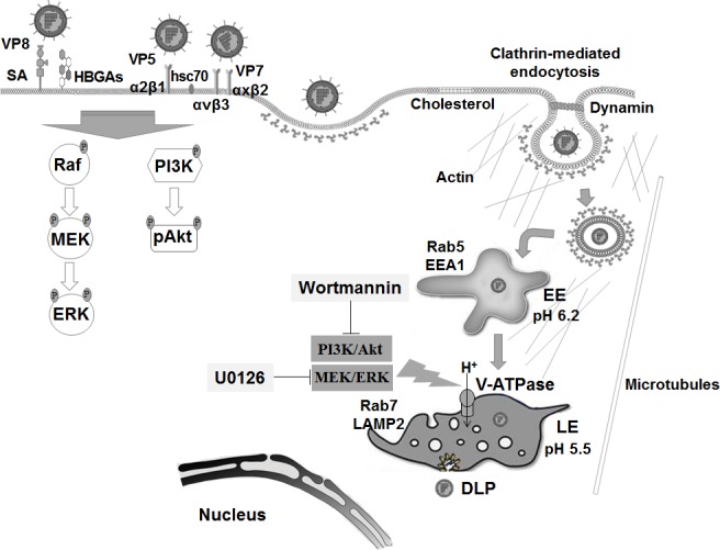
The cellular PI3K/Akt and/or MEK/ERK signaling pathways mediate the entry process or endosomal acidification during infection of many viruses. However, their roles in the early infection events of group A rotaviruses (RVAs) have remained elusive. Here, we show that late-penetration (L-P) human DS-1 and bovine NCDV RVA strains stimulate these signaling pathways very early in the infection. Inhibition of both signaling pathways significantly reduced production of viral progeny due to blockage of virus particles in the late endosome, indicating that neither of the two signaling pathways is involved in virus trafficking. However, immunoprecipitation assays using antibodies specific for pPI3K, pAkt, pERK and the subunit E of the V-ATPase co-immunoprecipitated the V-ATPase in complex with pPI3K, pAkt, and pERK. Moreover, Duolink proximity ligation assay revealed direct association of the subunit E of the V-ATPase with the molecules pPI3K, pAkt, and pERK, indicating that both signaling pathways are involved in V-ATPase-dependent endosomal acidification. Acidic replenishment of the medium restored uncoating of the RVA strains in cells pretreated with inhibitors specific for both signaling pathways, confirming the above results. Isolated components of the outer capsid proteins, expressed as VP4-VP8* and VP4-VP5* domains, and VP7, activated the PI3K/Akt and MEK/ERK pathways. Furthermore, psoralen-UV-inactivated RVA and CsCl-purified RVA triple-layered particles triggered activation of the PI3K/Akt and MEK/ERK pathways, confirming the above results. Our data demonstrate that multistep binding of outer capsid proteins of L-P RVA strains with cell surface receptors phosphorylates PI3K, Akt, and ERK, which in turn directly interact with the subunit E of the V-ATPase to acidify the late endosome for uncoating of RVAs. This study provides a better understanding of the RVA-host interaction during viral uncoating, which is of importance for the development of strategies aiming at controlling or preventing RVA infections.


Conflict of interest statement

The authors have declared that no competing interests exist

Figures

Fig 1
Fig 1. RVA-induced early activation of PI3K, Akt, and ERK signaling molecules in MA104 cells.
The human RVA DS-1 (A) and bovine RVA NCDV (B) strains (MOI = 10 FFU/cell) were inoculated into serum-starved MA104 cells. Cells were harvested at the indicated time points. Cell lysates were subjected to Western blot analysis to check the expression levels of phosphorylated PI3K (pPI3K), PI3K, pAkt, Akt, pERK, and ERK using the relevant antibody. GAPDH was used as a loading control. (C) MA104 cells were mock-treated or pretreated with wortmannin or U0126 at the indicated doses for 1 h at 37°C, followed by infection with strains DS-1 or NCDV. Cell lysates were harvested at 5 mpi and the expression levels of pAkt, Akt, pERK, and ERK were evaluated by Western blot analysis. GAPDH was used as a loading control. (D) MA104 cells were transfected with scrambled siRNA or siRNAs specific for PI3K p85α or MEK and then infected with the DS-1 and NCDV strains (MOI = 10 FFU/cell). Cell lysates were subjected to Western blot analysis to check the expression levels of pAkt, Akt, pERK, and ERK using the appropriate antibody. GAPDH was used as a loading control. The intensity of pPI3K, pAkt, and pERK relative to GAPDH was determined by densitometric analysis and is indicated above each lane.
Fig 2
Fig 2. Rotavirus entry and infection depend on Rab5 and Rab7.
(A) MA104 cells were transfected with either scrambled siRNA or siRNAs against Rab5 or Rab7. Afterwards, cells were exposed to Alexa 594-labeled DS-1 (approximately 595 particles/cell) or NCDV (approximately 790 particles/cell) for 30 min at 4°C. Unbound virus was washed off and the cells were shifted to 37°C for 90 min. Cells were then fixed and processed for confocal microscopy. Cells treated in parallel were analyzed by Western blot analysis to ensure effective knockdown of protein levels. (B-E) MA104 cells transfected with siRNAs against Rab5 or Rab7 were infected with the DS-1 and NCDV strains (MOI = 10 FFU/cell). The total viral RNA (B), antigen-positive cells (using anti-RVA VP6 Mab) (C), and VP6 protein (D) were determined by real-time RT-PCR, immunofluorescence, and Western blot analyses, respectively. GAPDH was used as a loading control. The intensity of pPI3K, pAkt, and pERK relative to GAPDH was determined by densitometric analysis and is indicated above each lane. (E) The virus titer was determined by cell culture immunofluorescence assay using cell lysates produced by 3 cycles of freezing and thawing, and is expressed as FFU. All experiments were performed in triplicate and panels A and C show a representative set of results. Data are presented as means ± standard error of the mean from three independent experiments. Differences were evaluated using the One-Way ANOVA. *p<0.05; **p<0.001; ***p<0.0001. The scale bars in panels A and C correspond to 20 μm.
Fig 3
Fig 3. Rotavirus transits from the early to the late endosome during the internalization process.
MA104 cells were incubated with the DS-1 (A and C) and NCDV (B and D) strains for the indicated times at 37°C. The cells were then fixed, permeabilized, and processed for immunofluorescence assay. Virus particles were visualized using anti-VP8* primary antibody and secondary antibody labeled with Alexa Fluor 594. Endosomal markers were detected with specific antibodies against EEA1 (A and B) and LAMP2 (C and D), and the corresponding Alexa Fluor 647-conjugated secondary antibodies. Actin cytoskeleton was stained with Alexa Fluor 488-labeled phalloidin. All experiments were performed in triplicate and panels A to D show a representative set of results. The scale bars in each panel correspond to 5 μm.
Fig 4
Fig 4. Rotavirus requires endosomal acidification for efficient internalization.
(A) Mock-, 100 μM chloroquine-, or 100 mM ammonium chloride (NH4Cl)-pretreated MA104 cells were incubated with the DS-1 or NCDV strains for 30 min at 4°C and then shifted to 37°C for 2 h. The cells were then fixed and processed for immunofluorescence using anti-VP8* primary antibody in parallel with anti-LAMP2 antibody. (B-D) RVA DS-1 and NCDV strains (MOI = 10 FFU/cell) were inoculated into mock- or chloroquine- or NH4Cl-pretreated MA104 cells. The total viral RNA (B), antigen-positive cells (using anti-RVA VP6 Mab) (C), and VP6 protein (D) were determined by real-time RT-PCR, immunofluorescence, and Western blot analyses, respectively. GAPDH was used as a loading control. The intensity of pPI3K, pAkt, and pERK relative to GAPDH was determined by densitometric analysis and is indicated above each lane. (E) The virus titer was determined by immunofluorescence assay using cell lysates produced after 3 cycles of freezing and thawing, and it is expressed as FFU. All experiments were performed in triplicate and panels A and C show a representative set of results. Data are presented as means ± standard error of the mean from three independent experiments. Differences were evaluated using the One-Way ANOVA. *p<0.05; **p<0.001; ***p<0.0001. The scale bars in panels A and C correspond to 20 μm.
Fig 5
Fig 5. Involvement of PI3K/Akt and MEK/ERK signaling pathways in RVA uncoating.
(A-D) MA104 cells were pretreated with or without wortmannin or U0126 for 1 h at 37°C and then infected with the DS-1 or NCDV strains (MOI = 10 FFU/cell) for the indicated time. After fixation and permeabilization, the cells were prepared for confocal microscopy using anti-VP8* antibody, anti-EEA1 antibody (A and B), and anti-LAMP2 antibody (C and D), and the relevant secondary antibodies. Actin cytoskeleton was stained with AF488-labeled phalloidin. (E and F) MA104 cells were transfected with scrambled siRNA or siRNAs specific for PI3K p85α or MEK, and then infected with the DS-1 and NCDV strains (MOI = 10 FFU/cell). After fixation and permeabilization, the cells were prepared for confocal microscopy using anti-VP8* antibody, anti-EEA1 antibody (E), and anti-LAMP2 antibody (F), and the relevant secondary antibodies. Representative images are shown. The scale bars correspond to 5 μm.
Fig 6
Fig 6. Involvement of PI3K/Akt and MEK/ERK signaling pathways in late endosomal acidification.
MA104 cells were pretreated with or without chloroquine, wortmannin or U0126 for 1 h at 37°C, and subsequently infected with the strains DS-1 or NCDV (MOI = 10 FFU/cell) for 30 min at 37°C. Cells were then incubated for 30 min with CMFDA (10 μM) to visualize acidification of intracellular compartments followed by 30 min in serum-free media (A), or incubated with CMFDA followed by anti-LAMP2 antibody to check colocalization by confocal microscopy (B). (C and D) After chemical pretreatment and virus infection, cells were incubated with neutral (pH 7.2) (C) or acidic (pH 5) (D) buffers for 5 min at 37°C. The cells were then washed and incubated for further 2 h at 37°C and prepared for confocal microscopy to check colocalization of RVA VP8* with LAMP2. Representative images are shown. The scale bars correspond to 20 μm (A and B) and 5 μm (C and D).
Fig 7
Fig 7. Direct interaction of pPI3K, pAkt, and pERK with subunit E of the V-ATPase V1 domain for endosomal acidification.
(A-D) Serum-starved MA104 cells were inoculated with RVA DS-1 or NCDV (MOI = 10 FFU/cell) for the indicating time. Subsequently, the cell lysates were immunoprecipitated using antibodies specific for the V1 subunit E of the V-ATPase (A), pPI3K (B), pAkt (C), and pERK (D). The co-immunoprecipitated products were analyzed by Western blot analysis to detect pPI3K, pAkt, pERK, and the V1 subunit E using the relevant antibodies. GAPDH was used as a loading control.
Fig 8
Fig 8. Direct interaction of pPI3K, pAkt, and pERK with the subunit E of the V-ATPase V1 domain determined by Duolink proximity assay.
Serum-starved MA104 cells were either mock-inoculated or inoculated with the RVA strains DS-1, NCDV, and RRV (MOI = 10 FFU/cell). Subsequently, the cells were fixed, permeabilized and incubated with or without the primary antibodies (goat anti-V-ATPase E subunit and rabbit anti-pPI3K, pAkt, and pERK antibodies) overnight at 4°C. The Duolink PLA was performed as described in the Materials and Methods section and the signals are represented as red dots. Representative images are shown. The scale bars correspond to 20 μm.
Fig 9
Fig 9. Activation of the PI3K/Akt and MEK/ERK signaling pathways by RVA outer capsid surface proteins.
Serum-starved MA104 cells were incubated with recombinant GST-fused VP8* or his-tagged VP5* or VP7 proteins of the strains DS-1 (A) and NCDV (B) at 10 μg/ml for the indicated time points. The cell lysates were subjected to Western blot analysis for the detection of pPI3K, pAkt, pERK, PI3K, Akt, and ERK using the relevant antibodies. GAPDH was used as a loading control. The intensity of pPI3K, pAkt, and pERK relative to GAPDH was determined by densitometric analysis and is indicated above each lane.
Fig 10
Fig 10. PI3K/Akt and MEK/ERK signaling pathways were activated by psoralen-UV-inactivated RVA and CsCl-purified RVA TLPs but not by intracellularly transfected RVA DLPs at the immediate early stage.
(A-D) MA104 cells were independently infected with psoralen-UV-inactivated DS-1 and NCDV strains, or CsCl-purified RVA TLPs of DS-1 and NCDV strains, or transfected with RVA DLPs of DS-1 and NCDV strains, in a time dependent manner. Cell lysates were collected for Western blot analysis for the detection of PI3K, pPI3K, Akt, pAkt, ERK, and pERK. GAPDH was also analyzed and used as a loading control. The intensity of pPI3K, pAkt, and pERK relative to GAPDH was determined by densitometric analysis and is indicated above each lane.
Fig 11
Fig 11. Schematic diagram for endosomal acidification by RVA-induced early activation of PI3K, Akt, and ERK signaling molecules.
RVAs enter the cells by sequential multistep binding of outer capsid proteins (VP4-VP8*, VP4-VP5*, and VP7) to their cognate host cellular receptors and coreceptors, represented by SA, HBGAs, integrins, and Hsc70 protein, depending on the virus strain. Subsequently, RVA particles are internalized by clathrin-dependent or -independent endocytic pathways, depending on the virus strain. All RVA strains move to EEs expressing Rab5 and EEA1. In addition, some RVA strains travel to LEs (L-P viruses). Multistep binding of RVA outer capsid proteins (VP4-VP8*, VP4-VP5*, and VP7) to host cell surface receptors and coreceptors activates the PI3K/Akt and MEK/ERK signaling pathways. The phosphorylated signaling molecules, pPI3K, pAkt, and pERK, interact directly with the subunit E of the V1 domain of the V-ATPase to produce a proton gradient by ATP hydrolysis to acidify the LE for uncoating of RVAs.

References

    1. Estes MK, Greenberg HB. Rotaviruses In: Fields Virology, 6th ed., eds Knipe DM, Howley PM et al. (Lippincott Williams & Wilkins, Philadelphia: ); 2013. pp. 1917–1974.
    1. Desselberger U. Rotaviruses. Virus Res. 2014; 190:75–96. doi: 10.1016/j.virusres.2014.06.016 . - DOI - PubMed
    1. Walker CL, Rudan I, Liu L, Nair H, Theodoratou E, Bhutta ZA, et al. Global burden of childhood pneumonia and diarrhea. Lancet. 2013; 381(9875):1405–1416. doi: 10.1016/S0140-6736(13)60222-6 . - DOI - PMC - PubMed
    1. Arias CF, Silva-Ayala D, López S. Rotavirus entry: a deep journey into the cell with several exits. J Virol. 2015; 89(2):890–893. doi: 10.1128/JVI.01787-14 . - DOI - PMC - PubMed
    1. López S, Arias CF. Multistep entry of rotavirus into cells: a Versaillesque dance. Trends Microbiol. 2004; 12(6):271–278. doi: 10.1016/j.tim.2004.04.003 . - DOI - PubMed

Publication types

MeSH terms

Substances