Review
. 2018 Aug;10(4):1187-1199.
doi: 10.1007/s12551-017-0395-y. Epub 2018 Jan 20.

The multiple roles of titin in muscle contraction and force production

Affiliations
Review

The multiple roles of titin in muscle contraction and force production

Walter Herzog. Biophys Rev. 2018 Aug.

Abstract

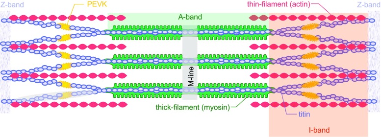
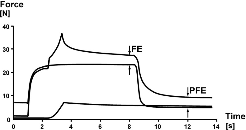
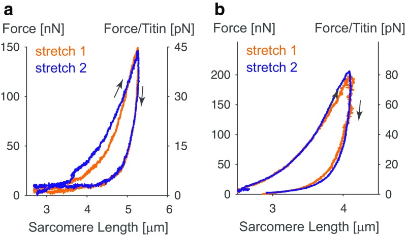
Titin is a filamentous protein spanning the half-sarcomere, with spring-like properties in the I-band region. Various structural, signaling, and mechanical functions have been associated with titin, but not all of these are fully elucidated and accepted in the scientific community. Here, I discuss the primary mechanical functions of titin, including its accepted role in passive force production, stabilization of half-sarcomeres and sarcomeres, and its controversial contribution to residual force enhancement, passive force enhancement, energetics, and work production in shortening muscle. Finally, I provide evidence that titin is a molecular spring whose stiffness changes with muscle activation and actin-myosin-based force production, suggesting a novel model of force production that, aside from actin and myosin, includes titin as a "third contractile" filament. Using this three-filament model of sarcomeres, the stability of (half-) sarcomeres, passive force enhancement, residual force enhancement, and the decrease in metabolic energy during and following eccentric contractions can be explained readily.

Keywords: Active/passive force regulation; Cross-bridge theory; Force production; Mechanical functions; Mechanisms of muscle contraction; Molecular spring; Muscle energetics; Muscle shortening; Three filament sarcomere model; Titin.


Conflict of interest statement

Walter Herzog declares that he has no conflict of interest.

Ethics approvals for all experiments described in this study were obtained by the Life Sciences and Animal Research Ethics Commitee of the University of Calgary.

Figures

Fig. 1
Fig. 1
Schematic two-dimensional illustration of a sarcomere bordered by Z-bands at either end. Thick, myosin-based filaments are in the center of the sarcomere (green), thin, actin-based filaments insert into the Z-band at either end of the sarcomere (red), and titin filaments (blue) run from the M-line in the middle of the sarcomere to the Z-band. Adapted from Granzier and Labeit (2007) with permission
Fig. 2
Fig. 2
Rabbit psoas myofibril comprised of six sarcomeres that is stretched while activated from an average sarcomere length of about 2.4 μm to about 3.0 μm. After active stretching, all individual sarcomeres are on the descending limb of the force–length relationship, but there is no apparent overstretching or popping (quick sarcomere elongations beyond actin–myosin filament overlap: 3.9 μm) as has been proposed by proponents of the sarcomere length non-uniformity theory. Rather, sarcomeres seem to remain at an essentially constant length following the active stretch
Fig. 3
Fig. 3
Residual force enhancement observed in whole muscle (cat soleus; a), single myofibrils (rabbit psoas; b), and single sarcomeres (rabbit psoas; c). Note the increase in force enhancement (FE; a) with increasing stretch magnitude, and the increased passive force [passive force enhancement (PFE)] following deactivation of the muscles after an active stretch (a, b). Note also the vast FE in a single sarcomere (c) and the substantially greater force after active stretching compared to the isometric, steady-state force prior to stretching which occurred at the plateau of the force–length relationship (2.4 μm)
Fig. 4
Fig. 4
Steady-state isometric forces obtained in single, mechanically isolated sarcomeres (rabbit psoas) at sarcomere lengths of 2.4 μm [optimal length = 100% force (filled brown circle)] and 3.4 μm [approximately 50% of maximal isometric force at the plateau length (filled blue diamonds and filled black square = mean force). Also shown are the isometric steady-state forces of these isolated sarcomeres following a stretch from 2.4 to 3.4 μm (filled green triangles and filled black circle). FE Mean force enhancement observed in these sarcomeres, OFE the mean force above the maximal, isometric plateau forces for these sarcomeres. Note the enormous force enhancement and the consistently greater forces in the enhanced state compared to the plateau force. Adapted from Leonard et al. (2010) with permission
Fig. 5
Fig. 5
Force–time histories of cat soleus muscle stretched passively (lowest trace at 6 s), stretched actively (highest trace at 6 s), and activated isometrically at the final stretch length (middle trace at 6 s). Note the increased passive force following muscle deactivation (at 12 s) for the actively stretched muscle, compared to the passively stretched muscle and the muscle activated isometrically at the final stretch length. The increase in passive force following active muscle stretching (here seen at 12 s) was termed passive force enhancement (PFE). Adapted from Herzog and Leonard (2002) with permission
Fig. 6
Fig. 6
Unfolding force of the first five (out of 8 identical) cardiac I27 immunoglobulin (Ig) domains of titin. Note that unfolding of the I27 Ig domains in the absence of calcium (Control) required about 20% less force than in the presence of calcium (Calcium). Adapted from DuVall et al. (2013) with permission
Fig. 7
Fig. 7
Passive (a) and active (b) stretching of proximal titin segments labeled using an antibody [F146 that binds to the PEVK region (diamond symbols)] region that allows for measurements of proximal and distal titin segment elongations during passive and active stretching of single rabbit psoas myofibrils. Figures on the left show elongation of the half-sarcomere [top traces (circular symbols) using an M-line label) and elongations of the proximal titin segment (bottom traces: from X-band to F146 label) for two representative sarcomeres from two different myofibrils. Note in a (passive stretching) that the two proximal titin segments elongate continuously with half-sarcomere elongations, reaching final lengths of approximately 0.95 μm (at a sarcomere length of 4.0 μm) and about 0.6 μm (at a sarcomere length of about 3.5 μm). In contrast, when the myofibrils are stretched while activated, the proximal segments elongate similarly to the elongations observed in the passive condition, but then stop elongating and remain substantially shorter than in the passive case (i.e., with a length of about 0.6 and 0.35 μm, respectively). The panels on the right illustrate schematically what we believe might be happening. In the passive stretch (a), the proximal and distal titin segments elongate in accordance with their stiffness properties. In the active stretch (b), titin is thought to attach to actin at some point, thereby shortening titin’s free spring length, increasing its stiffness, eliminating elongation of proximal titin, and increasing titin-based force
Fig. 8
Fig. 8
Stress (force/cross-sectional area) versus sarcomere length relationship for single rabbit psoas myofibrils stretched from an average sarcomere length of 2.0 μm to 6.0 μm. Myofibrils were stretched passively (Passive), actively (Active), actively from an average sarcomere length of 3.4 μm (Half-Force), and after deletion of titin (No Titin). Actin myosin filament overlap is lost at an average sarcomere length of about 4.0 μm (indicated by the shaded area). According to the cross-bridge theory, one would expect the forces beyond actin myosin filament overlap (non-shaded area) to be purely passive and the same for all conditions with intact titin filaments. However, the forces in the actively stretched myofibrils were substantially greater than those for the passively stretched myofibrils in the area where myofilament overlap was lost. Deactivation of selected myofibrils at an average sarcomere length of 5.0 μm did not result in a loss of force (results not shown), indicating that there was no remnant cross-bridge-based force at these lengths. Elimination of titin from the myofibrils abolished all passive and all active force in myofibrils, indicating that titin is not only an essential protein for passive force production but is absolutely essential for active force transmission from the cross-bridges to the Z-bands and for centering the myosin filaments in the sarcomere. Adapted from Leonard and Herzog (2010) with permission
Fig. 9
Fig. 9
Examples of two separate rabbit psoas myofibrils that were repeatedly stretched to sarcomere lengths beyond actin myosin filament overlap. Note that in both cases repeat stretches did not result in a decrease in peak force or loading energy, thus indicating that even stretching to lengths up to 5.0 μm did not result in permanent damage and loss of force. a Myofibril stretched to an average sarcomere length of approximately 5.2 μm (stretch 1), then shortened and rested for 10 min at an average sarcomere length of 2.6 μm and re-stretched (stretch 2). Two sets of three stretch-shortening cycles were performed and the third stretch of the first set (stretch 1), and the first stretch of the second set (stretch 2) are shown b Myofibril stretched to an average sarcomere length of approximately 4.2 μm, then shortened and rested for 10 min at an average sarcomere length of approximately 1.8 μm. Two sets of three stretch-shortening cycles were performed (with a 10-min rest in between) and the first cycles (stretch 1) of the first and second set (stretch 2) are shown. Adapted from Herzog et al. (2014) with permission

References

    1. Abbott BC, Aubert XM. The force exerted by active striated muscle during and after change of length. J Physiol. 1952;117:77–86. - PMC - PubMed
    1. Allinger TL, Epstein M, Herzog W. Stability of muscle fibers on the descending limb of the force- length relation. A theoretical consideration. J Biomech. 1996;29:627–633. - PubMed
    1. Anderson BR, Bogomolovas J, Labeit S, Granzier HLM. The effects of PKCalpha phosphorylation on the extensibility of titin’s PEVK element. J Struct Biol. 2010;170:270–277. - PMC - PubMed
    1. Astier C, Raynaud F, Lebart MC, Roustan C, Benyamin Y. Binding of a native titin fragment to actin is regulated by PIP2. FEBS Lett. 1998;429:95–98. - PubMed
    1. Bartoo ML, Linke WA, Pollack GH. Basis of passive tension and stiffness in isolated rabbit myofibrils. Am J Phys. 1997;273:C266–C276. - PubMed

LinkOut - more resources