. 2018 Jan 5:8:1108.
doi: 10.3389/fphys.2017.01108. eCollection 2017.

Deficiency of Carbonic Anhydrase II Results in a Urinary Concentrating Defect

Affiliations

Deficiency of Carbonic Anhydrase II Results in a Urinary Concentrating Defect

Devishree Krishnan et al. Front Physiol. .

Abstract

Carbonic anhydrase II (CAII) is expressed along the nephron where it interacts with a number of transport proteins augmenting their activity. Aquaporin-1 (AQP1) interacts with CAII to increase water flux through the water channel. Both CAII and aquaporin-1 are expressed in the thin descending limb (TDL); however, the physiological role of a CAII-AQP1 interaction in this nephron segment is not known. To determine if CAII was required for urinary concentration, we studied water handling in CAII-deficient mice. CAII-deficient mice demonstrate polyuria and polydipsia as well as an alkaline urine and bicarbonaturia, consistent with a type III renal tubular acidosis. Natriuresis and hypercalciuria cause polyuria, however, CAII-deficient mice did not have increased urinary sodium nor calcium excretion. Further examination revealed dilute urine in the CAII-deficient mice. Urinary concentration remained reduced in CAII-deficient mice relative to wild-type animals even after water deprivation. The renal expression and localization by light microscopy of NKCC2 and aquaporin-2 was not altered. However, CAII-deficient mice had increased renal AQP1 expression. CAII associates with and increases water flux through aquaporin-1. Water flux through aquaporin-1 in the TDL of the loop of Henle is essential to the concentration of urine, as this is required to generate a concentrated medullary interstitium. We therefore measured cortical and medullary interstitial concentration in wild-type and CAII-deficient mice. Mice lacking CAII had equivalent cortical interstitial osmolarity to wild-type mice: however, they had reduced medullary interstitial osmolarity. We propose therefore that reduced water flux through aquaporin-1 in the TDL in the absence of CAII prevents the generation of a maximally concentrated medullary interstitium. This, in turn, limits urinary concentration in CAII deficient mice.

Keywords: aquaporin-1; carbonic anhydrase II; metabolic; polyuria; urine concentration.


Figures

Figure 1
Figure 1
CAII-deficient mice are polydipsic and polyuric. (A) Weight (B) Chow consumed (C) Urine volume in 24 h and (D) Water consumed in wild-type (WT) and CAII deficient mice (CAII def). *Represents p < 0.05.
Figure 2
Figure 2
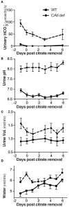
CAII-deficient mice remain polyuric when not fed alkali. (A) Urine bicarbonate excretion, (B) Urine pH, (C) Urine volume and (D) Water drank in wild-type and CAII-deficient mice before and for 6 days after removal of citrate from the drinking water.
Figure 3
Figure 3
CAII-deficient mice display a dilute urine. (A) Urinary sodium excretion, (B) Urinary creatinine excretion, (C) Urinary calcium excretion and (D) Urine osmolarity in wild-type and CAII-deficient mice. *Represents p < 0.05.
Figure 4
Figure 4
CAII-deficient mice have a urine concentration defect. (A) Weight and (B) Urine osmolarity post removal of water from the metabolic cage in wild-type and CAII-deficient mice. (C) Urine osmoarity before and after ddAVP administration to CAII-deficient mice only. *Represents p < 0.05.
Figure 5
Figure 5
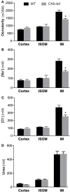
CAII-deficient mice display a dilute medullary interstitium. Renal cortical (Cortex), inner stripe of the outer medulla (ISOM) and inner medulla (IM) (A) Osmolarity, (B) Sodium concentration, (C) Chloride concentration and (D) Urea concentration. *Represents p < 0.05.
Figure 6
Figure 6
CAII-deficient mice have altered Aquaporin-1 expression. (A) Representative immunoblots and (B) Protein quantification listed, all samples were normalized to beta actin and the results presented as a % of wild-type. (C) mRNA quantification, *Represents p < 0.05.
Figure 7
Figure 7
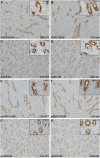
No alterations in Aquaporin-2 localization in CAII deficient mice. Renal sections from wild-type (A,C,E,G) or CAII-deficient (B,D,F,H) immunostained for aquaporin-2 (AQP2) A-D or serine 256 phosphorylated aquaporin-2 (pAQP2). Representative images from the inner stripe of the outer medulla (ISOM) (A,B,E,F) and from the inner medulla (IM) (C,D,G,H) are displayed. The scale bar represents 20 μM on the low magnification image and 50 μM on the higher magnification inset image.
Figure 8
Figure 8
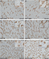
Neither NKCC2 nor aquaporin-1 distribution is altered in CAII deficient mice. Renal sections from wild-type (A,C,E) or CAII-deficient mice (B,D,F) immunostained for aquaporin-1 (AQP1) (A–D) or NKCC2 (E,F). Representative images from the inner stripe of the outer medulla (ISOM) (A,B,E,F) and from the inner medulla (C,D) are displayed. The scale bar represents 20 μM on the low magnification image and 50 μM on the higher magnification inset image.

References

    1. Alexander R. T., Beggs M. R., Zamani R., Marcussen N., Frische S., Dimke H. (2015). Ultrastructural and immunohistochemical localization of plasma membrane Ca2+-ATPase 4 in Ca2+-transporting epithelia. Am. J. Physiol. Renal Physiol. 309, F604–F616. 10.1152/ajprenal.00651.2014 - DOI - PubMed
    1. Becker H. M., Deitmer J. W. (2007). Carbonic anhydrase II increases the activity of the human electrogenic Na+/HCO3- cotransporter. J. Biol. Chem. 282, 13508–13521. 10.1074/jbc.M700066200 - DOI - PubMed
    1. Beggs M. R., Appel I., Svenningsen P., Skjødt K., Alexander R. T., Dimke H. (2017). Expression of transcellular and paracellular calcium and magnesium transport proteins in renal and intestinal epithelia during lactation. Am. J. Physiol. Renal Physiol. 313, F629–F640. 10.1152/ajprenal.00680.2016 - DOI - PubMed
    1. Bouley R., Palomino Z., Tang S. S., Nunes P., Kobori H., Lu H. A., et al. . (2009). Angiotensin II and hypertonicity modulate proximal tubular aquaporin 1 expression. Am. J. Physiol. Renal Physiol. 297, F1575–F1586. 10.1152/ajprenal.90762.2008 - DOI - PMC - PubMed
    1. Brown B. F., Quon A., Dyck J. R., Casey J. R. (2012). Carbonic anhydrase II promotes cardiomyocyte hypertrophy. Can. J. Physiol. Pharmacol. 90, 1599–1610. 10.1139/y2012-142 - DOI - PubMed