

Electroencephalogram-Electromyography Coupling Analysis in Stroke Based on Symbolic Transfer Entropy
- PMID: 29354091
- PMCID: PMC5758532
- DOI: 10.3389/fneur.2017.00716
Electroencephalogram-Electromyography Coupling Analysis in Stroke Based on Symbolic Transfer Entropy
Abstract
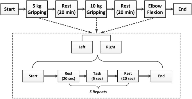
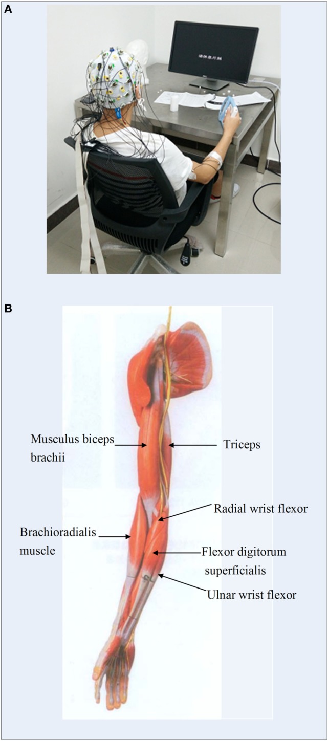
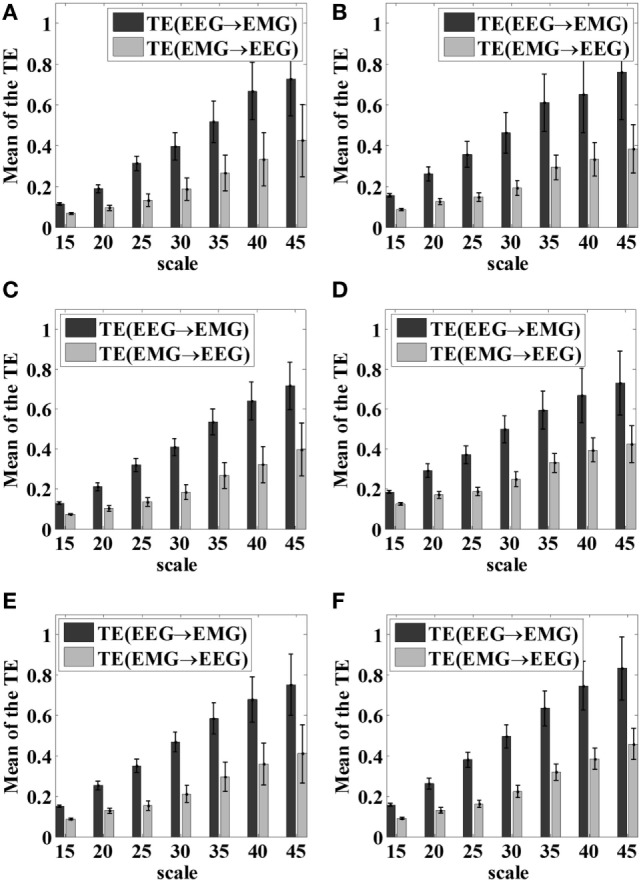
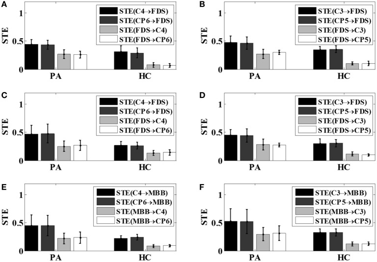
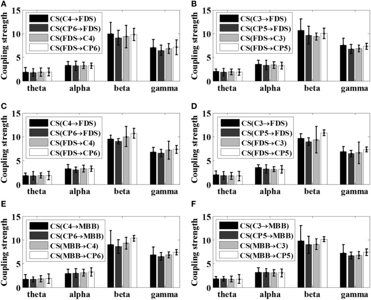
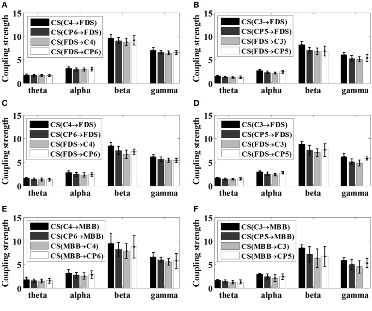
The coupling strength between electroencephalogram (EEG) and electromyography (EMG) signals during motion control reflects the interaction between the cerebral motor cortex and muscles. Therefore, neuromuscular coupling characterization is instructive in assessing motor function. In this study, to overcome the limitation of losing the characteristics of signals in conventional time series symbolization methods, a variable scale symbolic transfer entropy (VS-STE) analysis approach was proposed for corticomuscular coupling evaluation. Post-stroke patients (n = 5) and healthy volunteers (n = 7) were recruited and participated in various tasks (left and right hand gripping, elbow bending). The proposed VS-STE was employed to evaluate the corticomuscular coupling strength between the EEG signal measured from the motor cortex and EMG signal measured from the upper limb in both the time-domain and frequency-domain. Results showed a greater strength of the bi-directional (EEG-to-EMG and EMG-to-EEG) VS-STE in post-stroke patients compared to healthy controls. In addition, the strongest EEG-EMG coupling strength was observed in the beta frequency band (15-35 Hz) during the upper limb movement. The predefined coupling strength of EMG-to-EEG in the affected side of the patient was larger than that of EEG-to-EMG. In conclusion, the results suggested that the corticomuscular coupling is bi-directional, and the proposed VS-STE can be used to quantitatively characterize the non-linear synchronization characteristics and information interaction between the primary motor cortex and muscles.
Keywords: corticomuscular coupling; electroencephalogram; electromyography; stroke; symbolic transfer entropy.
Figures
References
-
- Liang TJ, Long YB. Modified constraint-induced movement therapy on lower extremity dyskinesia of stroke patients. Chin J Rehabil (2011) 26(5):339–41. 10.3870/zgkf.2011.05.006 - DOI
-
- Conway BA, Halliday DM, Shahani U, Maas P, Weir AI, Rosenberg JR, et al. Common frequency components identified from correlations between magnetic recordings of cortical activity and the electromyogram in man. J Physiol (1995) 483:35–69.
LinkOut - more resources
Full Text Sources
Other Literature Sources
Research Materials