Review
. 2018 Jan 4:8:2629.
doi: 10.3389/fmicb.2017.02629. eCollection 2017.

Insights into Structural and Mechanistic Features of Viral IRES Elements

Affiliations
Review

Insights into Structural and Mechanistic Features of Viral IRES Elements

Encarnacion Martinez-Salas et al. Front Microbiol. .

Abstract

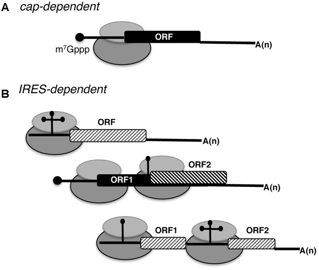
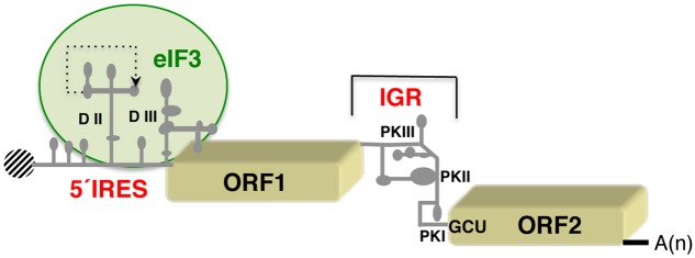
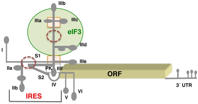
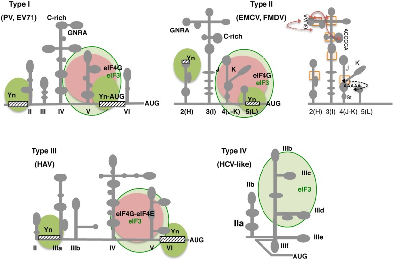
Internal ribosome entry site (IRES) elements are cis-acting RNA regions that promote internal initiation of protein synthesis using cap-independent mechanisms. However, distinct types of IRES elements present in the genome of various RNA viruses perform the same function despite lacking conservation of sequence and secondary RNA structure. Likewise, IRES elements differ in host factor requirement to recruit the ribosomal subunits. In spite of this diversity, evolutionarily conserved motifs in each family of RNA viruses preserve sequences impacting on RNA structure and RNA-protein interactions important for IRES activity. Indeed, IRES elements adopting remarkable different structural organizations contain RNA structural motifs that play an essential role in recruiting ribosomes, initiation factors and/or RNA-binding proteins using different mechanisms. Therefore, given that a universal IRES motif remains elusive, it is critical to understand how diverse structural motifs deliver functions relevant for IRES activity. This will be useful for understanding the molecular mechanisms beyond cap-independent translation, as well as the evolutionary history of these regulatory elements. Moreover, it could improve the accuracy to predict IRES-like motifs hidden in genome sequences. This review summarizes recent advances on the diversity and biological relevance of RNA structural motifs for viral IRES elements.

Keywords: IRES elements; RNA structure; RNA viruses; RNA-binding proteins; conserved RNA motifs.


Figures

FIGURE 1
FIGURE 1
Schematic representation of eukaryotic mRNAs. (A) Basic features of a conventional monocistronic mRNA translated via a cap-dependent mechanism. The black circle at the 5′ end depicts the cap (m7Gppp). The ribosome is represented by gray filled ovals; the black filled rectangle depicts the open reading frame (ORF), and A(n) the poly(A) tail. (B) Distinct types of mRNAs translated using IRES-dependent mechanisms. RNA regions adopting different stem-loop structures promote internal initiation of translation; the cap at the 5′ end and the poly(A) tail at the 3′ end can be present or absent. Monocistronic and dicistronic mRNAs (containing overlapping ORFs or independent ORFs) are depicted.
FIGURE 2
FIGURE 2
Schematic representation of the dicistrovirus RNA genome. The IRES on the 5′ UTR of the cricket paralysis virus (CrPV) RNA and the intergenic region (IGR) promoting translation of the open reading frames (ORF1 and ORF2, respectively) are indicated. Gray lines depict domains (DII, DIII) and pseudoknots (PKI, II, III) referred to as in the text. A dashed-line indicates the tertiary interaction predicted in domain II of the 5′ end IRES. A green circle denotes the region involved in interaction with the multi-subunit initiation factor eIF3. A black shaded circle depicts the viral protein (Vpg) covalently linked to the 5′ end of the genome; A(n) denotes the poly(A) tract at the 3′ end.
FIGURE 3
FIGURE 3
Schematic representation of the hepatitis C genome. Secondary structure of the HCV virus IRES flanked by stem loop I at the 5′ end, and hairpins V and VI at the 3′ end. Subdomains IIa,b, IIIa,b,c,d,e, and domain IV of the IRES are indicated. S1 and S2 denote short stems surrounding the pseudoknot (PK). A green circle denotes the domain III region involved in eIF3 interaction; a rectangle depicts a four-way junction in domain III; brown circles denote regions of local flexibility. The 3′ UTR contains several stem-loops, but is not polyadenylated.
FIGURE 4
FIGURE 4
Secondary structure and conserved RNA motifs of picornavirus IRES elements classified as types I, II, III, and IV. Representative members of type I are PV and EV71, members of type II are EMCV and FMDV. Type III is present in HAV, and type IV, also termed HCV-like, is represented by teschovirus IRES. Gray lines depict IRES domains: II–VI in type I; 2 to 5 (or H to L) in type II; II–VI in type III, and IIa to IIIf including a pseudoknot (PK) in type IV. Conserved motifs (GNRA, C-rich, Yn, A-rich) are indicated. Approximate binding sites of eIF3 in all types (light green circle), eIF4G in types I and II, or eIF4G-eIF4E complex in type III (pink circle) are indicated; the recognition of the pyrimidine-rich Yn motif by the polypyrimidine binding protein (PTB) is depicted by solid green circles. Other factors interacting with the IRES are mentioned in the text. For type II IRES elements, brown arrows denote tertiary interactions within the apical region of domain 3; a black arrow depicts the rearrangement of subdomains J-K and stem (St) of domain 4 as a consequence of eIF4G binding (right panel). Rectangles denote regions of local flexibility identified by RNA footprint using dimetallic compounds.
FIGURE 5
FIGURE 5
Secondary structure of the 5′ UTR of Halastavi arva virus (HaIV) and HIV-1 RNAs. (A) Stem-loops (1–17) of HaIV 5′ UTR are indicated; A-rich unstructured regions flank the functional AUG codon. (B) Structural motifs of the HIV-1 5′ UTR are schematically represented. The minimal IRES element overlaps the primer binding site (PBS), dimerization initiation site (DIS), and splice donor (SD) stem-loops. RNA structural motifs within the 5′ UTR flanking the minimal IRES are the trans-activating region (TAR) and the polyadenylation signal (PBS) at the 5′ end, and the packaging signal (Psi) downstream of the IRES element.
FIGURE 6
FIGURE 6
Distinct role of initiation factors (eIFs) and RNA-binding proteins (RBPs) in IRES-dependent translation. (A) Direct interaction of the IRES element with the ribosome promotes internal initiation of translation. (B) One or more eIFs (blue and pink squares) alone, or eIFs acting in concerted action with several RBPs (green and yellow hexagons) contribute to recruit the ribosome. (C) RBPs acting as chaperones (brown circle) stabilize the RNA structure facilitating the interaction with other factors (eIFs or RBPs, square or hexagons, respectively) helping to recruit ribosomal subunits.

References

    1. Abaeva I. S., Pestova T. V., Hellen C. U. (2016). Attachment of ribosomal complexes and retrograde scanning during initiation on the Halastavi arva virus IRES. Nucleic Acids Res. 44 2362–2377. 10.1093/nar/gkw016 - DOI - PMC - PubMed
    1. Abeyrathne P. D., Koh C. S., Grant T., Grigorieff N., Korostelev A. A. (2016). Ensemble cryo-EM uncovers inchworm-like translocation of a viral IRES through the ribosome. Elife 5:e14874. 10.7554/eLife.14874 - DOI - PMC - PubMed
    1. Aldaz-Carroll L., Tallet B., Dausse E., Yurchenko L., Toulme J. J. (2002). Apical loop-internal loop interactions: a new RNA-RNA recognition motif identified through in vitro selection against RNA hairpins of the hepatitis C virus mRNA. Biochemistry 41 5883–5893. 10.1021/bi0121508 - DOI - PubMed
    1. Ali I. K., McKendrick L., Morley S. J., Jackson R. J. (2001). Activity of the hepatitis A virus IRES requires association between the cap-binding translation initiation factor (eIF4E) and eIF4G. J. Virol. 75 7854–7863. 10.1128/JVI.75.17.7854-7863.2001 - DOI - PMC - PubMed
    1. Andreev D. E., Fernandez-Miragall O., Ramajo J., Dmitriev S. E., Terenin I. M., Martinez-Salas E., et al. (2007). Differential factor requirement to assemble translation initiation complexes at the alternative start codons of foot-and-mouth disease virus RNA. RNA 13 1366–1374. 10.1261/rna.469707 - DOI - PMC - PubMed