. 2018 Jan 5:8:1930.
doi: 10.3389/fimmu.2017.01930. eCollection 2017.

Early Microglia Activation Precedes Photoreceptor Degeneration in a Mouse Model of CNGB1-Linked Retinitis Pigmentosa

Affiliations

Early Microglia Activation Precedes Photoreceptor Degeneration in a Mouse Model of CNGB1-Linked Retinitis Pigmentosa

Thomas Blank et al. Front Immunol. .

Abstract

Retinitis pigmentosa (RP) denotes a family of inherited blinding eye diseases characterized by progressive degeneration of rod and cone photoreceptors in the retina. In most cases, a rod-specific genetic defect results in early functional loss and degeneration of rods, which is followed by degeneration of cones and loss of daylight vision at later stages. Microglial cells, the immune cells of the central nervous system, are activated in retinas of RP patients and in several RP mouse models. However, it is still a matter of debate whether activated microglial cells may be responsible for the amplification of the typical degenerative processes. Here, we used Cngb1-/- mice, which represent a slow degenerative mouse model of RP, to investigate the extent of microglia activation in retinal degeneration. With a combination of FACS analysis, immunohistochemistry and gene expression analysis we established that microglia in the Cngb1-/- retina were already activated in an early, predegenerative stage of the disease. The evidence available so far suggests that early retinal microglia activation represents a first step in RP, which might initiate or accelerate photoreceptor degeneration.

Keywords: cyclic nucleotide-gated channel; innate immune response; microglia; retinal degeneration; retinitis pigmentosa.


Figures
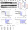
Figure 1
Figure 1
Activation of immunological pathways in Cngb1−/− retinas at 12 and 28 days after birth. (A) Representative optical coherence tomography (OCT) images from Cngb1−/− (upper panel) and wild-type retinas (lower panel), which display the slow progression (P28-P312) of outer nuclear layer (ONL) thinning over time. (B) Quantification of ONL thickness from OCT data at P28 (****p < 0.0001, n = 6 for each genotype). (C) Performance in the visual cliff test for the study of visual depth perception (**p < 0.01, n = 3 for each genotype). (D–G) Biological functions and canonical pathways were significantly altered in Cngb1−/− mice compared to age-matched controls. A high number of genes were related to the immune system or to immune responses. (H) The indicated upstream regulators for proinflammatory cytokines and the NF-κB pathway were predicted to have a significantly higher activation state in Cngb1−/− retinas when compared to age-matched wild-type controls (p < 0.05).
Figure 2
Figure 2
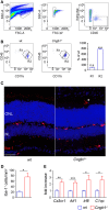
Strong increase of retinal microglia cells in Cngb1−/− retinas 28 days after birth. (A) Gating strategy for identifying microglia cells in retina lysates by size (left panel), single cells (middle panel), and living CD45+ cells (right panel) expression. (B) Representative gating and quantification of CD11b+CD45low retinal microglia (R2) and CD11b+CD45hi cells (R1) from wild-type or Cngb1−/− retinas. (Statistical significance was determined vs. percentage of wild-type cell counts, ns = non significant; ***p < 0.001, n = 3 for each genotype.) (C) Immunofluorescence of Iba-1 (red) in the retina of Cngb1−/− and age-matched wt demonstrating migration of microglia into the photoreceptor layer of Cngb1−/− mice (asterisk). Nuclei were counterstained with DAPI. ONL, outer nuclear layer, INL, inner nuclear layer, scale bar 50 µm. (D) Quantification of Iba-1-positive cells in Cngb1−/− mice compared to wt animals (*p < 0.05, n = 4 for each genotype). (E) Gene expression of microglia-specific genes in Cngb1−/− and wt retinas. Significant increase in the expression of Cx3cr1, Aif1, Irf8, and C1qc in retinas of Cngb1-deficient mice (*p < 0.05, **p < 0.01, ***p < 0.001, n = 4 for each genotype).
Figure 3
Figure 3
Amoeboid microglia morphology in Cngb1−/− is accompanied by the expression of activation markers. (A) Whole mount images of Iba-1-stained microglia in the outer nuclear layer (ONL) of Cngb1−/− mice induced morphological changes from a resting to an amoeboid phenotype at P28. Nuclei were counterstained with DAPI. Upper panel: maximum projection. Lower two panels: single images with an increment of 1.3 µm. Scale bar. 10 µm. (B) Flow cytometric analysis of retinal microglia for the expression of F4/80, CD44, and MHC class II (left panel). Quantification of the numbers (middle panel) and geometric mean fluorescent intensities (gMFI, right panel) of F4/80, CD44, and MHC class II are depicted. Results were obtained from two independent experiments with at least three replicates (*p < 0.05, **p < 0.01, ***p < 0.001, n = 3 for each genotype, blue line = wt, red line = Cngb1−/−, grey = isotype control). (C) Misplaced microglia in the ONL in Cngb1−/− coexpressed activation marker Lamp-2. Scale bar 25 µm. (D) Confirmation of MHC II expression by costaining of Iba-1 (red) and MHC class II (green) in wt and Cngb1−/− retinas. Scale bar 25 µm.
Figure 4
Figure 4
Time course of early photoreceptor apoptosis and microglia activation. (A) Costaining of Iba-1-positive microglia (pink) in the ONL and INL of Cngb1−/− and wt mice at P12, P19 and P28 with the activation marker Lamp-2 (green) and MHC class II (red). (B) Immunofluorescence of cleaved caspase 3 (red) in Cngb1−/− and wt mice. Nuclei were counterstained with DAPI (blue). (C) Quantification of Iba-1-positive cells and the percentage of Iba-1+ MHC class II− (D) or Iba-1+ Lamp-2-positive cells (E) in Cngb1−/− mice compared to wt animals. (F) Cleaved caspase 3-positive cells in Cngb1−/− and wt mice at indicated time points. ONL, outer nuclear layer, INL, inner nuclear layer, scale bar 20 µm; insert 10 µm (*p < 0.05, **p < 0.01, ***p < 0.001, n = 5 for each genotype).
Figure 5
Figure 5
Activation of proinflammatory signaling in Cngb1−/− mice. Immunofluorescence of Iba-1 (red) and pIκBα (green) in Cngb1−/− and wt mice at P28. Quantification of double-positive cells in wt and KO revealed exclusive presence of active NF-κB-signaling in Cngb1−/− mice (n = 3 for each genotype). Insert: Iba-1-positive resting microglia in the OPL. Nuclei were counterstained with DAPI. ONL, outer nuclear layer, IS, inner segment, OS, outer segment, OPL outer plexiform layer, scale bar 20 µm, nd = not detectable.

References

    1. Kreutzberg GW. Microglia: a sensor for pathological events in the CNS. Trends Neurosci (1996) 19:312–8.10.1016/0166-2236(96)10049-7 - DOI - PubMed
    1. Koizumi S, Shigemoto-Mogami Y, Nasu-Tada K, Shinozaki Y, Ohsawa K, Tsuda M, et al. UDP acting at P2Y6 receptors is a mediator of microglial phagocytosis. Nature (2007) 446:1091–5.10.1038/nature05704 - DOI - PMC - PubMed
    1. Tay TL, Mai D, Dautzenberg J, Fernandez-Klett F, Lin G, Sagar, et al. A new fate mapping system reveals context-dependent random or clonal expansion of microglia. Nat Neurosci (2017) 20:793–803.10.1038/nn.4547 - DOI - PubMed
    1. Prinz M, Priller J. Microglia and brain macrophages in the molecular age: from origin to neuropsychiatric disease. Nat Rev Neurosci (2014) 15:300–12.10.1038/nrn3722 - DOI - PubMed
    1. Karlstetter M, Scholz R, Rutar M, Wong WT, Provis JM, Langmann T. Retinal microglia: just bystander or target for therapy? Prog Retin Eye Res (2015) 45:30–57.10.1016/j.preteyeres.2014.11.004 - DOI - PubMed