. 2018 Jan;4(1):27-40.
doi: 10.1136/bmjstel-2017-000234. Epub 2017 Oct 21.

3D printing materials and their use in medical education: a review of current technology and trends for the future

Affiliations

3D printing materials and their use in medical education: a review of current technology and trends for the future

Justine Garcia et al. BMJ Simul Technol Enhanc Learn. 2018 Jan.

Abstract

3D printing is a new technology in constant evolution. It has rapidly expanded and is now being used in health education. Patient-specific models with anatomical fidelity created from imaging dataset have the potential to significantly improve the knowledge and skills of a new generation of surgeons. This review outlines five technical steps required to complete a printed model: They include (1) selecting the anatomical area of interest, (2) the creation of the 3D geometry, (3) the optimisation of the file for the printing and the appropriate selection of (4) the 3D printer and (5) materials. All of these steps require time, expertise and money. A thorough understanding of educational needs is therefore essential in order to optimise educational value. At present, most of the available printing materials are rigid and therefore not optimum for flexibility and elasticity unlike biological tissue. We believe that the manipuation and tuning of material properties through the creation of composites and/or blending materials will eventually allow for the creation of patient-specific models which have both anatomical and tissue fidelity.

Keywords: 3d printing; medical simulation; simulators; surgical training; tissue fidelity.


Conflict of interest statement

Competing interests: None declared.

Figures

Figure 1
Figure 1
Steps required in the creation of a 3D printed model in healthcare education.
Figure 2
Figure 2
Representation of geometry of the thoracic aortic model for teaching purposes.
Figure 3
Figure 3
(A) Illustration of the planar 2D images of an area of interest captured by most medical imaging techniques, (B) segmentation of the object cross-section (black circles) extracted, and (C) interpolation required to fill in the missing volume between segments.
Figure 4
Figure 4
Two distinct objects made of different materials or printed in different colours creating an assembly.
Figure 5
Figure 5
(A) Point cloud created from two 2D images and an example of a (B) point cloud from the inner lumen of an ascending aorta of a patient from the Royal Victoria Hospital (Montreal, Canada) with a 0.625 mm thickness between the slices.
Figure 6
Figure 6
(A) Artefacts from the point cloud of an ascending aorta of a patient from the Royal Victoria Hospital (Montreal, Canada) and an (B) artefact before and after the mesh smoothing.
Figure 7
Figure 7
Spheres created to approximate the shape of a patient-specific aortic root, open source Meshlab (MeshLab, Italy).
Figure 8
Figure 8
(A) Most frequent Boolean operations to create objects (union and subtraction of volumes) and (B) volume overlapping that should be avoided in any circumstances.
Figure 9
Figure 9
Mesh errors of (A) non-connecting triangle, (B) overlapping triangles and with an (C) extra body that is not part of the main geometry.
Figure 10
Figure 10
Multicolor 3D printed liver with a tumour (pink) of a patient from the Royal Victoria Hospital (Montreal, Canada) for surgical planning of a diseased liver. The print contain the portal vein, the hepatic vein as well as the tumour.
Figure 11
Figure 11
Aortic model in process for being printed on a fused deposition modelling 3D printer by successive layers of acrylonitrile butadiene styrene plastic (0.3 mm thickness).
Figure 12
Figure 12
Rapid prototyping methods with red arrows indicating the directions of motion (x, y, z axes).
Figure 13
Figure 13
Structure with an overhang filled with a lattice structure, support material or non-cured material.
Figure 14
Figure 14
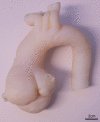
Model of the aorta with root aneurysm for teaching purposes.
Figure 15
Figure 15
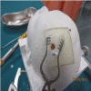
Head model made of one material to practise the drilling in medical simulation.
Figure 16
Figure 16
Head model made of a combination of materials to practise the drilling in medical simulation. (with permission Rockwater Inc.)
Figure 17
Figure 17
Accurate 3D model of an aortic aneurysm captured from CT imaging.

References

    1. Negi S, Dhiman S, Kumar Sharma R. Basics and applications of rapid prototyping medical models. Rapid Prototyp J 2014;20:256–67. 10.1108/RPJ-07-2012-0065 - DOI
    1. Rengier F, Mehndiratta A, von Tengg-Kobligk H, et al. . 3D printing based on imaging data: review of medical applications. Int J Comput Assist Radiol Surg 2010;5:335–41. 10.1007/s11548-010-0476-x - DOI - PubMed
    1. Malik HH, Darwood AR, Shaunak S, et al. . Three-dimensional printing in surgery: a review of current surgical applications. J Surg Res 2015;199:512–22. 10.1016/j.jss.2015.06.051 - DOI - PubMed
    1. Friedman T, Michalski M, Goodman TR, et al. . 3D printing from diagnostic images: a radiologist’s primer with an emphasis on musculoskeletal imaging-putting the 3D printing of pathology into the hands of every physician. Skeletal Radiol 2016;45:307–21. 10.1007/s00256-015-2282-6 - DOI - PubMed
    1. Matsumoto JS, Morris JM, Foley TA, et al. . Three-dimensional physical modeling: applications and experience at mayo clinic. Radiographics 2015;35:1989–2006. 10.1148/rg.2015140260 - DOI - PubMed