. 2017 Aug 7;6(4):e1263-e1270.
doi: 10.1016/j.eats.2017.05.004. eCollection 2017 Aug.

Acromioclavicular Joint Dislocation: Repair Through Open Ligament Transfer and Nonabsorbable Suture Fixation

Affiliations

Acromioclavicular Joint Dislocation: Repair Through Open Ligament Transfer and Nonabsorbable Suture Fixation

Ricardo Canquerini da Silva et al. Arthrosc Tech. .

Abstract

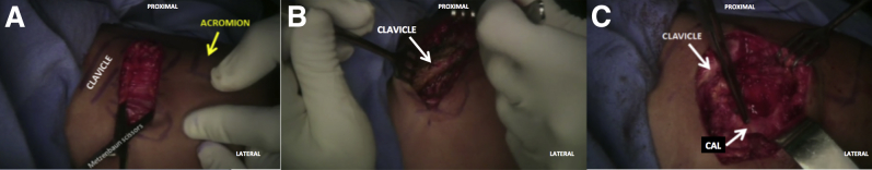
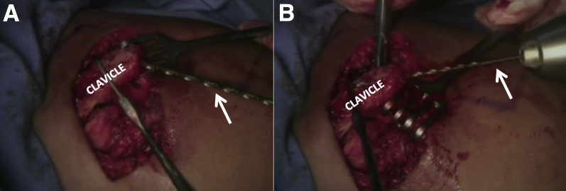
Acromioclavicular (AC) joint instability is a fairly common and particularly limiting injury that may result in persistent pain and reduced quality of life. In most cases, conservative management is successful. However, in the case of a severe AC joint dislocation, surgical intervention may be warranted. Previous surgical techniques for treatment of AC joint instability include screw fixation between the coracoid and clavicle, coracoacromial ligament transfer from its acromial insertion to the clavicle, and reconstruction of the coracoacromial and/or coracoclavicular ligaments. The purpose of this Technical Note is to describe our preferred technique for the treatment of a high-grade AC dislocation through coracoacromial ligament transfer to the lateral clavicle and nonabsorbable suture fixation between the coracoid process and clavicle.


Figures

Fig 1
Fig 1
The patient is positioned in a beach chair position with the left arm prepped in a standard fashion. The coracoid process, acromion, and clavicle are palpated and marked using a surgical pen. Based on Langer's lines, a line is drawn from the coracoid process to the clavicle (A). A scalpel is then used for the surgical approach (B), starting at the coracoid process and extending to the superior border of the clavicle.
Fig 2
Fig 2
The surgical approach for exposure of the acromioclavicular joint in the left shoulder is completed. Once the skin is incised, blunt dissection of the subcutaneous tissue is performed using Metzenbaum scissors (A). The deltotrapezoidal fascia is incised in line with the skin incision. Then, the superior and anterior aspect of the clavicle is exposed with release of all surrounding soft tissue (B). After this, exposure of the coracoacromial ligament (CAL) is completed (C).
Fig 3
Fig 3
The coracoacromial ligament is released from its acromial insertion in the left shoulder through the use of a scalpel. The lateral border of the ligament is then prepared using a No. 5 Ethibond suture (Ethicon, Somerville, NJ) by way of the Kessler suture technique (A, B). (CAL, coracoacromial ligament; CHL, coracohumeral ligament.)
Fig 4
Fig 4
After the coracoacromial ligament is released from its acromial insertion in the left shoulder, a surgical ruler is used to measure a 3 cm distance from the lateral end of the clavicle on the superior surface of the clavicle (A). The clavicle is secured and a 2.5-mm drill (white arrow) is used to create 2 tunnels in a superior-to-inferior direction (B). The first tunnel is located 3 cm from the acromioclavicular joint and the other is found 1 cm lateral from the first tunnel. Afterward, 1 cm of the most lateral end of the clavicle is resected using a rongeur (green arrow, C). (Yellow arrow, most lateral aspect of the clavicle.)
Fig 5
Fig 5
Once the lateral end of the left clavicle is resected, a 2.5-mm drill (white arrows, A and B) is used to create 2 intramedullary tunnels. The first tunnel starts at the anterior aspect of the intramedullary canal and is constructed toward the posterior aspect of the clavicle. The second tunnel begins at the posterior aspect of the canal and is constructed toward the anterior aspect of the clavicle.
Fig 6
Fig 6
In the left shoulder, 4 No. 5 Ethibond sutures (Ethicon, Somerville, NJ) are passed underneath the coracoid process (yellow arrow, A) from lateral to medial. To facilitate the passage of all the sutures at once, a coracoid suture passer (Hospitalia Cirurgica, Florianopolis, Brazil) is used from medial to lateral with a total of 4 suture limbs on each side of the coracoid process. Two suture limbs on the lateral side are then passed through the most medial tunnel in the clavicle and 2 suture limbs on the medial side are passed through the most lateral tunnel one pair over the other (B).
Fig 7
Fig 7
Once the clavicle of the left shoulder is reduced and kept in position using the nonabsorbable sutures, the sutures previously passed through the lateral end of the coracoacromial ligament (CAL, yellow arrow, (A)) are inserted through the intramedullary tunnels and tied and secured on the superior aspect of the clavicle. This fixation introduces the CAL into the intramedullary canal (B).
Fig 8
Fig 8
Postoperative imaging showing the preoperative (A) and postoperative (B) views of the affected left shoulder and contralateral shoulder. Note the complete dislocation of the acromioclavicular joint on the left side (yellow arrow, A) compared with the noninjured, contralateral side (white arrows) as well as the reduction of the joint after our surgical technique (yellow arrow, B).

References

    1. Pallis M., Cameron K.L., Svoboda S.J., Owens B.D. Epidemiology of acromioclavicular joint injury in young athletes. Am J Sports Med. 2012;40:2072–2077. - PubMed
    1. Beim G.M. Acromioclavicular joint injuries. J Athl Train. 2000;35:261–267. - PMC - PubMed
    1. Garretson R.B., III, Williams G.R., Jr. Clinical evaluation of injuries to the acromioclavicular and sternoclavicular joints. Clin Sports Med. 2003;22:239–254. - PubMed
    1. Ernberg L.A., Potter H.G. Radiographic evaluation of the acromioclavicular and sternoclavicular joints. Clin Sports Med. 2003;22:255–275. - PubMed
    1. Saccomanno M.F., DE Ieso C., Milano G. Acromioclavicular joint instability: Anatomy, biomechanics and evaluation. Joints. 2014;2:87–92. - PMC - PubMed