. 2017 Sep 4;6(5):e1471-e1476.
doi: 10.1016/j.eats.2017.06.039. eCollection 2017 Oct.

Reconstruction of the Medial Patellofemoral Ligament

Affiliations

Reconstruction of the Medial Patellofemoral Ligament

Juan C Monllau et al. Arthrosc Tech. .

Abstract

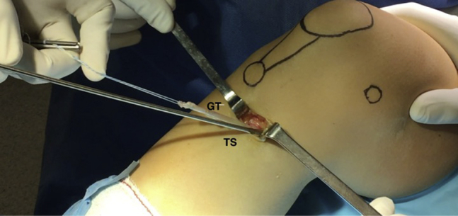
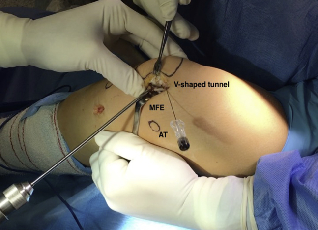
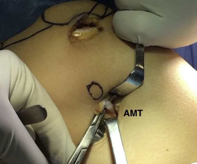
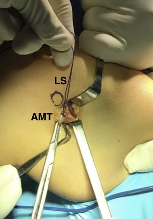
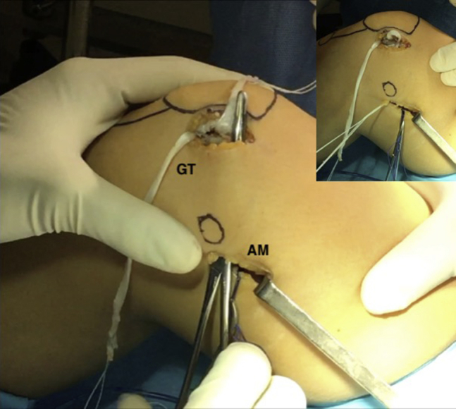
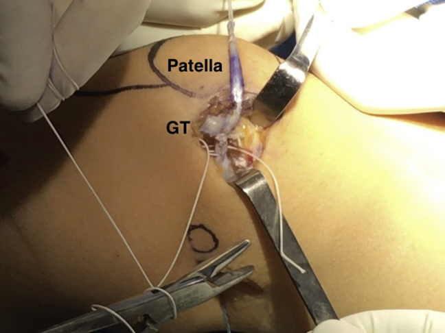
Patellar instability has been shown to be associated with different major factors. However, studies have demonstrated that soft tissue reconstructions are adequate enough to reestablish patellar constraint. In recent years, the medial patellofemoral ligament has been recognized as the primary passive restraint for lateral translation of the patella. Their reconstruction has gain popularity as the procedure is quite simple and fast. Although several surgical techniques have been described for their reconstruction, no clear consensus has been reached as to which is best. We present an implant-free, medial patellofemoral ligament reconstruction technique that uses a gracilis tendon autograft, 2 bone convergent tunnels at the original patellar attachment, and looping the graft around the adductor magnus tendon that is used as a pulley for femoral fixation.


Figures

Fig 1
Fig 1
Right knee, anteromedial view. The patient is placed in a supine position on the operating table, with the knee at 90° of flexion. Anteromedial skin incision to harvest the gracilis tendon (GT) using closed tendon stripper (TS).
Fig 2
Fig 2
Right knee, medial view. A V-shaped tunnel is drilled in the medial aspect of the patella, using a 4.5-mm reamer, leaving a cortical bone bridge of 10 mm between them to avoid a fracture. The medial femoral epicondyle (MFE) and adductor tubercle (AT) are marked.
Fig 3
Fig 3
Right knee, medial view. Skin incision made along the adductor magnus tendon (AMT). The tendon is identified and dissected.
Fig 4
Fig 4
Right knee, medial view. Looped suture (LS) placed around the adductor magnus tendon (AMT) for graft passage.
Fig 5
Fig 5
Right knee, medial view. The gracilis tendon (GT) is introduced in the patellar tunnel. Place the graft in the interval between layers 2 and 3 of the medial retinaculum. The graft should not be deeper than layer 3 so that it remains in the extra-articular environment.
Fig 6
Fig 6
Right knee, closer medial view. Graft passed through the patellar V-shaped tunnel, between layers 2 and 3 of the medial retinaculum and looped around the AM tendon back to the patella.
Fig 7
Fig 7
Both grafts were sutured together at 30° of flexion with no. 0 high-resistance nonabsorbable sutures. Tension was calculated on the basis that the patella could still be manually lateralized some 10 mm to avoid overconstraint.

References

    1. Fithian D.C., Paxton E.W., Stone M.L. Epidemiology and natural history of acute patellar dislocation. Am J Sports Med. 2004;32:1114–1121. - PubMed
    1. Becher C., Kley K., Lobenhoffer P., Ezechieli M., Smith T., Ostermeier S. Dynamic versus static reconstruction of the medial patellofemoral ligament for recurrent lateral patellar dislocation. Knee Surg Sports Traumatol Arthrosc. 2014;22:2452–2457. - PubMed
    1. Haupert A., Lorbach O. Anatomic reconstruction of the medial patellofemoral ligament using the fascia lata as an autograft. Arthrosc Tech. 2015;4:e57–e63. - PMC - PubMed
    1. Zaffagnini S., Dejour D., Grasse A. Patellofemoral anatomy and biomechanics: Current concepts. Joints. 2013;24:15–20. - PMC - PubMed
    1. Neri T., Philippot R., Carnesecchi O., Boyer B., Farizon F. Medial patellofemoral ligament reconstruction: Clinical and radiographic results in a series of 90 cases. Orthop Traumatol Surg Res. 2015;101:65–69. - PubMed

LinkOut - more resources