. 2017 Oct 12;6(5):e1567-e1572.
doi: 10.1016/j.eats.2017.06.031. eCollection 2017 Oct.

Circumferential Suture Repair of Isolated Horizontal Meniscal Tears Augmented With Fibrin Clot

Affiliations

Circumferential Suture Repair of Isolated Horizontal Meniscal Tears Augmented With Fibrin Clot

Michael S Laidlaw et al. Arthrosc Tech. .

Abstract

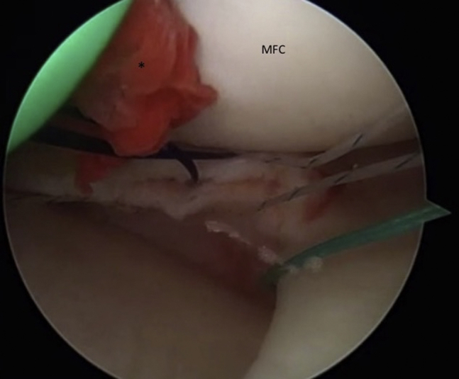
Traditionally, horizontal cleavage meniscus tears have been associated with osteoarthritis, indicative of the degenerative process. Recent treatment measures have focused on maintaining as much meniscal tissue as possible, despite the routine extension of these tears into the central white-white zones. In the absence of tunnel drilling for cruciate ligament reconstructions, the use of an exogenous fibrin clot is a useful adjunct to increase the local growth factors at the tear repair to aid in healing. This surgical technique is to describe the use of an all-arthroscopic tied circumferential suture repair of horizontal meniscal tears augmented with exogenous fibrin clot to treat all locations of horizontal meniscal tears.


Figures

Fig 1
Fig 1
Image of fibrin clot preparation. Thirty milliliters of venous blood is obtained in sterile fashion from an anesthesia IV draw and stirred slowly with a frosted glass rod (from a standard spinal tap set) until a fibrin clot starts to form (arrow). (IV, intravenous.)
Fig 2
Fig 2
Image of the fibrin clot. The fibrin clot (*) is removed from the glass rod carefully, placed on a 4 × 4 gauze, and stored on the back table.
Fig 3
Fig 3
Arthroscopic image right knee medial meniscus, anteromedial viewing portal, and anterolateral working portal. A standard 4.0-mm shaver is placed through the working portal, and the meniscal cleft (*) is debrided, as close to the periphery as possible to help prepare the cleft for fibrin clot introduction and debride degenerative medium. (MFC, medial femoral condyle.)
Fig 4
Fig 4
Arthroscopic image right knee medial meniscus, anteromedial viewing portal and anterolateral working portal. A no. 0 ethibond suture has been placed through the superior leaflet of the horizontal meniscus tear and is used to shuttle the smooth no. 0 PDS in through the superior leaflet. This will ultimately be used to shuttle the fibrin clot from outside-in and into the cleft (*). (MFC, medial femoral condyle.)
Fig 5
Fig 5
Arthroscopic image right knee medial meniscus, anteromedial viewing portal and anterolateral working portal. A Ceterix Novostitch device (*) is used to pass the 2-0 UHMPE suture circumferentially around the meniscus, at its periphery. (MTP, medial tibial plateau.)
Fig 6
Fig 6
Arthroscopic image right knee medial meniscus, anteromedial viewing portal and anterolateral working portal. Preplaced circumferential sutures have been placed through the periphery of the medial meniscus (*). Note, to help deconflict the passage of the fibrin clot, the most anterior sutures exit through the viewing portal and the posterior suture and no. 0 PDS exit through the working portal. (MFC, medial femoral condyle.)
Fig 7
Fig 7
Image of a right knee with fibrin clot woven onto the no. 0 PDS suture by Keeth needle and a Mulberry knot tied behind it to help guide it into the joint while shuttling it into the meniscal cleft. Of note, the cannula is in the working portal, and the external fluid valve has been removed (arrow) at this point so that it does not interfere with the passage of the clot.
Fig 8
Fig 8
Arthroscopic image right knee medial meniscus, anteromedial viewing portal and anterolateral working portal. The fibrin clot (*) is shuttled into the horizontal meniscal cleft by pulling on the no. 0 PDS. Note the green cannula is introduced into the medial compartment. (MFC, medial femoral condyle.)
Fig 9
Fig 9
Arthroscopic image right knee medial meniscus, anteromedial viewing portal and anterolateral working portal. Final arthroscopic image reveals the horizontal medial meniscus cleavage tear (*) has been repaired with exogenous fibrin clot with circumferential sutures tied with the arthroscopic knots on the tibial articular surface of the meniscus. (MFC, medial femoral condyle.)

References

    1. Voloshin A.S., Wosk J. Shock absorption of meniscectomized and painful knees: A comparative in vivo study. J Biomed Eng. 1983;5:157–161. - PubMed
    1. Cox J.S., Nye C.E., Schaefer W.W., Woodstein I.J. The degenerative effects of partial and total resection of the medial meniscus in dogs' knees. Clin Orthop Relat Res. 1975;109:178–183. - PubMed
    1. Newman A.P., Daniels A.U., Burks R.T. Principles and decision making in meniscal surgery. Arthroscopy. 1993;9:33–51. - PubMed
    1. Noble J., Hamblen D.L. The pathology of the degenerate meniscus lesion. J Bone Joint Surg Br. 1975;57:180–186. - PubMed
    1. Koh J.L., Yi S.J., Ren Y., Zimmerman T.A., Zhang L.Q. Tibiofemoral contact mechanics with horizontal cleavage tear and resection of the medial meniscus in the human knee. J Bone Joint Surg Am. 2016;98:1829–1836. - PubMed

LinkOut - more resources