. 2017 Oct 12;6(5):e1573-e1580.
doi: 10.1016/j.eats.2017.06.051. eCollection 2017 Oct.

Meniscal Allograft Transplantation With Concomitant Osteochondral Allograft Transplantation

Affiliations

Meniscal Allograft Transplantation With Concomitant Osteochondral Allograft Transplantation

Eric J Cotter et al. Arthrosc Tech. .

Abstract

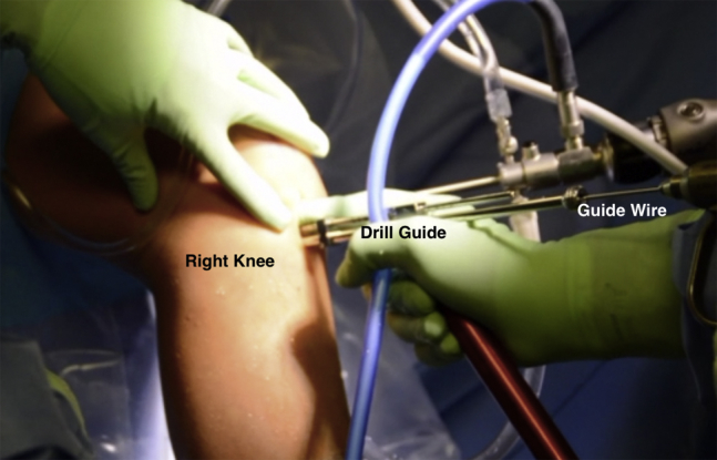
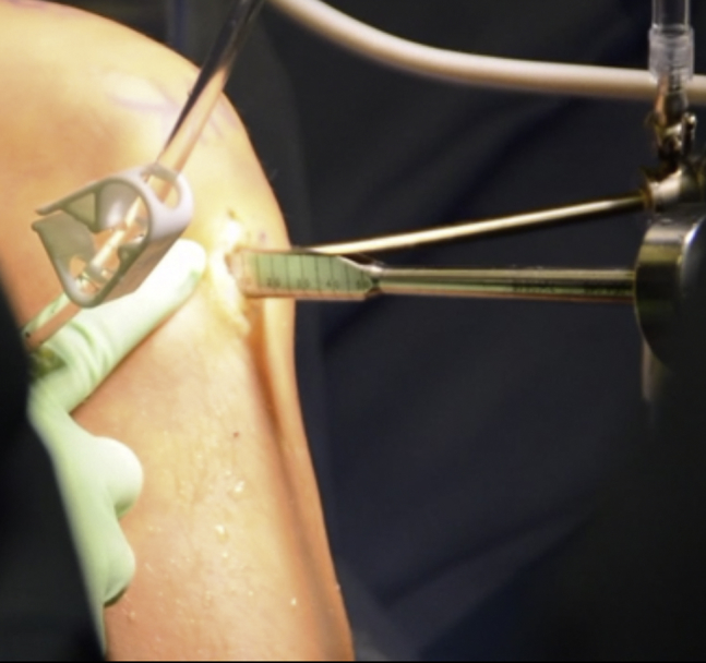
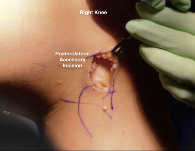
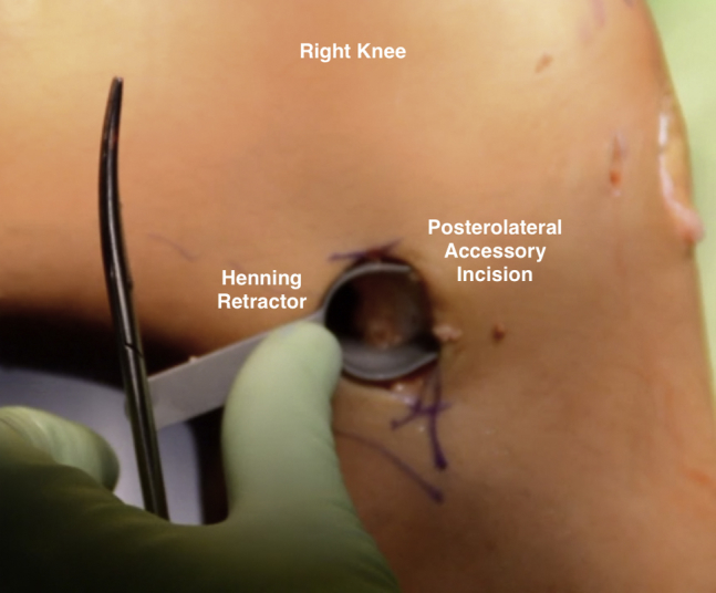
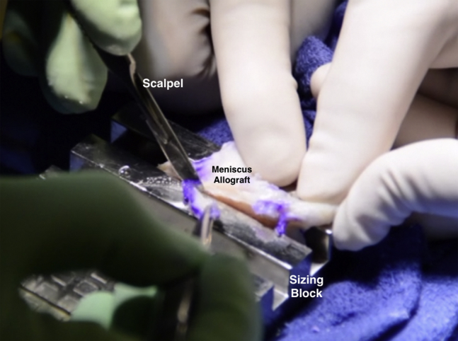
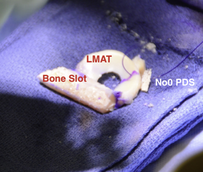
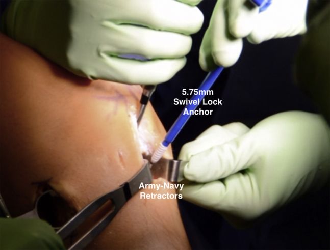
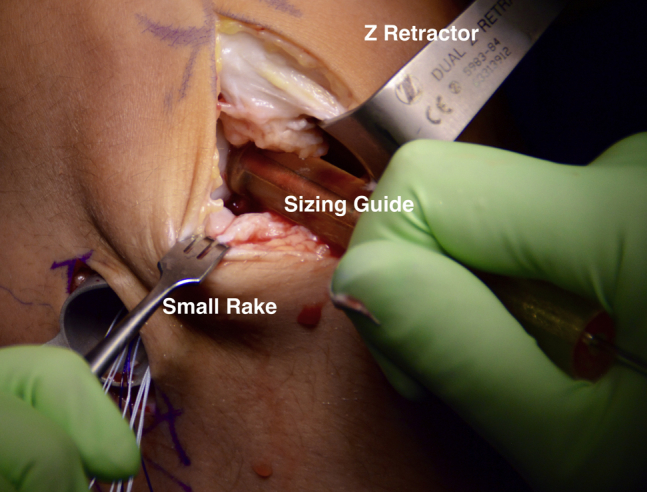
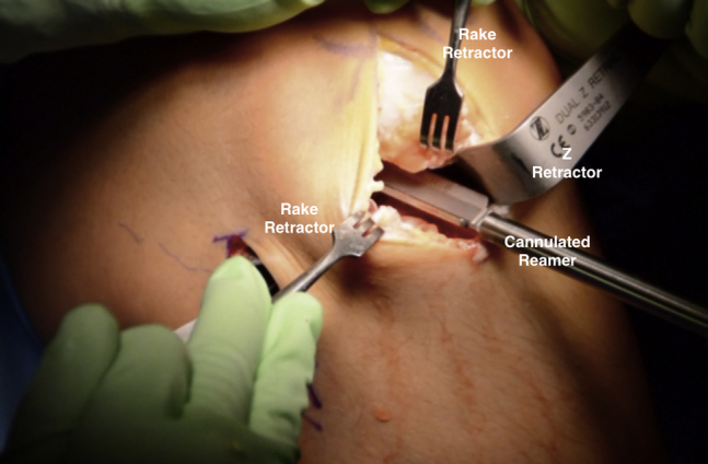
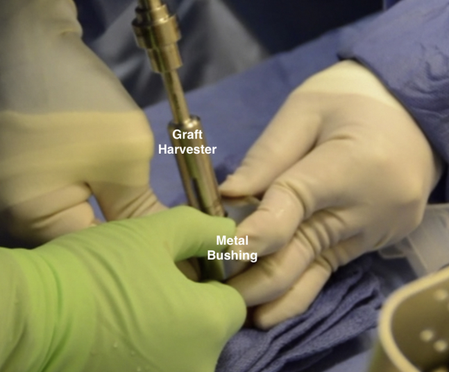
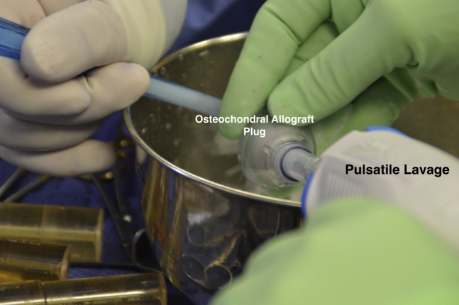
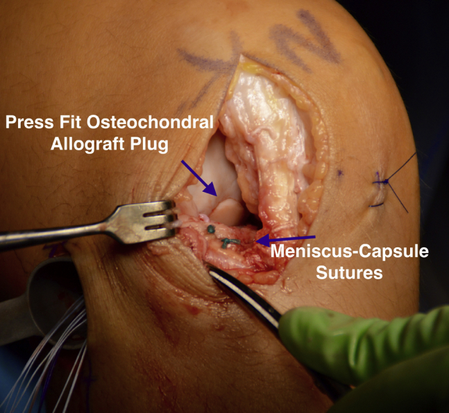
Surgical strategies for knee joint preservation are numerous, with the procedure(s) of choice for a given patient dependent on the status of the articular cartilage, meniscus, overall alignment, and ligamentous stability. For patients with large, isolated, osteochondral defects of the articular cartilage of the femoral condyle, osteochondral allograft transplantation (OCA) is often performed in an effort to reduce pain and improve function. Similarly, for appropriately indicated patients with symptomatic meniscus deficiency, meniscus allograft transplantation (MAT) is an excellent surgical solution. Often patients require concomitant MAT and OCA as part of a joint preservation strategy. In this Technical Note, we describe the surgical technique for performing arthroscopic-assisted concomitant lateral MAT and lateral femoral condyle OCA as part of a knee joint preservation strategy.


Figures

Fig 1
Fig 1
Intraoperative photograph of a patient positioned supine on the operating table with the right leg placed in a standard leg holder. The right knee is prepped and a well-padded thigh tourniquet has been placed.
Fig 2
Fig 2
Intraoperative arthroscopic photograph of a supine positioned patient's right knee. The arthroscopic view through the lateral portal demonstrating the lateral meniscus debrided to a peripheral rim of remaining tissue.
Fig 3
Fig 3
Intraoperative arthroscopic photograph of a supine positioned patient's right knee. The arthroscopic view through the medial portal of the 4-mm burr used to create an index slot along the tibial plateau through which a reamer and box cutter will be used to fully define the slot for the meniscal allograft bone bridge.
Fig 4
Fig 4
Intraoperative arthroscopic photograph of a supine positioned patient's right knee. The arthroscopic view through the medial portal demonstrating placement of the hooked depth gauge along the reference slot on the lateral tibial plateau.
Fig 5
Fig 5
Intraoperative photograph of a patient in the supine position, demonstrating use of the drill guide to place a guide pin along the reference slot in the lateral tibial plateau of the right knee.
Fig 6
Fig 6
Intraoperative photograph of a supine positioned patient. A large rasp being inserted into the bony slot within the lateral tibial plateau of the right knee. This rasp is used to smooth out the dimensions of the bony slot prior to insertion of the meniscus allograft.
Fig 7
Fig 7
Intraoperative photograph of a patient in the supine position. The photograph depicts the posterolateral accessory incision created on the right knee through which inside-out meniscus repair sutures will be passed to secure the meniscus allograft in place.
Fig 8
Fig 8
Intraoperative photograph of a patient in the supine position, demonstrating placement of a Henning retractor within the posterolateral accessory incision created on the right knee.
Fig 9
Fig 9
Intraoperative photograph of the meniscal allograft (MAT) being prepared within the meniscus sizing block. A scalpel is used to cut away extraneous tissue adjacent to the bone bridge.
Fig 10
Fig 10
Intraoperative photograph of the lateral meniscal allograft (LMAT). The bony slot and meniscal tissue can be clearly delineated. A no. 0 PDS suture is placed through the junction of the middle and posterior third of the LMAT.
Fig 11
Fig 11
Intraoperative photograph of a patient in the supine position, demonstrating placement of a 5.75-mm SwiveLock anchor into the meniscal allograft bony component to secure it to the right-sided tibia.
Fig 12
Fig 12
Intraoperative photograph of a patient in the supine position, demonstrating sizing of the lateral femoral condyle osteochondral defect of the right knee using a cannulated cylindrical sizing guide. A Z-retractor and small rack are used to retract the surrounding soft tissue.
Fig 13
Fig 13
Intraoperative photograph of a patient in the supine position, demonstrating use of a cannulated cutting reamer placed over the previously inserted guide pin into the center of the focal cartilage defect on the lateral femoral condyle of the right knee. This reamer is used to core the defect to a depth of approximately 6 to 8 mm.
Fig 14
Fig 14
Intraoperative photograph showing an assistant holding a metal bushing with a 15-mm diameter opening over the lateral femoral condyle of a donor hemicondyle while the surgeon uses a graft harvester to drill through the full extent of the hemicondyle to obtain the osteochondral allograft plug.
Fig 15
Fig 15
Intraoperative photograph of the osteochondral allograft plug undergoing pulsatile lavage with 2 L of saline to remove any remaining tissue debris and marrow elements.
Fig 16
Fig 16
Intraoperative photograph of a patient in the supine position, demonstrating a flush fit of the lateral femoral condyle osteochondral allograft to the surrounding articular cartilage of the right knee. In addition, 2 sutures attaching the anterior aspect of the meniscal allograft to the capsule are visualized.
Fig 17
Fig 17
Intraoperative photograph of a patient in the supine position, demonstrating the inside-out meniscus repair sutures being tied down through the posterolateral accessory portal in the right knee. The sutures are evaluated to ensure not to include tissue from the iliotibial band.

References

    1. Messner K., Gao J. The menisci of the knee joint. Anatomical and functional characteristics, and a rationale for clinical treatment. J Anatomy. 1998;193:161–178. - PMC - PubMed
    1. King D. The healing of semilunar cartilages. 1936. Clin Orthop Relat Res. 1990:4–7. - PubMed
    1. Sohn D.H., Toth A.P. Meniscus transplantation: current concepts. J Knee Surg. 2008;21:163–172. - PubMed
    1. Fairbank T.J. Knee joint changes after meniscectomy. J Bone Joint Surg Br. 1948;30:664–670. - PubMed
    1. Raber D.A., Friederich N.F., Hefti F. Discoid lateral meniscus in children. Long-term follow-up after total meniscectomy. J Bone Joint Surg Am. 1998;80:1579–1586. - PubMed

LinkOut - more resources