Review
. 2017 Dec 18;2(3-4):101-139.
doi: 10.3233/TRD-170015.

Disorders of metal metabolism

Affiliations
Review

Disorders of metal metabolism

Carlos R Ferreira et al. Transl Sci Rare Dis. .

Abstract

Trace elements are chemical elements needed in minute amounts for normal physiology. Some of the physiologically relevant trace elements include iodine, copper, iron, manganese, zinc, selenium, cobalt and molybdenum. Of these, some are metals, and in particular, transition metals. The different electron shells of an atom carry different energy levels, with those closest to the nucleus being lowest in energy. The number of electrons in the outermost shell determines the reactivity of such an atom. The electron shells are divided in sub-shells, and in particular the third shell has s, p and d sub-shells. Transition metals are strictly defined as elements whose atom has an incomplete d sub-shell. This incomplete d sub-shell makes them prone to chemical reactions, particularly redox reactions. Transition metals of biologic importance include copper, iron, manganese, cobalt and molybdenum. Zinc is not a transition metal, since it has a complete d sub-shell. Selenium, on the other hand, is strictly speaking a nonmetal, although given its chemical properties between those of metals and nonmetals, it is sometimes considered a metalloid. In this review, we summarize the current knowledge on the inborn errors of metal and metalloid metabolism.

Keywords: Birk-Landau-Perez syndrome; Huppke-Brendel syndrome; MEDNIK syndrome; Menkes disease; SBP2 deficiency; SEPSECS deficiency; SLC39A8 deficiency; Transition metals; Wilson disease; hemochromatosis; hypermanganesemia with dystonia; neurodegeneration with brain iron accumulation, acrodermatitis enteropathica; spondylocheirodysplastic Ehlers-Danlos syndrome; transient neonatal zinc deficiency.


Figures

Fig.1
Fig.1
Appearance of Kayser-Fleischer ring in Wilson disease. Dark pigment at periphery of the cornea is due to deposition of copper.
Fig.2
Fig.2
Wilson disease. (A) Microscopic section of liver in inflammatory reaction in the portal area. The hepatocytes contain copper pigment. (B) High-power view showing Mallory body (arrow).
Fig.3
Fig.3
Microscopic section of liver in Wilson disease showing copper pigmentation of hepatocytes stained with Rhodamine stain.
Fig.4
Fig.4
Gross appearance of cirrhotic liver in Wilson disease.
Fig.5
Fig.5
Wilson disease. Coronal section of brain showing small cystic cavitation and light brown discoloration in the basal ganglia, particularly in the putamen.
Fig.6
Fig.6
Electron micrograph of hepatocyte in Wilson disease. The mitochondria vary in size and shape, with dilated and microcystic cristae. (Courtesy Dr. Roma Chandra.)
Fig.7
Fig.7
Menkes syndrome child with typical coarse kinky hair.
Fig.8
Fig.8
Section of cerebral white matter in Menkes disease showing telangiectatic capillary.
Fig.9
Fig.9
Menkes disease. (A) Microscopic section of the aorta showing disruption and piling up of elastic fibrils of internal elastic lamella. (B) Fluorescence photomicrograph of longitudinal section of peripheral nerve. Note torpedo-like swelling of intense fluorescence-positive axon. Inset shows cross section of peripheral nerve with nonspecific fluorescence of myelin sheath (formaldehyde-induced fluorescence). (Courtesy of Dr. Hideo Uno.)
Fig.10
Fig.10
Neuroaxonal leukodystrophy. There are axonal spheroids in white matter associated with myelin degeneration.
Fig.11
Fig.11
Infantile neuroaxonal dystrophy. A portion of one of the unmyelinated axons (asterisk) in this conjuctival perivascular nerve is distended by an accumulation of tubulo-membranous structures, degenerate organelles, and amorphous electron-dense materials. With these, distinctive axonal “spheroids” become more condensed and display characteristic clefts. (Courtesy of Dr. Gary Mireau.)
Fig.12
Fig.12
Hereditary hemochromatosis. Gross appearance of micronodular cirrhotic liver.
Fig.13
Fig.13
Microscopic appearance of liver in hereditary hemochromatosis showing iron in hepatocytes. Prussian blue stain.
Fig.14
Fig.14
Microscopic appearance of the pancreas in hereditary hemochromatosis showing iron pigment in pancreatic acinar and islet cells. Prussian blue stain.
Fig.15
Fig.15
Gross appearance of the heart in hereditary hemochromatosis showing iron pigmentation.
Fig.16
Fig.16
Microscopic section of the heart in hereditary hemochromatosis showing iron pigment in the myocardial cells.
Fig.17
Fig.17
Microscopic appearance of skin in acrodermatitis enteropathica showing epidermal vacuolar degeneration.
Fig.18
Fig.18
Acrodermatitis enteropathica showing skin rash. (A) Before treatment. (B) After zinc treatment. (Courtesy of Dr. Bernard Cohen.)

References

    1. Danks D.M., Disorders of copper transport In: Scriver CR, Beaudet AL, Sly WS, Valle DL, editors. Metabolic and Molecular Bases of Inherited Diseases. 7th ed New York: McGraw Hill; 1995
    1. Culotta V.C. and Gitlin J.D., Disorders of Copper Transport In: Valle DL, Beaudet AL, Vogelstein B, Kinzler KW, Antonarakis SE, Ballabio A, et al. , editors. The Online Metabolic and MolecularBases of Inherited Disease [Internet] New York NY: The McGraw-Hill Comanies, Inc; 2014. [cited 2015 Jul 27]. Available from: http://mhmedical.com/content.aspx?aid=1102891541
    1. Palmiter R.D., The elusive function of metallothioneins, Proc Natl Acad Sci U S A 95(15) (1998), 8428–8430. - PMC - PubMed
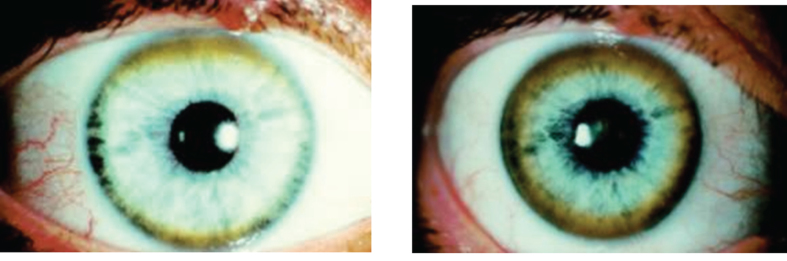
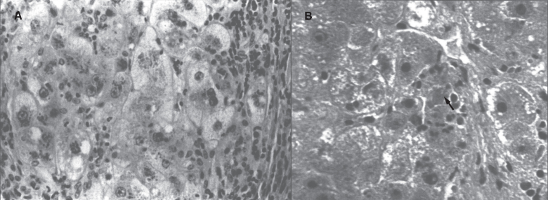
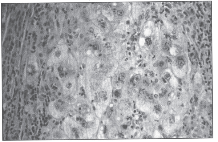
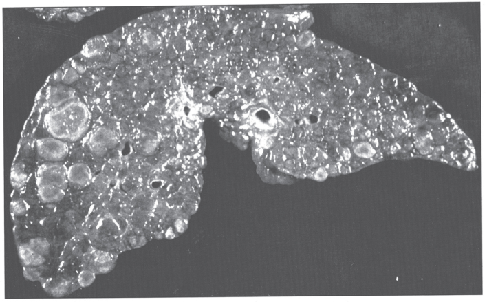
    1. Wilson S., Progressive lenticular degeneration: A familial nervous disease associated with cirrhosis of the liver, Brain (1912) 34, 295–509. - PubMed
    1. Weiss K.H., Wilson Disease In: Pagon RA, Adam MP, Ardinger HH, Wallace SE, Amemiya A, Bean LJ, et al. , editors. GeneReviews(®) [Internet] Seattle (WA): University of Washington, Seattle; 1993. [cited 2015 Aug 6]. Available from: http://www.ncbi.nlm.nih.gov/books/NBK1512/

LinkOut - more resources