

Retinal vasculature in glaucoma: a review
- PMID: 29354699
- PMCID: PMC5721639
- DOI: 10.1136/bmjophth-2016-000032
Retinal vasculature in glaucoma: a review
Erratum in
-
Correction: Retinal vasculature in glaucoma: a review.BMJ Open Ophthalmol. 2018 Jul 7;3(1):bmjophth-2016-000032corr1. doi: 10.1136/bmjophth-2016-000032corr1. eCollection 2018. BMJ Open Ophthalmol. 2018. PMID: 30123841 Free PMC article.
Abstract
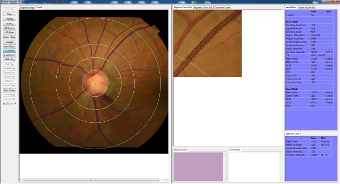
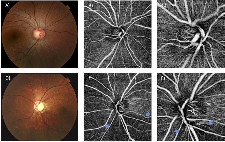
Despite the critical impact of glaucoma on global blindness, its aetiology is not fully characterised. Elevated intraocular pressure is highly associated with glaucomatous optic neuropathy. However, visual field loss still progresses in some patients with normal or even low intraocular pressure. Vascular factors have been suggested to play a role in glaucoma development, based on numerous studies showing associations of glaucoma with blood pressure, ocular perfusion pressure, vasospasm, cardiovascular disease and ocular blood flow. As the retinal vasculature is the only part of the human circulation that readily allows non-invasive visualisation of the microcirculation, a number of quantitative retinal vascular parameters measured from retinal photographs using computer software (eg, calibre, fractal dimension, tortuosity and branching angle) are currently being explored for any association with glaucoma and its progression. Several population-based and clinical studies have reported that changes in retinal vasculature (eg, retinal arteriolar narrowing and decreased fractal dimension) are associated with optic nerve damage and glaucoma, supporting the vascular theory of glaucoma pathogenesis. This review summarises recent findings on the relationships between quantitatively measured structural retinal vascular changes with glaucoma and other markers of optic nerve head damage, including retinal nerve fibre layer thickness. Clinical implications, recent new advances in retinal vascular imaging (eg, optical coherence tomography angiography) and future research directions are also discussed.
Keywords: Glaucoma; Retinal Photography; Retinal Vasculature.
Conflict of interest statement
Competing interests: None declared.
Figures
References
-
- Kass MA, Heuer DK, Higginbotham EJ, et al. The Ocular Hypertension Treatment Study: a randomized trial determines that topical ocular hypotensive medication delays or prevents the onset of primary open-angle glaucoma. Arch Ophthalmol 2002;120:701–13. discussion 829–30. - PubMed
-
- Satilmis M, Orgül S, Doubler B, et al. Rate of progression of glaucoma correlates with retrobulbar circulation and intraocular pressure. Am J Ophthalmol 2003;135:664–9. doi:10.1016/S0002-9394(02)02156-6 - DOI - PubMed
-
- Klein BE, Klein R, Sponsel WE, et al. Prevalence of glaucoma. The Beaver Dam Eye Study. Ophthalmology 1992;99:1499–504. - PubMed
-
- Gupta N, Weinreb RN. New definitions of glaucoma. Curr Opin Ophthalmol 1997;8:38–41. doi:10.1097/00055735-199704000-00007 - DOI - PubMed
-
- Tielsch JM, Katz J, Singh K, et al. A population-based evaluation of glaucoma screening: the Baltimore Eye Survey. Am J Epidemiol 1991;134:1102–10. doi:10.1093/oxfordjournals.aje.a116013 - DOI - PubMed
Publication types
LinkOut - more resources
Full Text Sources
Other Literature Sources
Research Materials