. 2018 Apr;9(2):185-198.
doi: 10.1007/s12975-018-0610-6. Epub 2018 Jan 21.

Delayed Recanalization Promotes Functional Recovery in Rats Following Permanent Middle Cerebral Artery Occlusion

Affiliations

Delayed Recanalization Promotes Functional Recovery in Rats Following Permanent Middle Cerebral Artery Occlusion

Devin William McBride et al. Transl Stroke Res. 2018 Apr.

Abstract

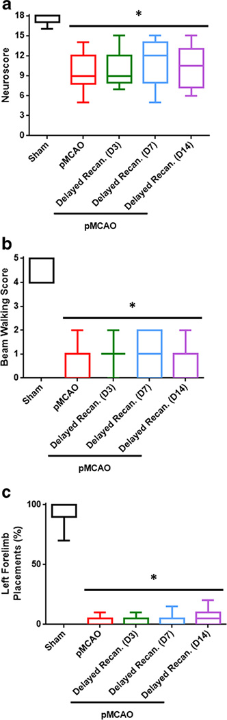
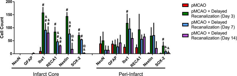
Most large vessel stroke patients have permanent occlusion, for which there are no current treatment options. Recent case studies have indicated delayed recanalization, that is recanalization outside of the 6-h treatment window, may lead to improved outcome. We hypothesized that delayed recanalization will restore cerebral blood flow, leading to improved function in rats. Male SD rats were subjected to pMCAO or sham surgery. Delayed recanalization was performed on either day 3, 7, or 14 after pMCAO in a subset of animals. Cerebral blood flow was monitored during suture insertion, during recanalization, and then at sacrifice. Neurological function was evaluated for 1 week after delayed recanalization and at 4 weeks post-ictus. After sacrifice, cerebral morphology was measured. Compared to no treatment, delayed recanalization restored cerebral blood flow, leading to sensorimotor recovery, improved learning and memory, reduced infarct volume, and increased neural stem/progenitor cells within the infarction. The data indicate that earlier delayed recanalization leads to better functional and histological recovery. Yet, even restoring cerebral blood flow 14 days after pMCAO allows for rats to regain sensorimotor function. This exploratory study suggests that delayed recanalization may be a viable option for treatment of permanent large vessel stroke.

Keywords: Delayed recanalization; Ischemia; Permanent middle cerebral artery occlusion; Stroke.


Conflict of interest statement

Compliance with Ethical Standards

Conflict of Interest The authors declare that they have no conflicts of interest.

Figures

Fig. 1
Fig. 1
a Kaplan-Meier survival curve. Dashed lines indicate day 1 and day 3. n = 15–41/group. b Cerebral blood flow change during suture insertion (n = 15–41/group), pre-recanalization (7–12/group), post-recanalization (n = 7–12/group), and at the time of sacrifice (n = 7–19/group). #p <0.05 vs pMCAO, N.S. indicates no significance
Fig. 2
Fig. 2
a Modified Garcia neuroscore 1 day after pMCAO (n = 12–34/group). b Beam walking score 24 hours after pMCAO (n = 12–34/group). c Forelimb placement test 1 day post-ictus (n = 12–34/group). *p <0.05 vs Sham
Fig. 3
Fig. 3
Functional Recovery after delayed recanalization assessed using the modified Garcia neuroscore. a Recanalization on day 3 (n = 6–15/group). b Recanalization on day 7 (n = 7–15/group). c Recanalization on day 14 (n = 6–15/group). *p < 0.05 Sham vs pMCAO, and p < 0.05 Sham vs pMCAO + delayed recanalization, #p <0.05 pMCAO vs pMCAO + delayed recanalization
Fig. 4
Fig. 4
Functional recovery after delayed recanalization assessed using the beam walking test. a Recanalization on day 3 (n = 6–15/group). b Recanalization on day 7 (n = 7–15/group). c Recanalization on day 14 (n = 6–15/group). *p <0.05 Sham vs pMCAO, and p <0.05 Sham vs pMCAO + delayed recanalization, #p < 0.05 pMCAO vs pMCAO + delayed recanalization
Fig. 5
Fig. 5
Functional recovery after delayed recanalization assessed using the forelimb placement test. a Recanalization on day 3 (n = 6–15/group). b Recanalization on day 7 (n = 7–15/group). c Recanalization on day 14 (n = 6–15/group). *p <0.05 Sham vs pMCAO, and p <0.05 Sham vs pMCAO + delayed recanalization, #p <0.05 pMCAO vs pMCAO + delayed recanalization
Fig. 6
Fig. 6
Neurological function on day 32. a Modified Garcia neuroscore. b Beam walking score. c Left forelimb placement test. n = 12–34/group. *p < 0.05 vs Sham
Fig. 7
Fig. 7
Behavioral performance on rotarod, open field, and Morris water maze 4 weeks after injury. a Rotarod (n = 7–19/group). b Open field (n = 7–19/group). c Morris water maze memory test (6–19/group) (median with interquartile range is plotted). d Morris mater maze probe test (6–19/group). Dotted line indicates random chance of time (25%) spent in target quadrant. Rotarod, open field, Morris water maze probe test: *p < 0.05 vs Sham, #p < 0.05 vs pMCAO, and p < 0.05 vs pMCAO + delayed recanalization (day 3). Morris water maze memory test: @p < 0.05 Sham vs pMCAO, p < 0.05 Sham vs pMCAO + delayed recanalization (day 3), p <0.05 Sham vs pMCAO + delayed recanalization (day 7) and Sham vs pMCAO + delayed recanalization (day 14), $p <0.05 pMCAO vs pMCAO + delayed recanalization (day 3), §p < 0.05 pMCAO + delayed recanalization (day 3) vs pMCAO + delayed recanalization (day 7) and pMCAO + delayed recanalization (day 3) vs pMCAO + delayed recanalization (day 14)
Fig. 8
Fig. 8
Cerebral morphology 32 days after pMCAO. a Representative Nissl stained brains. b Infarct volume (%). c White matter and ventricular volumes (mm3). d Hippocampus area (mm2). n = 7/group. *p < 0.05 vs Sham, #p < 0.05 vs pMCAO, and p < 0.05 vs pMCAO + delayed recanalization (day 3)
Fig.9
Fig.9
Neurons and astrocytes are absent within the infarction. Neurons (NeuN, green) and astrocytes (GFAP, red) are not observed in the infarct core in untreated pMCAO rats, or pMCAO rats treated with delayed recanalization. Scale bar: 20 μm
Fig. 10
Fig. 10
Delayed recanalization increases endothelial cells and microglia. Endothelial cells (RECA1, green) and microglia (Iba1, red) increase in number for pMCAO rats treated with delayed recanalization compared to untreated pMCAO rats. Arrowheads indicate some RECA1-positive cells. Arrows indicate some Iba1-positive cells. Scale bar: 20 μm
Fig. 11
Fig. 11
Delayed recanalization on day 3 promotes proliferation of neural stem cells and neural progenitor cells. Within the ischemic core, delayed recanalization induced proliferation of neural progenitor cells (Nestin-positive, green) and neural stem cells (Nestin-positive and SOX-2-positive (red)). Arrowheads indicate Nestin-positive staining. Arrows indicate SOX-2-positive staining. Scale bar: 20 μm
Fig. 12
Fig. 12
Cell counting within the infarct core and peri-infarct zones. n = 2–4/group. #p < 0.05 vs pMCAO, and p < 0.05 vs pMCAO + delayed recanalization (day 3), @p < 0.05 vs pMCAO + delayed recanalization (day 7)

References

    1. Rai AT. Red pill, blue pill: reflections on the emerging large vessel stroke ‘market’. J Neurointerventional Surg. 2015;7(9):623–5. 10.1136/neurintsurg-2015-011971. - DOI - PubMed
    1. Gonzalez RG, Furie KL, Goldmacher GV, Smith WS, Kamalian S, Payabvash S, et al. Good outcome rate of 35% in IV-tPA-treated patients with computed tomography angiography confirmed severe anterior circulation occlusive stroke. Stroke. 2013;44(11):3109–13. 10.1161/STROKEAHA.113.001938. - DOI - PMC - PubMed
    1. Yoshimura S, Sakai N, Okada Y, Kitagawa K, Kimura K, Tanahashi N, et al. Efficacy of endovascular treatment for acute cerebral large-vessel occlusion: analysis of nationwide prospective registry. J Stroke Cerebrovasc Dis. 2014;23(5):1183–90. 10.1016/j.jstrokecerebrovasdis.2013.10.014. - DOI - PubMed
    1. Saver JL. Improving reperfusion therapy for acute ischaemic stroke. Thromb Haemost. 2011;9(Suppl 1):333–43. - PubMed
    1. McBride DW, Zhang JH. Precision stroke animal models: the permanent MCAO model should be the primary model, not transient MCAO. Transl Stroke Res. 2017;8(5):397–404. 10.1007/s12975-017-0554-2. - DOI - PMC - PubMed

Publication types

MeSH terms

Substances

LinkOut - more resources