. 2018 Apr:302:181-195.
doi: 10.1016/j.expneurol.2018.01.012. Epub 2018 Jan 24.

Zn2+-induced disruption of neuronal mitochondrial function: Synergism with Ca2+, critical dependence upon cytosolic Zn2+ buffering, and contributions to neuronal injury

Affiliations

Zn2+-induced disruption of neuronal mitochondrial function: Synergism with Ca2+, critical dependence upon cytosolic Zn2+ buffering, and contributions to neuronal injury

Sung G Ji et al. Exp Neurol. 2018 Apr.

Abstract

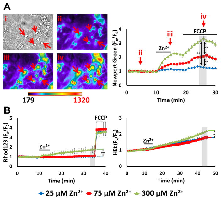
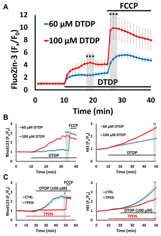
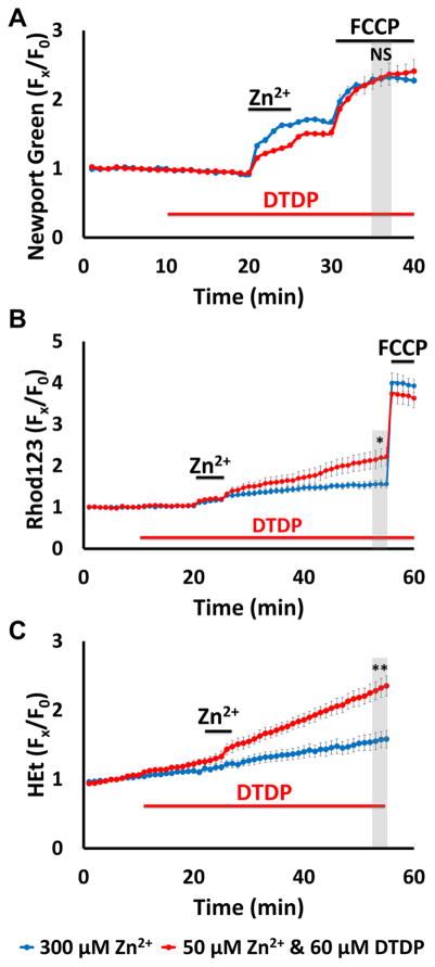
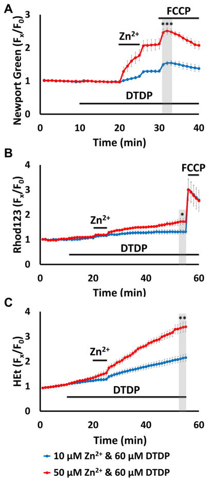
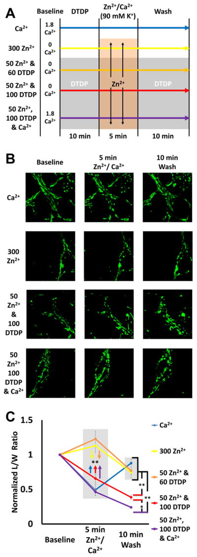
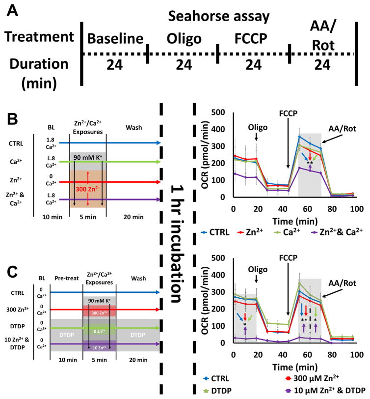
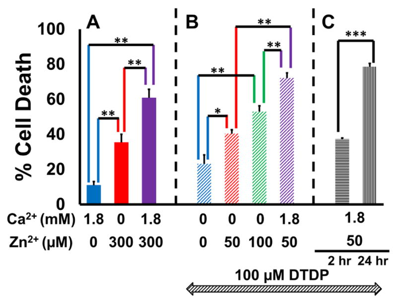
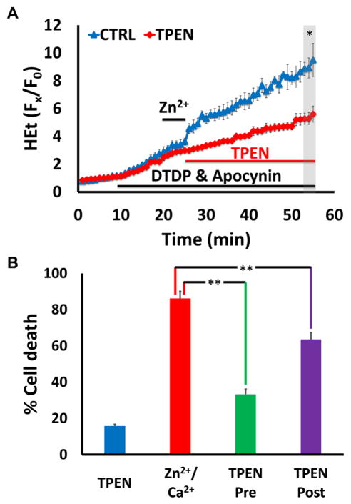
Excitotoxic Zn2+ and Ca2+ accumulation contributes to neuronal injury after ischemia or prolonged seizures. Synaptically released Zn2+ can enter postsynaptic neurons via routes including voltage sensitive Ca2+ channels (VSCC), and, more rapidly, through Ca2+ permeable AMPA channels. There are also intracellular Zn2+ binding proteins which can either buffer neuronal Zn2+ influx or release bound Zn2+ into the cytosol during pathologic conditions. Studies in culture highlight mitochondria as possible targets of Zn2+; cytosolic Zn2+ can enter mitochondria and induce effects including loss of mitochondrial membrane potential (ΔΨm), mitochondrial swelling, and reactive oxygen species (ROS) generation. While brief (5 min) neuronal depolarization (to activate VSCC) in the presence of 300 μM Zn2+ causes substantial delayed neurodegeneration, it only mildly impacts acute mitochondrial function, raising questions as to contributions of Zn2+-induced mitochondrial dysfunction to neuronal injury. Using brief high (90 mM) K+/Zn2+ exposures to mimic neuronal depolarization and extracellular Zn2+ accumulation as may accompany ischemia in vivo, we examined effects of disrupted cytosolic Zn2+ buffering and/or the presence of Ca2+, and made several observations: 1. Mild disruption of cytosolic Zn2+ buffering-while having little effects alone-markedly enhanced mitochondrial Zn2+ accumulation and dysfunction (including loss of ∆Ψm, ROS generation, swelling and respiratory inhibition) caused by relatively low (10-50 μM) Zn2+ with high K+. 2. The presence of Ca2+ during the Zn2+ exposure decreased cytosolic and mitochondrial Zn2+ accumulation, but markedly exacerbated the consequent dysfunction. 3. Paralleling effects on mitochondria, disruption of buffering and presence of Ca2+ enhanced Zn2+-induced neurodegeneration. 4. Zn2+ chelation after the high K+/Zn2+ exposure attenuated both ROS production and neurodegeneration, supporting the potential utility of delayed interventions. Taken together, these data lend credence to the idea that in pathologic states that impair cytosolic Zn2+ buffering, slow uptake of Zn2+ along with Ca2+ into neurons via VSCC can disrupt the mitochondria and induce neurodegeneration.

Keywords: Ca(2+) channel; Calcium; Excitotoxicity; Ischemia; Metallothionein; Mitochondria; Neuronal cultures; Reactive oxygen species; VSCC; Zinc.


Figures

Figure 1
Figure 1. High K+/Zn2+ exposures induce dose-dependent mitochondrial Zn2+ uptake but only mild acute dysfunction
A. High K+/Zn2+ exposures cause dose-dependent mitochondrial Zn2+ accumulation. Cultures were loaded with the low affinity cytosolic Zn2+ indicator Newport Green (Kd ~ 1 μM), exposed to 90 mM K+ (high K+) with Zn2+ (25, 75, 300 μM) for 5 min in 0 Ca2+ HSS, followed by wash into 0 Ca2+ HSS for additional 5 min prior to application of FCCP (1 μM). Left: Representative images: Brightfield image (i) shows appearance of neurons at baseline, and pseudocolor images show Newport Green fluorescence from the same field at baseline (ii), 5 min after high K+/300 μM Zn2+ exposure (iii), and 5 min after FCCP (iv). (Arrows highlight the same neurons in these images.) Right: Traces show time course of Newport Green ΔF (background subtracted and normalized to baseline [Fx/F0]; arrows indicate the time points illustrated in the images), and represent means ± standard error of the mean (SEM) of 6 experiments, ≥ 120 neurons. Grey bar indicates time points of comparison (** indicates p < 0.01 by one-way ANOVA with Tukey post hoc). Note the Zn2+ exposure concentration-dependent mitochondrial Zn2+ accumulation, indicated by the increase in ΔF after FCCP B). These exposures only induce mild mitochondrial dysfunction: Cultures were loaded with the ΔΨmito sensitive indicator, Rhod123, or the superoxide preferring ROS indicator, HEt, and exposed to high K+/Zn2+ for 5 min, followed by wash into 0 Ca2+ HSS, as above. After 20 min, FCCP (1 μM) was applied to Rhod123-loaded cultures to induce full loss of ΔΨm. Traces show time course of Rhod123 ΔF (left) or HEt ΔF (right), normalized to baseline values (after background subtraction, as above; Fx/F0), and represent mean ± SEM 6 experiments, ≥ 120 neurons. Grey bars indicate time points of comparison (* indicates p < 0.05, ** indicates p < 0.01, by one-way ANOVA with Tukey post hoc). Note that only 300 μM Zn2+ exposure induced discernable effects.
Figure 2
Figure 2. Disruption of cytosolic Zn2+ buffering leads to Zn2+-dependent mitochondrial dysfunction
Cultures were loaded with the high affinity Zn2+ indicator FluoZin-3 (Kd ~ 15 nM), Rhod123 or HEt in 0 Ca2+ HSS, then exposed to DTDP (60 or 100 μM; to disrupt cytosolic Zn2+ buffering), with FCCP (1 μM) or TPEN (20 μM) applied as indicated. Traces represent mean ± SEM Fx/F0 values for each indicator and represents ≥ 5 experiments consisting of ≥ 100 neurons. Grey bars indicate time points of comparison (* indicates p < 0.05, ** indicates p < 0.01, *** indicates p < 0.001, by two-tailed t-test). A). DTDP induces dose-dependent cytosolic Zn2+ release and mitochondrial Zn2+ accumulation: FluoZin-3 loaded neurons were exposed to DTDP followed by addition of FCCP, as indicated. Note the dose dependent effects of DTDP, with 100 μM causing both greater cytosolic Zn2+ rise and mitochondrial Zn2+ loading (as indicated by the FCCP-induced ΔF) than 60 μM. B). Disruption of buffering via DTDP can induce mitochondrial dysfunction: Rhod123- (left) or HEt- (right) loaded neurons were subjected to the indicated DTDP and FCCP exposures. Note that the 100 μM DTDP exposure resulted in substantial loss of ΔΨmito within 25 min (as indicated by the minimal response to FCCP) and significant ROS production, while 60 μM DTDP had far smaller effects. C). DTDP effects on mitochondria are Zn2+-dependent: Rhod123- (left) or HEt- (right) loaded neurons were exposed to 100 μM DTDP ± Zn2+ chelator TPEN (applied 10 min before DTDP), followed by FCCP (only in Rhod123 loaded cultures), as indicated. Note that TPEN largely eliminated the DTDP induced loss of ΔΨmito (left) and markedly attenuated the ROS production (right).
Figure 3
Figure 3. Impaired cytosolic Zn2+ buffering markedly enhances the acute impact of Zn2+ exposures on mitochondria
Cultures were loaded with Newport Green, Rhod123, or HEt in 0 Ca2+ HSS, and exposed to high K+/300 μM Zn2+ alone (blue), or to high K+/50 μM Zn2+ with 60 μM DTDP (applied as indicated; red); FCCP (1 μM) was added as indicated. Traces represent mean ± SEM Fx/F0 values for each dye and represents 6 experiments consisting of ≥ 120 neurons. Grey bars indicate time points of comparison (NS indicates No Significance, * indicates p < 0.05, ** indicates p < 0.01, by two-tailed t-test). A). Low exogenous Zn2+ exposure to neurons with impaired buffering results in similar degrees of mitochondrial uptake as much higher Zn2+ exposure with intact buffering: Note the similar magnitudes of mitochondrial Zn2+ loading caused by the high K+/300 μM Zn2+ (blue) and the high K+/50 μM Zn2+/DTDP exposures. B, C). However, the lower Zn2+ exposure with impaired buffering results in greater mitochondrial dysfunction: Rhod123 (B) or HEt (C) loaded neurons were exposed as indicated. Note that the 50 μM Zn2+/DTDP exposure induced markedly greater loss of Δψm and ROS generation than 300 μM Zn2+ alone.
Figure 4
Figure 4. Zn2+ exposure dose-dependence of mitochondrial Zn2+ loading and acute dysfunction in neurons with impaired buffering
Cultures were loaded with Newport Green, Rhod123 or HEt in 0 Ca2+ HSS, and exposed to high K+ with 10 (blue) or 50 (red) μM Zn2+ in 60 μM DTDP. FCCP (1 μM) was added as indicated. Traces represent mean ± SEM Fx/F0 values for each dye and represents 6 experiments consisting of ≥ 120 neurons. Grey bars indicate time points of comparison (* indicates p < 0.05, ** indicates p < 0.01, *** indicates p < 0.001, by two-tailed t-test). A). Zn2+ exposure induces dose-dependent mitochondrial Zn2+ loading in neurons with disrupted buffering: Note the dose dependency of the cytosolic Zn2+ rise and mitochondrial Zn2+ uptake, with the 50 μM Zn2+ exposure causing far greater Zn2+ uptake than the 10 μM Zn2+. B, C). Mitochondrial dysfunction reflects the extent of Zn2+ accumulation: Rhod123 (B) or HEt (C) loaded neurons were exposed as indicated. Note that the 50 μM Zn2+ exposure induced far greater loss of Δψm and ROS generation than 10 μM Zn2+. Further note that despite causing relatively strong ROS generation, 50 μM Zn2+ still only caused modest loss of Δψm.
Figure 5
Figure 5. Ca2+ attenuates mitochondrial Zn2+ accumulation despite exacerbating the consequent dysfunction
Cultures were loaded with Newport Green, Rhod123 or HEt in 0 or 1.8 Ca2+ HSS, and exposed to high K+ with 300, 50,10 or 0 μM Zn2+ (as indicated, along with 10 μM MK-801, to inhibit Ca2+-entry via NMDA receptor activation); DTDP (60 μM), FCCP (1 μM) and/or apocynin (500 μM) were added as indicated. Traces represent mean ± SEM Fx/F0 values for each dye and represents ≥ 5 experiments consisting of ≥ 120 neurons. Grey bars indicate time points of comparison (NS indicates No Significance, * indicates p < 0.05, ** indicates p < 0.01, *** indicates p < 0.001, by two-tailed t-test [A, C] or by one-way ANOVA with Tukey post hoc [B]). A). Presence of Ca2+ decreases neuronal and mitochondrial Zn2+ uptake: Note that presence of Ca2+ attenuated both cytosolic Zn2+ rise during the exposure and FCCP-induced mitochondrial Zn2+ release. B). Ca2+ and Zn2+ synergistically induce mitochondrial dysfunction: Neurons loaded with Rhod123 (left) or HEt (right) were exposed to high K+/DTDP/MK-801 with 10 μM Zn2+ (blue), 1.8 mM Ca2+ (red) or with both Zn2+ and Ca2+ (purple) for 5 min, then washed as indicated. Apocynin was added to HEt-loaded neurons (right) to inhibit contributions from Ca2+-dependent NOX activation. Note that despite relatively little effects from Ca2+ and Zn2+ individually, together they induced significant mitochondrial dysfunction. C). Overwhelming mitochondrial Zn2+ loading induces rapid mitochondrial depolarization: Neurons loaded with Rhod123 (left) or HEt (right) in 1.8 Ca2+ HSS were exposed to high K+/DTDP/MK-801/Ca2+, with 50 (blue) or 300 μM (red) Zn2+ for 5 min, followed by wash as indicated. Note that 300 μM Zn2+ induced greater loss of ΔΨm than 50 μM Zn2+, despite both inducing similar levels of ROS generation.
Figure 6
Figure 6. Effects of Ca2+, Zn2+, and disruption of cytosolic Zn2+ buffering on mitochondrial morphology
A). Experiment schematic: Neurons loaded with the mitochondrial dye MitoTracker Green (200 nM) were placed in 0 or 1.8 Ca2+ HSS, then exposed to DTDP (60 or 100 μM; where indicated), high K+/MK-801 with Zn2+ (0, 50, or 300 μM) and/or 1.8 mM Ca2+, followed by wash into HSS ± DTDP, as described. B). Representative images: Confocal images (1000x) were taken at baseline, 5 min after Zn2+ and/or Ca2+ exposure, and 10 min after wash. C). Zn2+ and Ca2+ induce different patterns of morphology change: The length and width of individual mitochondria were measured blindly, and length/width (L/W) ratios calculated and normalized to baseline. Values for baseline, 5 min after exposure, and after 10 min wash are displayed. Traces show mean ± SEM normalized L/W ratio for each time point, each representing ≥ 5 experiments consisting of ≥ 50 mitochondria (* indicates p < 0.05, ** indicates p < 0.01, by one-way ANOVA with Tukey post hoc). Note that while the Ca2+ induces a rapid but transient morphologic change, Zn2+ triggers more progressive changes (that increase with the degree of Zn2+ loading).
Figure 7
Figure 7. Zn2+-induced inhibition of mitochondrial respiration: synergy with Ca2+ and effects of disrupted buffering
A). Schematic of experiment: Neurons were exposed to a series of treatments (detailed in B and C, left), incubated for 1 hr, then placed in the Seahorse assay, which measures O2 consumption rate (OCR) at baseline and after sequential application of oligomycin (Oligo; 1 μM), FCCP (2 μM), and antimycin A & rotenone (AA/Rot; both 1 μM) to characterize various respiratory parameters. Traces (B and C, right) show time course of OCR and represent mean ± SEM of 3 separate experiments, each consisting of 3 – 4 wells of cultured neurons, with arrows indicating time point at which mitochondrial inhibitors were added. Grey bars indicate time points of comparison (* indicates p < 0.05, ** indicates p < 0.01, by one-way ANOVA with Tukey post hoc). B). Ca2+ and Zn2+ synergistically inhibit mitochondrial respiration: Neurons were placed in 0 or 1.8 Ca2+ HSS, exposed to high K+/MK-801 with 300 μM Zn2+, 1.8 mM Ca2+ or both Zn2+ and Ca2+ as described (left). After 1 hr incubation OCR was measured (right). Note that simultaneous exposure to Zn2+ and Ca2+ induced significant inhibition of mitochondrial respiration, despite the ions having minimal effects individually. C). Disrupted Zn2+ buffering significantly exacerbates Zn2+ effects on mitochondrial respiration: Neurons were placed in 0 Ca2+ HSS, exposed to DTDP (100 μM; where indicated), high K+/MK-801 with Zn2+ (300, 10 or 0 μM, as indicated; left). After 1 hr incubation OCR was measured (right). Note the near complete inhibition of mitochondrial respiration by 10 μM Zn2+ exposure with DTDP.
Figure 8
Figure 8. Mitochondrial Zn2+ accumulation contributes to neuron death
Neurons were exposed to a sequence of 10 min DTDP (100 μM; as indicated in B and C), 5 min high K+/MK-801 exposures with Zn2+ and/or Ca2+ (concentration as shown), washed for 10 min (with DTDP in B and C), transferred to MEM + 25 mM glucose and returned to the incubator for 24 hrs (or for only 2 hrs where indicated in C), prior to assessing cell death via LDH efflux assay. Bars show % cell death (see Material and methods), and represent mean ± SEM of 3 independent experiments, each consisting of 4 wells of cultured neurons (* indicates p < 0.05, ** indicates p < 0.01, *** indicates p < 0.001 by one-way ANOVA with Tukey post hoc [A and B] or by two-tailed t-test [C]). A). Ca2+ and Zn2+ synergistically induce cell death: Note that while 300 μM Zn2+ induced more cell death than 1.8 mM Ca2+, its impact was further exacerbated by the presence of Ca2+. B). Dose-dependency of Zn2+-induced cell death under conditions of strongly disrupted buffering: Note the dose-dependent increase in cell death with increasing Zn2+ exposures, that was further enhanced by the presence of Ca2+. C). Zn2+-induced cell death progresses gradually over hours: Note the significantly greater cell death at 24 hrs compared to 2 hrs.
Figure 9
Figure 9. Delayed Zn2+ chelation attenuates mitochondrial ROS generation and neuron death
Neurons were exposed to high K+/50 μM Zn2+/MK-801, with DTDP (100 μM), and apocynin (500 μM, A only). TPEN was applied as indicated below. Traces (A) represent HEt Fx/F0 and bars (B) represent % cell death after 24 hr; all values are mean ± SEM and represents ≥ 3 independent experiments. Grey bar (in A) indicates time points of comparison (* indicates p < 0.05, ** indicates p < 0.01, by two-tailed t-test [A] or by one-way ANOVA with Tukey post hoc [B]). A). Delayed Zn2+ chelation attenuates Zn2+ triggered ROS production: Note the rapid rise in HEt ΔF that was largely attenuated by TPEN (20 μM), added after the Zn2+ exposure. B). Zn2+ chelation attenuates cell death even when delivered after the Zn2+ exposure: Cultured neurons were exposed as described, with TPEN (10 μM) present either for 10 min before, during and 10 min after high K+/Zn2+ exposure (TPEN pre), or only for 10 min after Zn2+ exposure (TPEN post). Cultures were then transferred to MEM + 25 mM glucose and returned to the incubator for 24 hrs, prior to assessing cell death via LDH efflux assay. Note that both the TPEN pre- and post-treatments attenuated neuron death.

Similar articles

Cited by

References

    1. Aizenman E, Stout AK, Hartnett KA, Dineley KE, McLaughlin B, Reynolds IJ. Induction of neuronal apoptosis by thiol oxidation: putative role of intracellular zinc release. J Neurochem. 2000;75:1878–1888. - PubMed
    1. Aras MA, Aizenman E. Obligatory role of ASK1 in the apoptotic surge of K+ currents. Neurosci Lett. 2005;387:136–140. - PMC - PubMed
    1. Assaf SY, Chung SH. Release of endogenous Zn2+ from brain tissue during activity. Nature. 1984;308:734–736. - PubMed
    1. Bindokas VP, Jordan J, Lee CC, Miller RJ. Superoxide production in rat hippocampal neurons: selective imaging with hydroethidine. J Neurosci. 1996;16:1324–1336. - PMC - PubMed
    1. Bonanni L, Chachar M, Jover-Mengual T, Li H, Jones A, Yokota H, Ofengeim D, Flannery RJ, Miyawaki T, Cho CH, Polster BM, Pypaert M, Hardwick JM, Sensi SL, Zukin RS, Jonas EA. Zinc-dependent multi-conductance channel activity in mitochondria isolated from ischemic brain. J Neurosci. 2006;26:6851–6862. - PMC - PubMed

Publication types

MeSH terms