Review
. 2018 Mar;16(3):156-170.
doi: 10.1038/nrmicro.2017.161. Epub 2018 Jan 22.

Drug resistance in Plasmodium

Affiliations
Review

Drug resistance in Plasmodium

Kasturi Haldar et al. Nat Rev Microbiol. 2018 Mar.

Abstract

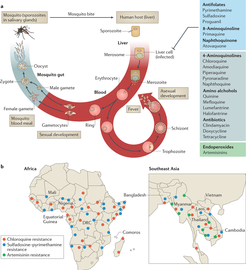
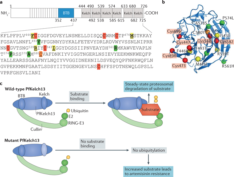
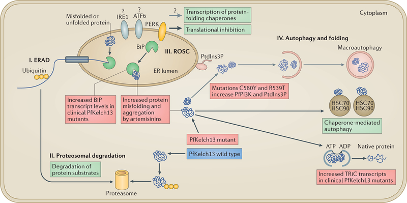
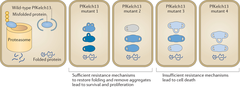
A marked decrease in malaria-related deaths worldwide has been attributed to the administration of effective antimalarials against Plasmodium falciparum, in particular, artemisinin-based combination therapies (ACTs). Increasingly, ACTs are also used to treat Plasmodium vivax, the second major human malaria parasite. However, resistance to frontline artemisinins and partner drugs is now causing the failure of P. falciparum ACTs in southeast Asia. In this Review, we discuss our current knowledge of markers and mechanisms of resistance to artemisinins and ACTs. In particular, we describe the identification of mutations in the propeller domains of Kelch 13 as the primary marker for artemisinin resistance in P. falciparum and explore two major mechanisms of resistance that have been independently proposed: the activation of the unfolded protein response and proteostatic dysregulation of parasite phosphatidylinositol 3- kinase. We emphasize the continuing challenges and the imminent need to understand mechanisms of resistance to improve parasite detection strategies, develop new combinations to eliminate resistant parasites and prevent their global spread.


Figures

Figure 1 |
Figure 1 |. Life cycle of Plasmodium falciparum and epidemiology of antimalarial drug resistance.
a | Infection begins when a mosquito bite releases sporozoites into the bloodstream of the vertebrate host. Sporozoites subsequently infect hepatocytes, in which they proliferate and develop into merozoites. Merozoites are released into blood, where they invade erythrocytes. The parasites develop into the ring form and subsequently develop into proliferative (and morphologically distinct) trophozoite and schizont stages. When the schizont lyses, new merozoites are released, which initiate a new asexual cycle in the blood. A small proportion of ring-form parasites develop into gametocytes (sexual stages), which are taken up by the mosquito during a blood meal. In the mosquito midgut, male and female gametes emerge and fuse to form zygotes, which further differentiate into oocysts. Each oocyst divides to produce and release thousands of haploid sporozoites into the mosquito body cavity. These sporozoites travel and invade the mosquito salivary glands, from where they are injected into the human host. Trophozoite and schizont stages are sequestered in host tissue to cause severe disease. All antimalarial classes shown (and comprehensively reviewed in TABLE 1) target the asexual trophozoite and schizont stages. Antifolates, primaquine (8-aminoquinoline) and atovaquone (naphthoquinone) also target liver-stage parasites. Endoperoxides (artemisinins) target all asexual and early sexual blood-stage parasites. Primaquine is the only drug that targets latency in the liver to prevent the relapsing infection characteristic of Plasmodium vivax. b | Detailed maps showing the distribution of P. falciparum resistance to chloroquine, sulfadoxine-pyrimethamine and artemisinin in Africa and southeast Asia,,,,,,–. Artemisinin resistance is based on two criteria: clearance and PfKelch13-associated mutation. Although the A578S mutation has been reported, no pfkelch13 mutations associated with resistance have been reported yet in Bangladesh (resistance is indicated at the Bangladesh-Myanmar border). Resistance to chloroquine and sulfadoxine- pyrimethamine (but not artemisinin) has emerged in the Amazon basin of South America (not shown). Resistance of P. vivax to chloroquine is emerging (not shown). Each dot represents a region of emergence of drug resistance. DRC, Democratic Republic of the Congo. Part a adapted from REF. .
Figure 2 |
Figure 2 |. Schematic and structural representation of Plasmodium falciparum Kelch 13 and its hypothesized function as a substrate adapter for a cullin E3 ligase.
a | Plasmodium falciparum Kelch 13 (PfKelchl3) contains a single BTB domain (common to all Broad-complex, Tramtrack and Bric-a-brac domain (BTB) family proteins). The BTB domain is a homodimerization domain at the amino terminus of proteins that contain multiple copies of Kelch repeats. PfKelch13 contains six β-propeller domains, also called Kelch domains, characteristic of families of Kelch proteins conserved in eukaryotes. The amino acid sequences of the PfKelch13 β-propeller domains are shown, with cysteine residues indicated in red (Cys447, Cys469, Cys473, Cys532, Cys542 and Cys696). C580Y (shown in orange) is the most prevalent resistance mutation in Cambodia. Cysteine mutations identified in Cambodia that have been validated in the laboratory (R539T and I543T) and a major mutation (F446I) found in Myanmar, as well as the M476I mutation (which confers high-level resistance in the laboratory) are indicated in yellow. Mutations that are not associated (A578S) or are less associated (Y493T) with resistance or that have been identified in clinical isolates in minor populations (R561H, Y493T and P574L) are indicated in green. b | A 3D model of PfKelch13 amino acid residues 338–726 generated by pyMOL from the Research Collaboratory for Structural Bioinformatics Protein Data Bank (RCSB PDB; ID: 4YY8) shows R539T, I543T, F446I and M476I proximal to mutations in cysteine residues primarily in β strands and A578S and Y493T mutations in loops. c | The single BTB domain of PfKelch13 is expected to bind the cullin-really interesting new gene (RING)-E3 ubiquitin ligase complex, whereas the β-propeller Kelch domain binds substrate to facilitate ubiquitin transfer from the E2 ligase, which is also present in the complex, for degradation by the 26S proteasome. Mutation in (β-propeller) Kelch domains reduces the affinity of PfKelch13 for its substrates, their ubiquitylation and proteasomal degradation, thereby increasing substrate half-life in the cytoplasm. The major southeast Asia artemisinin-resistance mutation, PfKelch13-C580Y, has been shown to decrease PfKelch13 binding and ubiquitylation of P. falciparum phosphatidylinositol 3-kinase (PfPI3K), leading to increased levels of the kinase itself and of its lipid product phosphatidylinositol-3-phosphate (PtdIns3P), which confers artemisinin resistance. The PfKelch13-R539T mutation was also found to increase PtdIns3P levels.
Figure 3 |
Figure 3 |. Model of protein quality control in the endoplasmic reticulum lumen and cytoplasm of eukaryotes illustrating mechanisms of artemisinin resistance in Plasmodium falciparum.
In eukaryotes, proteostasis pathways function in protein quality control in the endoplasmic reticulum (ER) and the cytoplasm and stimulate protein translation, translocation, vesicular export and additional chaperone functions to restore proper folding and function of proteins. They have been proposed to rescue Plasmodium falciparum cells from artemisinin-induced protein alkylation, aggregation and toxicity (or proteopathy) and death. There are four major pathways that maintain quality control of proteins in the ER and cytoplasm of eukaryotes, as indicated in the figure. In the ER-associated degradation (ERAD) pathway (I), misfolded proteins are unfolded by binding to the ER chaperone immunoglobulin heavy chain-binding protein (BiP), translocated from the ER to the cytoplasm, ubiquitylated and targeted for proteasomal degradation by the ubiquitin-26S proteasome pathway (II). Misfolded proteins in the ER are initially removed by ERAD, but when this pathway gets saturated, BiP continues to bind to misfolded proteins in the ER lumen. BiP is a key component of the reactive oxygen stress complex (ROSC) pathway (III) in the ER lumen, which is regulated by the unfolded protein response (UPR). Under equilibrium conditions, BiP binds to and keeps inactive transmembrane ER stress-response proteins activating cAMP-dependent transcription factor (ATF6), inositol-requiring protein 1 (IRE1) and protein-kinase R (PKR)-like ER kinase (PERK; which targets eukaryotic translation initiation factor 2A, eIF2A, to suppress translation). Because BiP has a high preference for unfolded proteins, accumulation of these proteins in the ER lumen decreases the binding of BiP to transmembrane stress-response proteins, leading to their activation. In clinical isolates of artemisinin-resistant P. falciparum, increased transcript levels of the gene encoding BiP have been detected, possibly indicating a mechanism of artemisinin resistance to remove misfolded, aggregated toxic proteins and to activate putative transmembrane ER stress-response proteins. Because P. falciparum orthologues of PERK and eIF2A exist, activation of PERK might yield a UPR mechanism of translational repression and explain why artemisinin-resistant ring parasites may temporarily slow down. However, P. falciparum does not encode for orthologues of transcription factors of ATF6 and IRE1, which are the main drivers of UPR in eukaryotes. Therefore, these major UPR pathways of transcriptional activation of redox survival (needed for resistance) have yet to be established in P. falciparum. Thus, how the parasite induces the UPR-induced transcriptional increase of ROSC and the compensatory global antioxidant responses needed for resistance is unclear. Cytoplasmic unfolded and misfolded proteins may also be removed by autophagy (IV), including by macroautophagy, which involves phosphatidylinositol-3-phosphate (PtdIns3P)-dependent expansion of the ER. However, macroautophagy has not been established in P. falciparum. Chaperone-mediated autophagy is a second type of autophagy that also assists in the removal of misfolded proteins via interactions with cytoplasmic heat shock protein 70 (HSC70) and HSC90. T-Complex protein 1 (TCP1) ring complex (TRiC) chaperones enable misfolded proteins to become properly folded in the cytoplasm (and hence, TRiC substrates do not undergo chaperone-mediated autophagy). Resistant clinical isolates have been shown to have increased transcript levels of genes encoding TRiC chaperones. Thus, a model has been proposed whereby the transcriptional increase of two major chaperone complexes, the ROSC linked to the UPR and the TRiC, might mitigate toxic protein aggregates in the ER and cytoplasm by accelerating their removal through the proteasome (via the ROSC) or by restoring proper folding (via the TRiC). PfKelch13 mutations C580Y and R539T increase levels of the P. falciparum phosphatidylinositol 3-kinase (PfPI3K) and its lipid product phosphatidylinositol-3-phosphate (PtdIns3P). Thus, stimulation of macroautophagy by elevation of PtdIns3P suggests a second mechanism to remove toxic protein aggregates from the parasite.
Figure 4 |
Figure 4 |. A working model for heterogeneity in levels of artemisinin resistance.
In wild-type parasites, in addition to properly folded proteins, misfolded or poorly folded proteins are also made. These ill-folded proteins are bound by Plasmodium falciparum Kelch 13 (PfKelch13), ubiquitylated and targeted to the proteasome for degradation, which keeps their levels low in the parasite. In PfKelch13 mutants, in the absence of ubiquitylation and protein quality control, PfKelch13 substrates (blue) accumulate with varying levels of folding and activity (shown by numbers of loops and intensity of blue colour, respectively). PfKelch13 substrates such as P. falciparum phosphatidylinositol 3-kinase (PfPI3K) may function in pathways that restore protein folding or remove aggregates to mitigate the toxicity induced by the drug. Therefore, genotypically identical parasites have heterogeneous resistance mechanisms to remove the toxic misfolded aggregates induced by artemisinins. As such, following artemisinin-induced toxicity due to protein alkylation and oxidation, only parasites with sufficient resistance mechanisms (which allow clearance of toxic intermediates and restoring of protein folding) survive and proliferate.

Similar articles

Cited by

References

    1. World Health Organization. World Malaria Report 2015 (WHO, 2015).
    1. World Health Organization. World Malaria Report 2016 (WHO, 2016).
    1. Guerra CA et al. The international limits and population at risk of Plasmodium vivax transmission in 2009. PLOS Negl. Trop. Dis 4, e774 (2010). - PMC - PubMed
    1. Haldar K, Murphy SC, Milner DA & Taylor TE Malaria: mechanisms of erythrocytic infection and pathological correlates of severe disease. Annu. Rev. Pathol 2, 217–249 (2007). - PubMed
    1. Milner DA Jr. et al. Quantitative assessment of multiorgan sequestration of parasites in fatal pediatric cerebral Malaria. J. Infect. Dis 212, 1317–1321 (2015). - PMC - PubMed

Publication types

MeSH terms

LinkOut - more resources