Review
. 2018 Feb;9(1):103-118.
doi: 10.1007/s13244-017-0586-x. Epub 2018 Jan 22.

Mechanical birth-related trauma to the neonate: An imaging perspective

Affiliations
Review

Mechanical birth-related trauma to the neonate: An imaging perspective

Apeksha Chaturvedi et al. Insights Imaging. 2018 Feb.

Abstract

Mechanical birth-related injuries to the neonate are declining in incidence with advances in prenatal diagnosis and care. These injuries, however, continue to represent an important source of morbidity and mortality in the affected patient population. In the United States, these injuries are estimated to occur among 2.6% of births. Although more usual in context of existing feto-maternal risk factors, their occurrence can be unpredictable. While often superficial and temporary, functional and cosmetic sequelae, disability or even death can result as a consequence of birth-related injuries. The Agency for Healthcare research and quality (AHRQ) in the USA has developed, through expert consensus, patient safety indicators which include seven types of birth-related injuries including subdural and intracerebral hemorrhage, epicranial subaponeurotic hemorrhage, skeletal injuries, injuries to spine and spinal cord, peripheral and cranial nerve injuries and other types of specified and non-specified birth trauma. Understandably, birth-related injuries are a source of great concern for the parents and clinician. Many of these injuries have imaging manifestations. This article seeks to familiarize the reader with the clinical spectrum, significance and multimodality imaging appearances of neonatal multi-organ birth-related trauma and its sequelae, where applicable. Teaching points • Mechanical trauma related to birth usually occurs with pre-existing feto-maternal risk factors.• Several organ systems can be affected; neurologic, musculoskeletal or visceral injuries can occur.• Injuries can be mild and transient or disabling, even life-threatening.• Imaging plays an important role in injury identification and triage of affected neonates.

Keywords: Cephalopelvic disproportion; Instrumental delivery; Macrosomia; Mechanical trauma; Neonate.


Figures

Fig. 1
Fig. 1
Illustration demonstrating the layers of scalp, skull, meninges and brain on a coronal section (a)
Fig. 2
Fig. 2
Illustration depicting hemorrhages by location within the different layers of the meninges (left of image) and scalp (right of image)
Fig. 3
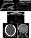
Fig. 3. Caput succedaneum and subgaleal hemorrhage
Grayscale ultrasound images (a-c) of the scalp in a newborn male demonstrate a fluid collection that crosses midline, is deep to the subcutaneous fat/galeal aponeurosis and superficial to the periosteum/calvarium (noted as thick echogenic interface) consistent with a subgaleal hemorrhage. Axial CT (d) image in a 1-day-old male with history of traumatic delivery demonstrate scalp soft tissue overlying bilateral parietal regions and crossing the sagittal suture (arrows). Follow-up coronal MR image (e) demonstrates a deep subaponeurotic scalp fluid collection crossing the sagittal suture and extending anteriorly into the right temporal region, consistent with subgaleal hematoma (arrows). A more superficial overlying fluid collection with a similar distribution also noted (arrowheads). This collection is within the subcutaneous fibrofatty tissues superficial to galea aponeurosis and is consistent with caput succedaneum
Fig. 4
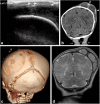
Fig. 4. Cephalhematoma
1-month-old male with history of traumatic delivery presenting with right parietooccipital soft tissue swelling. Transverse grayscale ultrasound (a) image of the left parietooccipital scalp shows a complex fluid collection (arrow), with punctate linear echogenic foci along the superficial aspect (arrowheads), suggestive of calcifications. Relationship with the adjacent left lambdoid suture was difficult to evaluate by ultrasound. B. Coronal non-contrast head CT (b) image demonstrates a lobulated fluid collection with thick septations and peripheral calcifications (arrowheads) that does not cross the adjacent sagittal or the lambdoid suture, suggestive of cephalhematoma. 3-D volume (c) rendered images in bone algorithm shows cortical irregularity along the left parietal bone at the site of cephalhematoma as well as peripheral calcifications along the superficial aspect of the cephalhematoma. Coronal T2 image (d) from an MR exam obtained one week later in the setting of patient’s seizures re-demonstrates the large subperiosteal complex fluid collection with thick septations and isointense fluid signal consistent with evolving blood products in the known left parietal cephalhematoma
Fig. 5
Fig. 5. Molding of the skull post vaginal delivery
Immediate post-delivery appearances of the skull on head CT. The occipital bone is slightly depressed with associated sutural overlap as seen on the axial and coronal CT images and the 3-D reconstructions (a-d). Lateral skull radiograph (e) demonstrates overlap of the occipital bone (white arrow)
Fig. 6
Fig. 6. Skull fractures in two neonates
Axial bony algorithm reconstruction from a head CT (a) in a 2-day-old demonstrates a non-displaced linear left parietal skull fracture (arrow) with overlying soft tissue swelling (marked by asterisk on a). Ultrasound and CT images on another 1-day-old male (b, c) with a history of traumatic delivery characterized by multiple attempts of vacuum extraction. Coronal gray scale ultrasound image (b) demonstrates a displaced left parietal fracture (arrow) with underlying extra-axial fluid collection (arrowheads). Axial non-contrast head CT image (c) shows a complex left parietal bone fracture with an angulated anterior component and an adjacent depressed “ping-pong” fracture component posteriorly (arrow). There is an associated overlying hyperdense fluid collection consistent with cephalhematoma (arrowhead). There is also an underlying large epidural hemorrhage with fluid/fluid levels. 3-D volume rendered image (d) re-demonstrates the complex left parietal bone fracture (black arrows)
Fig. 7
Fig. 7. Leptomeningeal cyst
Axial skull CT (a) in a now 6-year-old with a history of traumatic birth and subsequent cerebral palsy. He had a right posterior parietal calvarial fracture at birth, which did not heal, but enlarged secondary to entrapment of leptomeninges at the fracture site—an entity called growing fracture or leptomeningeal cyst [volume rendered (b), and MIP 3D reconstruction (c)]. Bony margins at the fracture site are scalloped and smooth
Fig. 8
Fig. 8. Subdural Hematoma
Brain MRI in a 6-day-old term neonate with history of vacuum-assisted delivery. Sagittal T1 (a) MR image demonstrates subdural hematomas tracking along bilateral occipital lobes and along the tentorium. Another 8-day-old term neonate with history of prolonged rupture of membranes shows subdural hemorrhage (b) layering along the occipital-parietal convexities and along the tentorium (arrows). Corresponding axial gradient-recalled echo (susceptibility-weighted) image (c) at a slightly more cephalad level reveals hemosiderin staining along the tentorium and posterior convexities of the brain (arrows)
Fig. 9
Fig. 9. Subarachnoid hematoma
Axial T1/GRE MRI in a 9-day-old term neonate with history of difficult, vacuum-assisted delivery. Right frontal blood is noted in a gyriform distribution, suggesting subarachnoid blood. Subdural blood was also noted tracking along the falx
Fig. 10
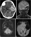
Fig. 10. Extraaxial, intraventricular and parenchymal hemorrhages in 3-day-old female post-complicated vaginal delivery presenting with seizures
Axial non-contrast head CT (a) and coronal T2 MR image (b) show a large left temporal parenchymal hemorrhage (arrows), with an overlying small subdural hemorrhage (white arrowheads). Small foci of subarachnoid hemorrhage noted along the posterior fossa (white arrowheads on a), with susceptibility artifact on axial susceptibility-weighted image (SWI) (c). Axial SWI MR image (d) through the left temporal hemorrhage also demonstrates blood products within occipital horns of bilateral lateral ventricles and trace subdural hemorrhage along the tentorium (white arrowheads). Follow-up MR at 3 months (not shown) did not demonstrate an underlying left temporal parenchymal mass or vascular malformation
Fig. 11
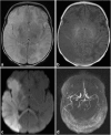
Fig. 11. Middle cerebral artery infarct in a 4-day-old male with history of prolonged delivery with nuchal cord presenting with seizures
Axial T2 (a), T1(b) and diffusion MR (c) images demonstrate an extensive region of hypointense T1 and hyperintense T2 signal involving right cerebral hemisphere in the distribution of the middle cerebral artery, with effacement of the sulci and right lateral ventricle, with corresponding restricted diffusion, consistent with right MCA distribution subacute ischemic infarct. Restricted diffusion also extends into the right thalamus, basal ganglia, cerebral peduncle and across the splenium of the corpus callosum. Axial MIP image (d) from a 2 D time-of-flight MR angiogram of the head without contrast shows normal intracranial arteries with no evidence of decreased or absent flow in the right MCA
Fig. 12
Fig. 12. Spinal cord injury in 4-day-old female with a history of shoulder dystocia presenting with right sided upper and lower extremity neurologic deficits
Axial T2 (a) and sagittal T1 (b) images of the cervical spine demonstrate a focal area of T1 hyperintensity and T2 hypointensity (arrows) in the high cervical cord at the level of C2–3 consistent with acute injury. Focal hemorrhage in the right cervical cord with susceptibility artifact seen on C (axial SWI). Finding was thought consistent with spinal cord injury secondary to stretching/traction
Fig. 13
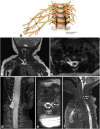
Fig. 13. Peripheral nerve/Brachial plexus injury in two neonates
Illustration (a) depicting normal brachial plexus. Image parts (b-f) High-resolution, steady state free precession (SSFP) MR images (b-d) in a 4-month-old boy with clinically suspected brachial plexus palsy shows a right-sided pseudomeningocele at C8-T1 level. The C8 nerve root was avulsed. Axial MR SSFP image of the cervical spine (e) in another 2-month-old female with left brachial plexus palsy demonstrated pseudomeningocele formation at the level of C8 (arrow) with disruption of the ventral nerve root (arrowhead). Sagittal MR SSFP image (f) shows disorganized soft tissue within the left C8 foramen with focal enlargement of the nerve at the exit of the neural foramen, consistent with neuroma formation (arrowhead). A smaller pseudomeningocele is also noted at C7 (arrow)
Fig. 14
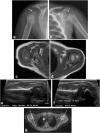
Fig. 14. Glenohumeral dysplasia in two neonates
Follow-up shoulder imaging in a 13-month-old child with clinically suspected brachial plexus palsy at birth (a-c). He subsequently developed glenohumeral dysplasia. The right humeral head is small, glenoid is shallow as seen on the radiograph (a); compare with the normally formed left humeral head and glenoid labrum (b). Axial T1 MR image (c) reveals posterior subluxation of humeral head; compare to normally aligned left humeral head (d). 2-month-old with history of right brachial plexus injury post-delivery (e-g). Ultrasound images from two exams performed 1 month apart (e and f) demonstrate progressive right glenohumeral dysplasia with interval increase in right humeral alpha angle from 33 degrees to 60 degrees. Axial DESS image from a follow-up MR exam (g) shows a shallow right glenoid with posterior humeral head subluxation. Normal left glenohumeral joint
Fig. 15
Fig. 15. Neonatal Phrenic Nerve Palsy
Chest radiograph of a 3-week-old with history of shoulder dystocia shows elevated right dome of diaphragm relative to left. Subsequently performed real-time ultrasound of the diaphragm (not included) revealed diminished excursion of the right hemidiaphragm relative to the left
Fig. 16
Fig. 16. Clavicle fracture, shoulder dystocia
Macrosomic infant of diabetic mother presented with mildly displaced fracture of the left clavicle after a delivery complicated by shoulder dystocia. Patient also had neonatal brachial plexus palsy from which he subsequently recovered
Fig. 17
Fig. 17. Humerus fractures as a consequence of traumatic birth
Frontal radiograph of the left upper extremity in a 2-day-old infant demonstrates mid shaft fracture of the left humerus (a). Illustration (b) depicting chondro-epiphyseal separation at the distal humerus. Upon separation of the distal humeral epiphysis from the bone, it no longer lines up with the distal humerus (black arrow), as seen in figure parts c-d. 10-day-old male twin infant with history of traumatic delivery, presenting with decreased right arm movements. Right elbow radiograph (c) demonstrates fragmentation of the distal right humeral metaphysis, with mild periosteal new bone formation (arrow). Sagittal STIR MR image of the distal right humerus (d) demonstrates increased STIR signal and enhancement surrounding and involving the distal right humeral metaphysis and epiphysis, with mild posterior angulation of the distal epiphysis relative to the metaphysis (arrows), suggestive of distal humeral fracture with chondroepiphyseal separation
Fig. 18
Fig. 18. Femur fractures
Frontal radiograph of the lower extremity in a 1-day-old infant (a) demonstrates displaced an oblique mid-shaft fracture of the right femur. This was a consequence of excessive traction on the femur. Lateral radiograph of the femur (b) in another 1-day-old breech infant demonstrates irregularity at the distal femoral metaphysis, which was proved to be a birth-trauma related physeal injury with chondroepiphyseal separation at the distal femur on the subsequently performed MR (images not included)
Fig. 19
Fig. 19. Fibromatosis colli
Neck ultrasound obtained in a 6-week-old revealed fusiform enlargement of the left sternocleidomastoid without discernible underlying masses. Patient was diagnosed with fibromatosis colli. This patient had a history of dytocic birth
Fig. 20
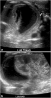
Fig. 20. Visceral Injury
Ultrasound of the left upper quadrant performed in a 3-day-old revealed a crescentic, near anechoic collection in the location of the left adrenal. Baby had a history of difficult birth characterized by prolonged 2nd stage of labor. Hematoma was followed by serial ultrasound and resolved at 13 weeks of age

Similar articles

Cited by

References

    1. Rabelo NN, Matushita H, Cardeal DD. Traumatic brain lesions in newborns. Arq Neuropsiquiatr. 2017;75(3):180–188. doi: 10.1590/0004-282x20170016. - DOI - PubMed
    1. Reichard R. Birth injury of the cranium and central nervous system. Brain Pathol. 2008;18(4):565–570. doi: 10.1111/j.1750-3639.2008.00205.x. - DOI - PMC - PubMed
    1. Hansen A, Chauhan SP. Shoulder dystocia: definitions and incidence. Semin Perinatol. 2014;38(4):184–188. doi: 10.1053/j.semperi.2014.04.002. - DOI - PubMed
    1. Gherman RB, et al. Shoulder dystocia: the unpreventable obstetric emergency with empiric management guidelines. Am J Obstet Gynecol. 2006;195(3):657–672. doi: 10.1016/j.ajog.2005.09.007. - DOI - PubMed
    1. Kichari JR, Gielkens H. Massive traumatic subgaleal haematoma. Emerg Med J. 2013;30(4):344. doi: 10.1136/emermed-2012-201729. - DOI - PubMed