Review
. 2017:1042:207-228.
doi: 10.1007/978-981-10-6955-0_10.

Architecture of the Saccharomyces cerevisiae Replisome

Affiliations
Review

Architecture of the Saccharomyces cerevisiae Replisome

Lin Bai et al. Adv Exp Med Biol. 2017.

Abstract

Eukaryotic replication proteins are highly conserved, and thus study of Saccharomyces cerevisiae replication can inform about this central process in higher eukaryotes including humans. The S. cerevisiae replisome is a large and dynamic assembly comprised of ~50 proteins. The core of the replisome is composed of 31 different proteins including the 11-subunit CMG helicase; RFC clamp loader pentamer; PCNA clamp; the heteroligomeric DNA polymerases ε, δ, and α-primase; and the RPA heterotrimeric single strand binding protein. Many additional protein factors either travel with or transiently associate with these replisome proteins at particular times during replication. In this chapter, we summarize several recent structural studies on the S. cerevisiae replisome and its subassemblies using single particle electron microscopy and X-ray crystallography. These recent structural studies have outlined the overall architecture of a core replisome subassembly and shed new light on the mechanism of eukaryotic replication.

Keywords: CMG; Cryo-EM; DNA polymerase; Eukaryotic DNA replication; Mcm; Replicative helicase; Replisome; Structural biology.


Figures
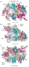
Fig. 10.1
Fig. 10.1
The CMG structure. The CMG structure is shown in cartoon view with each subunit a different color (PDB 3JC7). The surface representation of the structure is superimposed as a dim gray surface view. The structure is shown from the following angles: (a) top CTD ring view, (b) a side view, and (c) a bottom NTD view
Fig. 10.2
Fig. 10.2
Conformation changes proceeding from the inactive Mcm2-7 at an origin to Mcm2-7 in the active CMG helicase. The figure focuses on a side view of the Mcm2 and Mcm5 subunits in the Mcm2-7 hexamer in which one Mcm2-7 hexamer has been extracted from the inactive double hexamer (PDB 3JA8) (left side) and the Mcm2-7 hexamer has been extracted from the active CMG structure (PDB 3JC7) (middle). The two Mcm2-7 structures are then superimposed (right side). Structures are in cartoon view wrapped in a dim gray surface view. Mcm5 is gold and Mcm2 is blue. The Mcm2/5 interface is nearly opened in Mcm2-7 within CMG
Fig. 10.3
Fig. 10.3
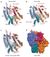
Comparison of the human and the yeast GINS structures. (a) Human GINS with each subunit shown in a different color (PDB 2Q9Q). An N-terminal insertion loop in Psf3 is highlighted by a semitransparent circle near the central pore region. (b) The yeast GINS structure, extracted from the CMG structure (PDB 3JC6), is shown in the same color scheme as in (a). (c) Superimposition of the human and the yeast GINS structures, revealing that the structural variation is focused in the central region. (d) Surface representation of the yeast GINS structure. The central pore is too small to thread ssDNA
Fig. 10.4
Fig. 10.4
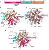
Comparison of the yeast and human Cdc45 structures. (a) The domain structure of Cdc45. (b) Cartoon view of the yeast Cdc45 structure extracted from the CMG structure (PDB 3JC6), with each domain colored according to the domain sketch in (a). PHM refers to protruding helical motif. (c) Superimposition of the yeast and human Cdc45 structures. Human Cdc45 is shown in light purple (PDB 5DGO). (d) Human Cdc45 point mutations identified in patients with Meier-Gorlin syndrome are highlighted as spheres
Fig. 10.5
Fig. 10.5
CMG helicase alternates between tilted (extended) and untilted (compact) conformations. (a) Side view of CMG conformer I (extended) in which the CTD motor ring is tilted relative to the NTD ring. (b) Side view of the CMG in conformer II (compact) with an untilted CTD ring. In panels (a, b), the cryo-EM density map is shown as a semitransparent surface rendering, and the atomic model is shown in cartoon (EMD-6535, EMD-6536, PDB 3JC5, and 3JC7). (c) An oil rig-like pump jack DNA unwinding model. Panel (c) is reproduced in part from Figure 7 in Yuan et al. (2016) with permission. Note that the CTD-tier ring pushing on the dsDNA is only for the purpose of illustration. The pump jack model would still function as a translocase if CMG were oriented with the NTD-tier ring pushing on the dsDNA
Fig. 10.6
Fig. 10.6
Architecture of the CMGE complex. (a) The Pol2 (P2) subunit of Pol ε is comprised of a catalytic N-terminal subdomain and an inactive B-family Pol in the C-terminal subdomain. (b) Negative stain 3D EM map of the CMGE in four different views (EMD-6465). The EM density is segmented and colored according to individual subunits except for the Pol ε where the complex is shown in green. Part of the Pol ε density is not visible in the EM and may be the N-terminal subdomain of Pol2 as proposed in Sun et al. (2015). (c) Cross-linking mass spectrometry identified multiple contacts between Pol2 and GINS, Cdc45, and the CTD regions of Mcm2, Mcm5, and Mcm6 (This figure is modified with permission from Figure 6 of Yuan et al. 2016)
Fig. 10.7
Fig. 10.7
Selected 2D class averages of several replisome complexes. (a) The Ctf4 trimer binds to the GINS from the bottom NTD side of CMG. (b) Pol α-primase appears as a fuzzy density suggesting a flexible association with the Ctf4 just under the NTD ring of Mcm2-7. (c) Simultaneous binding of Pol ε to the CTD side of Mcm2-7 on top and the Ctf4 trimer to the NTD side of CMG. (d) Pol ε binds to the CTD side of CMG in the presence of a primed forked DNA. (e) Pol ε binds to the CTD side of CMG, while Ctf4 and Pol α bind to the NTD side of CMG (This figure is modified with permission from Figure 5 of Sun et al. 2015)
Fig. 10.8
Fig. 10.8
The eukaryotic replisome positions Pols ε and α on opposite sides of CMG. Depending on whether the CTD motor ring or the NTD ring pushes against the forked junction, the polymerases are positioned either: (a) with Pol ε in front and Pol α-primase in back of CMG or (b) with Pol α-primase in front and Pol ε in back of CMG

Similar articles

Cited by

References

    1. Abid Ali F, Renault L, Gannon J, Gahlon HL, Kotecha A, Zhou JC, Rueda D, Costa A. Cryo-EM structures of the eukaryotic replicative helicase bound to a translocation substrate. Nat Commun. 2016;7:10708. - PMC - PubMed
    1. Asturias FJ, Cheung IK, Sabouri N, Chilkova O, Wepplo D, Johansson E. Structure of Saccharomyces cerevisiae DNA polymerase epsilon by cryo-electron microscopy. Nat Struct Mol Biol. 2006;13:35–43. - PubMed
    1. Banks GR, Boezi JA, Lehman IR. A high molecular weight DNA polymerase from Drosophila melanogaster embryos. Purification, structure, and partial characterization. J Biol Chem. 1979;254:9886–9892. - PubMed
    1. Bell SD, Botchan MR. The minichromosome maintenance replicative helicase. Cold Spring Harb Perspect Biol. 2013;5:a012807. - PMC - PubMed
    1. Bell SP, Labib K. Chromosome duplication in Saccharomyces cerevisiae. Genetics. 2016;203:1027–1067. - PMC - PubMed

MeSH terms

LinkOut - more resources