Review
. 2017 Dec 12;8(1):1-8.
doi: 10.1002/cphy.c170012.

Triglyceride Metabolism in the Liver

Affiliations
Review

Triglyceride Metabolism in the Liver

Michele Alves-Bezerra et al. Compr Physiol. .

Abstract

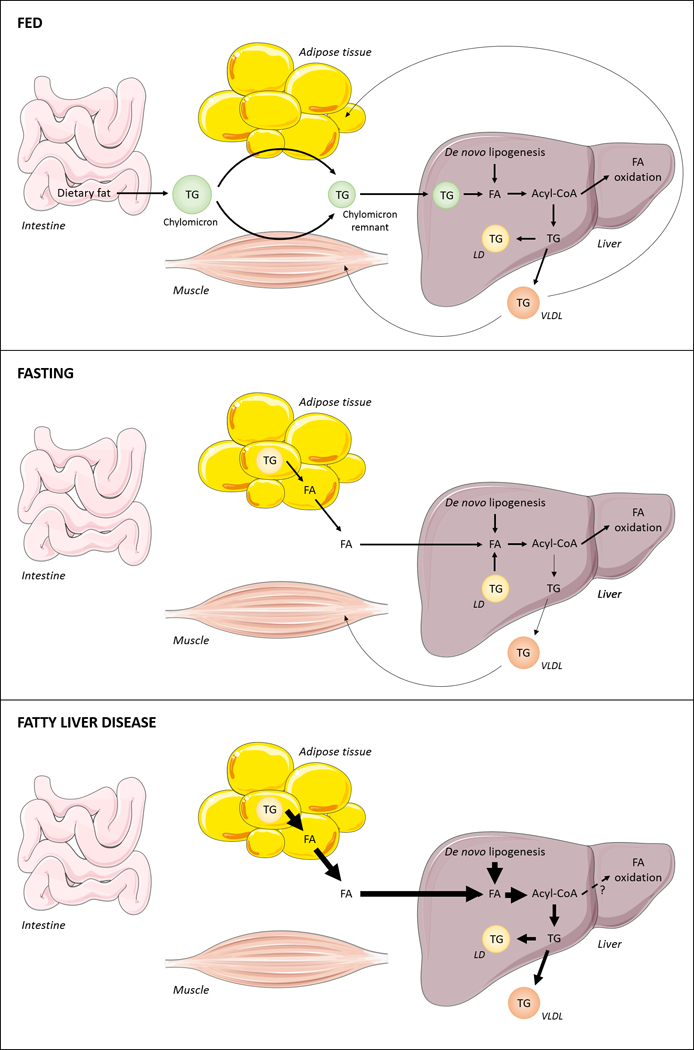
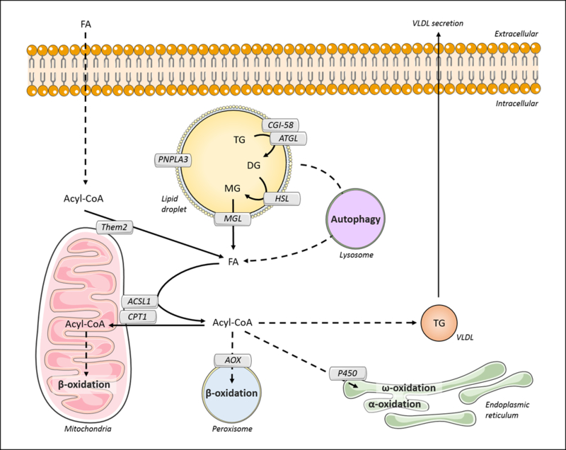
Triglyceride molecules represent the major form of storage and transport of fatty acids within cells and in the plasma. The liver is the central organ for fatty acid metabolism. Fatty acids accrue in liver by hepatocellular uptake from the plasma and by de novo biosynthesis. Fatty acids are eliminated by oxidation within the cell or by secretion into the plasma within triglyceride-rich very low-density lipoproteins. Notwithstanding high fluxes through these pathways, under normal circumstances the liver stores only small amounts of fatty acids as triglycerides. In the setting of overnutrition and obesity, hepatic fatty acid metabolism is altered, commonly leading to the accumulation of triglycerides within hepatocytes, and to a clinical condition known as nonalcoholic fatty liver disease (NAFLD). In this review, we describe the current understanding of fatty acid and triglyceride metabolism in the liver and its regulation in health and disease, identifying potential directions for future research. Advances in understanding the molecular mechanisms underlying the hepatic fat accumulation are critical to the development of targeted therapies for NAFLD. © 2018 American Physiological Society. Compr Physiol 8:1-22, 2018.


Figures

Figure 1.
Figure 1.. Major sources for hepatic fatty acids.
The three major sources for hepatic fatty acids (FA) are dietary lipids, adipose tissue derived-FA and de novo-synthesized FA. After a meal, dietary lipids are hydrolyzed within the intestinal lumen. Upon intestinal uptake, FA are re-esterified to form TG molecules, which are packaged into chylomicrons and delivered primarily to muscle and adipose tissue. The remaining TG present in chylomicron remnants is then transported to the liver and processed intracellularly, leading to FA release within hepatocytes. Carbohydrates, particularly glucose, are utilized in hepatic de novo lipogenesis (DNL) for the production of FA. In order to be metabolized, FAs are activated to form acyl-CoA molecules, which can undergo oxidation or be incorporated into complex lipids. Locally synthesized TG can be stored in intracellular lipid droplets (LDs) or packed into VLDL and secreted into the plasma. Upon fasting, intracellular TG stores are mobilized from adipocytes and hepatocytes to release FA products. Hepatic DNL may also contribute to form an acyl-CoA pool available for energy production, undergoing oxidation by mitochondria, or for VLDL-TG production. In the setting of overnutrition and insulin resistance, hepatic FA levels are increased due to enhanced lipolysis within adipocytes, which leads to increased circulating levels of FA in the bloodstream, and increased hepatic DNL. Excess FA cannot be consumed by oxidative pathways and FA are instead directed towards the synthesis of TG, leading to increased hepatic TG storage and VLDL overproduction. Arrow thickness denotes rates of metabolic activities.
Figure 2.
Figure 2.. Hepatic fatty acid transport and metabolism.
Within the plasma membrane, FA translocase (FAT)/CD36, plasma membrane FA-binding protein (FABPpm), and Caveolin-1 mediate the uptake of fatty acid (FA) that is bound to circulating albumin. Alternatively, hepatic FA can be obtained by the internalization of chylomicron remnant or by de novo lipogenesis. The latter occurs through the activity of three key enzymes: ATP-citrate lyase (ACLY), acetyl-CoA carboxylase (ACC), and fatty acid synthase (FAS). In the cytosol, FAs are bound to the fatty acid-binding protein-1 (FABP1) and sterol carrier protein-2 (SCP2), which may control their cellular distribution. FA transport proteins (FATP2, 4 and 5) and long-chain acyl-CoA synthetases (ACSL1, 3 and 5) mediate the activation of long-chain FA to acyl-CoA molecules and their channeling to metabolic pathways. Although associated with mitochondria, ACSL5 may function to promote triglyceride biosynthesis. In the cytosol, acyl-CoAs are bound to acyl-CoA-binding protein (ACBP) or SCP2. Acyl-CoA thioesterases (ACOT)/thioesterase superfamily members (Them1, 2 and 5) appear to counteract ACSL activity by catalyzing the hydrolysis of acyl-CoA molecules into FA and CoA. This may provide additional means of controlling the balance between FA and acyl-CoA within hepatocytes.
Figure 3.
Figure 3.. Hepatic triglyceride metabolism.
Acyl-CoA molecules can be esterified to glycerol-3-phosphate (G3P) by isoforms of glycerol-3-phosphate acyltransferase (GPAT). In hepatocytes, the isoforms are predominantly GPATs 1 and 4. The produced lysophosphatidic acid (LPA) is acylated by acylglycerol-3-phosphate acyltransferases (AGPATs) to form phosphatidic acid (PA), which can be dephosphorylated by phosphatidic acid phosphatase (PAP) to form diacylglycerol (DG). Both PA and DG can be directed towards phospholipid (PL) synthesis. Additionally, diacylglycerol acyltransferases (DGATs 1 and 2) synthesize triglyceride (TG) by acylation of DG. (A) Fas synthesized de novo are likely to be channeled to VLDL-TG production through DGAT1 activity. (B) Exogenous FA appear to be directed towards TG synthesis for storage in lipid droplets by the activity of DGAT2. In addition, lipid droplet-TG can undergo hydrolysis, with FA re-routed into VLDL by DGAT1.
Figure 4.
Figure 4.. Triglyceride storage and secretion in hepatocytes.
Triglyceride (TG) can be synthesized from diacylglycerol (DG) by diacylglycerol acyltransferases (DGAT1 and 2). DGAT1 preferably provides TG to VLDL during lipidation in the lumen of the endoplasmic reticulum. This process is mediated by microsomal triglyceride transfer protein (MTP), which facilitates the association between TG and apoB100. Transmembrane 6 superfamily member 2 (TM6SF2) may also contribute to VLDL lipidation via yet unknown mechanisms. The nascent VLDL particle is then transferred to the Golgi apparatus through the VLDL transfer vesicle (VTV), followed by a second MTP-mediated lipidation step. VLDL particles are secreted via a vesicle-mediated mechanism. TG can also be formed by the activity of DGAT2, which mainly contributes to storage in lipid droplets. Lipid droplets are delimitated by proteins and a phosphatidylcholine (PC)-enriched surfactant monolayer. Among the lipid-droplet associated proteins, perilipins (PLIN2, 3 and 5) and comparative gene identification-58 (CGI-58) contribute to lipid droplet structure and/or the regulation of lipid droplet-associated enzymes; CTP-phosphocholine cytidylyltransferase (CCT) and acyl-CoA synthetase 3 (ACSL3) may be required for the biosynthesis of lipids; DFF45-like effector (CIDE) proteins, Cidea, Cideb, and Fsp27, and the microsomal fat-inducing transmembrane protein 2 (FIT2) are required for lipid droplet formation, however their specific cellular roles are incompletely understood. Cideb also contributes to VLDL production and secretion via its interaction with Sar1 and Sec24, which are present in VTV.
Figure 5.
Figure 5.. Lipolysis and fatty acid oxidation in hepatocytes.
The initiation of hepatic lipolysis depends on the activation of adipose triglyceride lipase (ATGL) through the binding to the comparative gene identification-58 (CGI-58). ATGL catalyzes the hydrolysis of triglyceride (TG), releasing diacylglycerol (DG). This lipid is further hydrolyzed by hormone sensitive lipase (HSL), releasing monoacylglycerol (MG). Monoacylglycerol lipase (MGL) mediates the breakdown of MG into fatty acid (FA) and glycerol. Alternatively, autophagic pathways, such as macroautophagy and chaperone-mediated autophagy (CMA), promote the hydrolysis of LD. FA can also be generated from acyl-CoA by the activity of acyl-CoA thioesterase 13 (ACOT 13; synonym: thioesterase superfamily member 2, Them2). In the mitochondria outer membrane, long-chain acyl-CoA synthetase 1 (ACSL1) converts long-chain FAs to acyl-COAs. ACSL1 interacts physically with carnitine palmitoyltransferase 1 (CPT1). It is likely that ACSL1 channels acyl-CoA to CPT1 to control the availability of substrates for mitochondrial β-oxidation. Additionally, acyl-CoAs can be directed to β-oxidation in the peroxisome, where fatty acyl-CoA oxidase (AOX) represents a rate-limiting step, or ω-oxidation and α-oxidation in the endoplasmic reticulum, mediated by P450 4A family members. Also, part of the acyl-CoA pool can be directed for TG synthesis and VLDL assembly, sustaining the transfer of hepatic lipids to other organs.

References

    1. Abo-Hashema KAH, Cake MH, Power GW, and Clarke D. Evidence for triacylglycerol synthesis in the lumen of microsomes via a lipolysis-esterification pathway involving carnitine acyltransferases. J Biol Chem 274: 35577–35582, 1999. - PubMed
    1. Abu-Elheiga L, Matzuk MM, Abo-Hashema KA, and Wakil SJ. Continuous fatty acid oxidation and reduced fat storage in mice lacking acetyl-CoA carboxylase 2. Science 291: 2613–2616, 2001. - PubMed
    1. Abu-Elheiga L, Oh WK, Kordari P, and Wakil SJ. Acetyl-CoA carboxylase 2 mutant mice are protected against obesity and diabetes induced by high-fat/high-carbohydrate diets. Proc Natl Acad Sci U S A 100: 10207–10212, 2003. 10.1073/pnas.1733877100 - DOI - PMC - PubMed
    1. Achouri Y, Hegarty BD, Allanic D, Becard D, Hainault I, Ferre P, and Foufelle F. Long chain fatty acyl-CoA synthetase 5 expression is induced by insulin and glucose: Involvement of sterol regulatory element-binding protein-1c. Biochimie 87: 1149–1155, 2005. - PubMed
    1. Ahmed A, Wong RJ, and Harrison SA. Nonalcoholic Fatty Liver Disease Review: Diagnosis, Treatment, and Outcomes. Clin Gastroenterol Hepatol 13: 2062–2070, 2015. 10.1016/j.cgh.2015.07.029 - DOI - PubMed

Publication types

MeSH terms

LinkOut - more resources