Review
. 2017 Dec 12;8(1):407-450.
doi: 10.1002/cphy.c170028.

Apelinergic System Structure and Function

Affiliations
Review

Apelinergic System Structure and Function

Kyungsoo Shin et al. Compr Physiol. .

Abstract

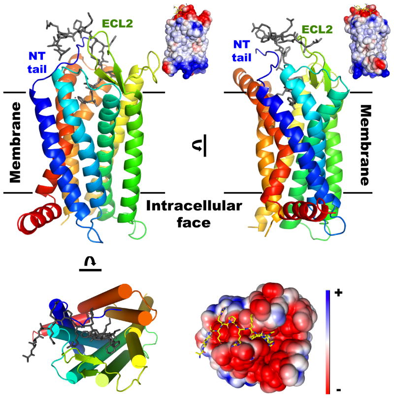
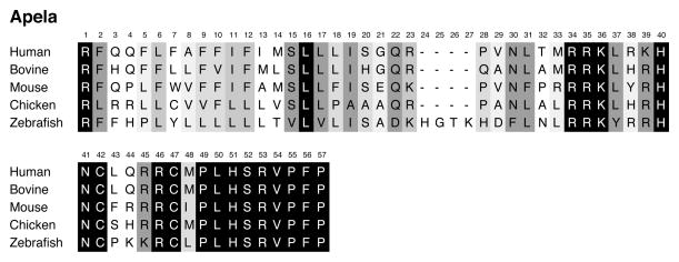
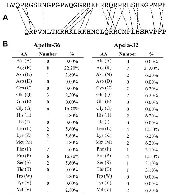
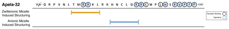
Apelin and apela (ELABELA/ELA/Toddler) are two peptide ligands for a class A G-protein-coupled receptor named the apelin receptor (AR/APJ/APLNR). Ligand-AR interactions have been implicated in regulation of the adipoinsular axis, cardiovascular system, and central nervous system alongside pathological processes. Each ligand may be processed into a variety of bioactive isoforms endogenously, with apelin ranging from 13 to 55 amino acids and apela from 11 to 32, typically being cleaved C-terminal to dibasic proprotein convertase cleavage sites. The C-terminal region of the respective precursor protein is retained and is responsible for receptor binding and subsequent activation. Interestingly, both apelin and apela exhibit isoform-dependent variability in potency and efficacy under various physiological and pathological conditions, but most studies focus on a single isoform. Biophysical behavior and structural properties of apelin and apela isoforms show strong correlations with functional studies, with key motifs now well determined for apelin. Unlike its ligands, the AR has been relatively difficult to characterize by biophysical techniques, with most characterization to date being focused on effects of mutagenesis. This situation may improve following a recently reported AR crystal structure, but there are still barriers to overcome in terms of comprehensive biophysical study. In this review, we summarize the three components of the apelinergic system in terms of structure-function correlation, with a particular focus on isoform-dependent properties, underlining the potential for regulation of the system through multiple endogenous ligands and isoforms, isoform-dependent pharmacological properties, and biological membrane-mediated receptor interaction. © 2018 American Physiological Society. Compr Physiol 8:407-450, 2018.


Figures

Figure 1
Figure 1
Human apelin receptor (AR) sequence illustrated in “snake plot” format, with seven transmembrane (TM) helices delineated. Additional structural features observed in the AR crystal structure (166) are illustrated: a short β-sheet in the second extracellular loop; the 8th helix, immediately C-terminal to TM7; and, the two extracellular domain disulfide linkages (denoted by distinct dagger symbols). Residues shown by mutagenesis to have functional importance are shown by filled circles. Important motifs common to class A GPCRs (106, 257) are shown by red circles: the Trp toggle (CWXP) in helix 6; the ionic lock (DRY) in helix 3; and, the NPXXY motif in TM7. Membrane interface positioning is as estimated by TMDET (258). Teaching points: This figure demonstrates the topology of the apelin receptor. It has the canonical G-protein-coupled receptor (GPCR) topology with seven transmembrane helices, an extracellular N-terminal tail, intracellular C-terminal tail, and three loops connecting TM helices on each side of the membrane. Additional structural features determined crystallographically are also shown, including a β-sheet in the second extracellular loop and a short “8th helix” immediately following transmembrane helical segment 7. A number of studies have now also demonstrated functionally important residues through mutagenesis and functional study, as are highlighted.
Figure 2
Figure 2
Sequence comparison of the seven transmembrane (TM) regions of human apelin receptor (AR; colored red) with 7 other GPCRs (human unless specified): bovine rhodopsin (Rh); fellow HIV-1 co-receptors CCR5 and CXCR4; angiotensin-II receptor isoform 1 (AT1); and, β2-adrenergic receptor (β2AdR), adenosine A2A receptor and turkey β1-adrenergic receptor (β1AdR). Alignments were performed using consensus residues defined by Baldwin et al. (17). Boxes surround conserved/homologous residues in >4 (or 4 including AR) of the GPCRs. Numbering above the alignments refer to the standard GPCR numbering used in the GPCRDB project (100) with green highlights showing putative TM region, while numbering at each end of the sequence refers to the sequential numbering for the full-length protein. Yellow highlights indicate residues in AR where mutagenesis has shown perturbation to either function or localization (113, 134, 143). Teaching points: The sequence of each of the seven apelin receptor transmembrane segments is compared to those known for several related class A GPCRs. Although sequence conservation is reasonably high, particularly for the angiotensin-II receptor isoform 1 (AT1), clear distinctions are apparent in all of these receptors. Correspondingly, although a canonical GPCR architecture is expected in each case, differences in structure, intramolecular dynamics, ligand binding, and activation mechanisms are not unexpected due to the variations in primary structuring.
Figure 3
Figure 3
Representations of the crystal structure of the apelin receptor in an inactive-like state in complex with the agonistic apelin-17 analog AMG3054 (PDB entry 5VBL; (166)). Cartoon and cylinder diagrams are colored from blue (N-terminus) to red (C-terminus), with the ligand shown as grey sticks. Surface representations colored (as indicated on lower right) from a charge of -2 as red to +2 as blue were generated using the PyMol (Schrödinger, Cambridge, MA) adaptive Poisson-Boltzmann Solver plugin. Membrane positioning is as estimated by TMDET (258). Teaching points: The first apelin receptor crystal structure is illustrated. The apelin-17 analog, AMG3054, was bound to the receptor, as shown. The GPCR architecture is clear in the structure and analysis of its features allowed determination of the fact that the state observed is an “inactive-like” conformation despite the fact that AMG3054 is an agonist. This was likely due to the mutagenesis required in order to obtain a stable construct for crystallographic studies. An important feature is the high degree of anionic character on the extracellular face of the receptor, with a number of unoccupied grooves being apparent. These grooves have been postulated to provide natural binding sites for longer apelin isoforms.
Figure 4
Figure 4
Structural and topological comparison of the apelin receptor to four other class A GPCRs crystallized in inactive-like conformations. Left column: Comparison of GPCR architecture (see, e.g., Hanson et al. (93) for a detailed discussion of these comparator GPCRs) colored from blue (N-terminus) to red (C-terminus), with the corresponding PDB entry codes provided with co-crystallized, bound agonist/antagonist molecules given in brackets; retinal is present in the rhodopsin structure shown. Each GPCR is shown in a cartoon representation in the same orientation and with the same color scheme. Right column: Comparison of topologies with TM helix kinks identified as bends (black circles) or disruptions (black lines) and TM helices shown as colored rectangles with position and angle correct relative to membrane boundaries (blue lines). Topologies were determined by the MC-HELAN algorithm (144) with TM orientation and membrane boundaries defined by the TMDET algorithm (258). Residues at start and end of each TM region and at kinks are indicated (see also Fig. 2). Loops connecting TM helices are shown as black lines (independent of length), while the N- and C-termini are not represented. Kink angles cannot be preserved in translation from 3D structure to 2D topology diagram, but are calculated correctly by MC-HELAN. Teaching points: The apelin receptor structure is compared to those of four other class A GPCRs, all crystallized in inactive-like conformations. Although the overall architecture is the same in all cases (cartoon illustrations in left-hand column), the topology of each GPCR in the transmembrane domain is distinct with some helices being more variable than others.
Figure 5
Figure 5
Comparison of β2-adrenergic receptor topology as a function of activation state. PDB entry is given, alongside its resolution and the nature of the bound ligand or effector protein. Topologies were predicted by MC-HELAN (144) with TM helix kinks identified as bends (black circles) or disruptions (black lines) and TM helices shown as colored rectangles with position and angle correct relative to membrane boundaries (blue lines) defined by the TMDET algorithm (258). Teaching points: The topologies of four distinct crystallographically-characterized states of the β2-adrenergic receptor, a relatively extensively characterized class A GPCR, are illustrated. Notably, even for a single GPCR, the topology in the transmembrane domain for the structural snapshots varies as a function of activation state.
Figure 6
Figure 6
Sequence conservation of apelin. Residues that are fully conserved over the six illustrated species are indicated with a black background; partially conserved with varying shades of grey; and, variable positions with white. Teaching points: The sequence alignment for pre(pro)apelin over a variety of species is shown. The N-terminal 22 residues are the predicted signal peptide, cleaved to produce a bioactive 55-residue form of apelin (apelin-55). Initially, apelin-55 was not believed to be bioactive and was referred to as proapelin. Alignment demonstrates very clearly that the C-terminal 12 residues are identical, directly corresponding to the requirement of these residues for receptor binding and activation. More variability is seen N-terminal to this region. Despite this variability, dibasic motifs associated with proprotein convertase processing are still frequently found, implying the potential of multiple isoforms being produced in all of the species compared.
Figure 7
Figure 7
Apelin processing pathways. A) Previously theorized “apelin-36 precursor” processing pathway. B) Current “myriad” processing pathway theory based on new publications, which also identifies proapelin as apelin-55 as an additional bioactive member of the apelinergic system. Note that this processing pathway does not show other post-translational modifications such as ACE-2-mediated C-terminal phenylalanine removal. Teaching points: Two apelin processing pathways are compared. In each case, the 77-residue pre(pro)protein is initially processed to produce a 55-residue form (apelin-55). Initially, this was believed to be an inactive proprotein, but subsequently apelin-55 secretion was demonstrated and it has now been shown to be bioactive. Despite this, apelin-55 may be processed intracellularly to shorter isoform(s) prior to secretion. Mechanisms by which shorter isoforms are produced remain ill characterized, with only two studies to date demonstrating that the proprotein convertase PCSK3 processes either apelin-55 or apelin-36. Further study is needed to flesh out these pathways.
Figure 8
Figure 8
Apelinergic system expression and isoform localization profile. A) Apelin, apela, and AR localization as a function of tissue/organ system. B) Predominant apelin isoform(s) detected, to date, in specific organs or body fluids. Teaching points: Localization of each of the components of the apelinergic system is quite widespread (panel A). This is better characterized for apelin and the AR, given the recent discovery of apela. Tissue- and fluid-specific isolation of apelin isoforms has also been demonstrated (panel B). This has implications in terms of physiological function, given that distinct apelin isoforms lead to differences in downstream signaling and receptor regulation.
Figure 9
Figure 9
Summary of structure-function correlations for apelin. Apelin-17 is illustrated as it has been the most widely-studied isoform in terms of biophysics and structure. Functional effects of mutagenesis or truncation are indicated directly on the peptide sequence (symbols denoted in the legend). For direct comparison, regions of apelin-17 exhibiting structural convergence (138) and membrane-interactive properties (141) are delineated alongside the segments of the apelin-17 analog AMG3054 that interact with the AR in the co-crystal structure (PDB entry 5VBL (166)). Teaching points: Motifs within the apelin-17 isoform are illustrated. The 12 residues in the C-terminal region of apelin that are widely conserved over many species demonstrate the most importance in terms of functional effects upon substitution or truncation, structural convergence, and membrane-interaction. The “RPRL” motif at the N-terminus of apelin-12 demonstrates structuring under a variety of conditions and is important in membrane interaction. In the AMG3054 analog, this region also interacts with an extracellular anionic groove. The C-terminal “GPMPF” motif exhibits distinct structural differences as a function of condition. In AMG3054, this region penetrates into the transmembrane domain of the AR, with a kink at the His. Also of note, the C-terminal Phe of apelin has a number of features ascribed in the literature. Signaling bias based upon its presence or absence appears to be the most likely source for these discrepancies.
Figure 10
Figure 10
Membrane catalysis hypothesis as applied to binding of apelin-17 to the apelin receptor. Sequentially, (1) apelin is proposed to bind to the membrane, increasing the likelihood of (2) its interaction with and recognition by an unliganded apelin receptor (AR) on a cell surface followed by (3) receptor binding and activation. Structures of apelin-17 in buffer (BMRB entry 20029 (138)) and bound to SDS micelles (BMRB entry 20082 (141)); and, of AR in absence of ligand with anionic patch residues E20 and D23 illustrated (143) and bound to apelin-17 analog AMG3054 (PDB entry: 5VBL (166)) were employed. Teaching points: The membrane catalysis hypothesis is illustrated using structural data from the apelinergic system. In the first step, the “RPRL” motif is modestly structured in solution and binds to membrane; this pre-structuring would lead to a decreased entropic penalty upon membrane binding. The receptor encounter likelihood of the membrane-bound apelin is then increased due to the reduction of the diffusional search from a 3D to a 2D process, the increased local concentration of ligand on the membrane, and the induction of structuring, particularly at the apelin C-terminus (the MPF within the “GPMPF” motif). Upon receptor interaction and recognition, an additional conformational change will take place during binding. An anionic patch on the apelin receptor (AR) N-terminal tail may also facilitate this step. Binding is likely to be a two-step process. An initial binding step (not illustrated) is likely whereby the apelin N-terminal region interacts with the AR extracellular domain. This would facilitate a “fly casting” mechanism by which the C-terminal tail of apelin is facilitated in finding and penetrate into the transmembrane domain of the AR. A subsequent, additional conformational change is then likely to go from an inactive bound state (as observed crystallographically) to an active bound state.
Figure 11
Figure 11
Sequence conservation of apela. Residues that are fully conserved over the five illustrated species are indicated with a black background; partially conserved with varying shades of grey; and, variable positions with white. Hyphens indicate residues absent from a given species. Teaching points: The sequence alignment for preapela over a variety of species is shown. The N-terminal 22 residues are the predicted signal peptide, cleaved to produce a bioactive 32-residue form of apela. Alignment demonstrates very clearly that the C-terminal 9 residues are identical, with a very high degree of conservation through the 13 C-terminal residues. As with apelin, this is indicative of the requirement of these residues for receptor binding and activation. Some additional variability is seen N-terminal to this region. Following the precedent of apelin, dibasic motifs associated with proprotein convertase processing are identical, implying the potential of the same sets of isoforms being produced in all of the species compared.
Figure 12
Figure 12
Comparison of apelin-36 and apela-32. A) Sequence comparison of apelin-36 (top) and apela-32 (bottom). Dashed lines represent residues falling in similar positions implying the potential for similar structural and/or functional roles. B) Comparison of amino acid composition. Teaching points: Apelin-36 and apela-32 exhibit similar, but non-identical, amino acid compositions. A number of key motifs observed for one peptide may be translated to the other, as indicated by dashed lines. These, however, are frequently translated in position.
Figure 13
Figure 13
Summary of structural and functional studies for apela. Apela-32 is illustrated, although it should be noted that a number of studies have focused on shorter isoforms. Functional effects of mutagenesis or truncation are indicated directly on the peptide sequence (symbols denoted in the legend). Regions exhibiting structuring in the presence of the indicted type of micelle (105) are delineated. Teaching points: Motifs within the apela-32 isoform are illustrated. Although substitution and truncation studies have been more limited, to date, relative to apelin, a number of key sites for both binding and signaling have been identified (as illustrated.) Distinct regions of apela-32 also become structured, depending upon the surface charge properties of membrane-mimetic micelles employed.
Figure 14
Figure 14
Implications of membrane catalysis for the regulation of signaling. A) Autocrine and B) paracrine or endocrine signaling of apelin or apela (denoted as “AP”) isoforms may be regulated by variation in preferential membrane headgroup association. Ligand-mediated apelin receptor (AR) activation is represented by G-protein binding and subsequent ERK phosphorylation (pERK), although many other signaling pathways are possible (Tables 13–14). Teaching points: This figure illustrates different possible fates of an apelin or apela (“AP”) ligand upon secretion. If there is a strong potential for interaction with the membrane of the cell from which it is secreted, this would bias toward autocrine signaling (A) through apelin receptor (AR) molecules on the same cell. Alternatively, the ligand may encounter and bind to a nearby cell (i.e., paracrine signaling) or more distant cell (i.e., endocrine signaling) as in panel B, exerting functional effects through AR on the cell in question. In any instance, further processing of the secreted peptide may take place prior to receptor interaction, either with cell surface localized proprotein convertases or other processing enzymes (autocrine, paracrine, or endocrine signaling) or with circulating enzymes (likely most applicable to endocrine signaling). The potential for post-secretion processing is not explicitly illustrated, but should be kept in mind as an important additional regulatory mechanism.

References

    1. Adam F, Khatib AM, Lopez JJ, Vatier C, Turpin S, Muscat A, Soulet F, Aries A, Jardin I, Bobe R, Stepanian A, de Prost D, Dray C, Rosado JA, Valet P, Feve B, Siegfried G. Apelin: an antithrombotic factor that inhibits platelet function. Blood. 2016;127:908–920. - PubMed
    1. Akcilar R, Turgut S, Caner V, Akcilar A, Ayada C, Elmas L, Ozcan TO. Apelin effects on blood pressure and RAS in DOCA-salt-induced hypertensive rats. Clin Exp Hypertens. 2013;35:550–557. - PubMed
    1. Akcilar R, Turgut S, Caner V, Akcilar A, Ayada C, Elmas L, Ozcan TO. The effects of apelin treatment on a rat model of type 2 diabetes. Adv Med Sci. 2015;60:94–100. - PubMed
    1. Alastalo TP, Li M, de Perez VJ, Pham D, Sawada H, Wang JK, Koskenvuo M, Wang L, Freeman BA, Chang HY, Rabinovitch M. Disruption of PPARγ/β-catenin-mediated regulation of apelin impairs BMP-induced mouse and human pulmonary arterial EC survival. J Clin Invest. 2011;121:3735–3746. - PMC - PubMed
    1. Andersen CU, Markvardsen LH, Hilberg O, Simonsen U. Pulmonary apelin levels and effects in rats with hypoxic pulmonary hypertension. Respir Med. 2009;103:1663–1671. - PubMed

Publication types

Grants and funding

LinkOut - more resources