

Altered Gradients of Glutamate and Gamma-Aminobutyric Acid Transcripts in the Cortical Visuospatial Working Memory Network in Schizophrenia
- PMID: 29357982
- PMCID: PMC5862743
- DOI: 10.1016/j.biopsych.2017.11.029
Altered Gradients of Glutamate and Gamma-Aminobutyric Acid Transcripts in the Cortical Visuospatial Working Memory Network in Schizophrenia
Abstract
Background: Visuospatial working memory (vsWM), which is impaired in schizophrenia, requires information transfer across multiple nodes in the cerebral cortex, including visual, posterior parietal, and dorsolateral prefrontal regions. Information is conveyed across these regions via the excitatory projections of glutamatergic pyramidal neurons located in layer 3, whose activity is modulated by local inhibitory gamma-aminobutyric acidergic (GABAergic) neurons. Key properties of these neurons differ across these cortical regions. Consequently, in schizophrenia, alterations in the expression of gene products regulating these properties could disrupt vsWM function in different ways, depending on the region(s) affected.
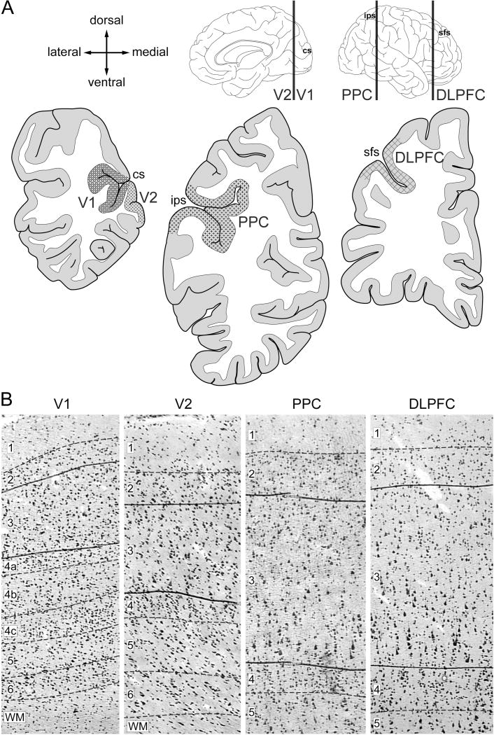
Methods: Here, we quantified the expression of markers of glutamate and GABA neurotransmission selectively in layer 3 of four cortical regions in the vsWM network from 20 matched pairs of schizophrenia and unaffected comparison subjects.
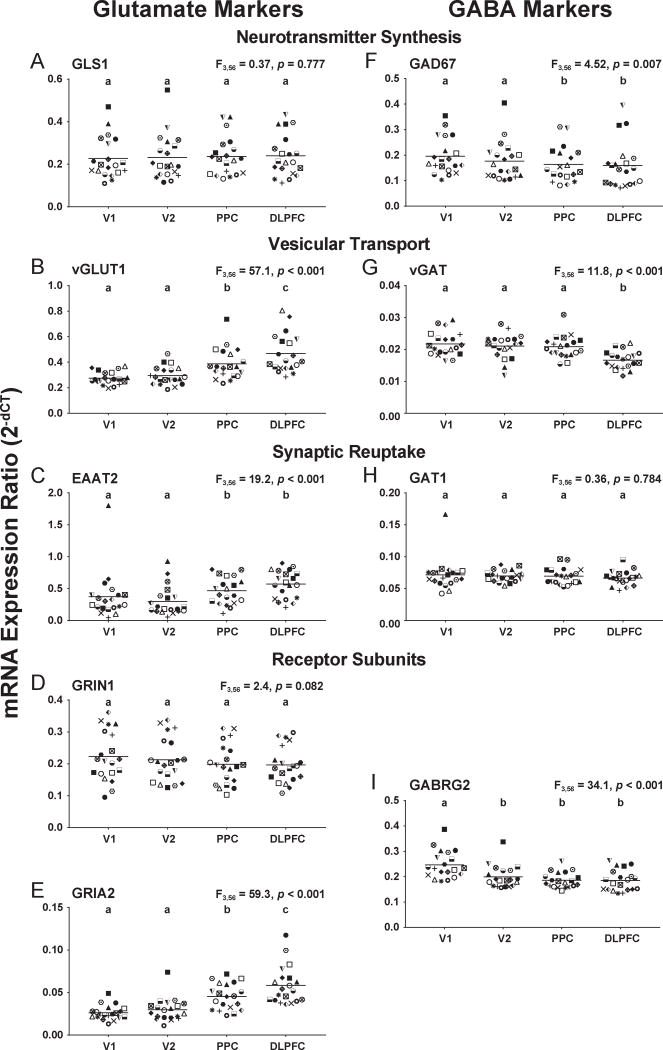
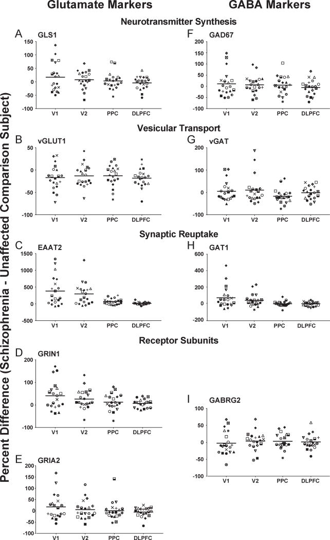
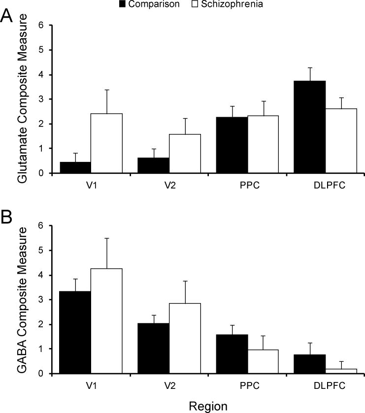
Results: In comparison subjects, levels of glutamate transcripts tended to increase, whereas GABA transcript levels tended to decrease, from caudal to rostral, across cortical regions of the vsWM network. Composite measures across all transcripts revealed a significant effect of region, with the glutamate measure lowest in the primary visual cortex and highest in the dorsolateral prefrontal cortex, whereas the GABA measure showed the opposite pattern. In schizophrenia subjects, the expression levels of many of these transcripts were altered. However, this disease effect differed across regions, such that the caudal-to-rostral increase in the glutamate measure was blunted and the caudal-to-rostral decline in the GABA measure was enhanced in the illness.
Conclusions: Differential alterations in layer 3 glutamate and GABA neurotransmission across cortical regions may contribute to vsWM deficits in schizophrenia.
Keywords: GABA; Glutamate; Prefrontal cortex; Schizophrenia; Visual cortex; Working memory.
Copyright © 2017 Society of Biological Psychiatry. Published by Elsevier Inc. All rights reserved.
Conflict of interest statement
Figures
Comment in
-
Toward Better Strategies for Understanding Disrupted Cortical Excitatory/Inhibitory Balance in Schizophrenia.Biol Psychiatry. 2018 Apr 15;83(8):632-634. doi: 10.1016/j.biopsych.2018.02.014. Biol Psychiatry. 2018. PMID: 29559094 No abstract available.
References
-
- Green MF. What are the functional consequences of neurocognitive deficits in schizophrenia? Am J Psychiatry. 1996;153(3):321–330. - PubMed
-
- Kahn RS, Keefe RS. Schizophrenia is a cognitive illness: time for a change in focus. JAMA Psychiatry. 2013;70(10):1107–1112. - PubMed
-
- Baddeley A. Working memory. Science. 1992;255(5044):556–559. - PubMed
Publication types
MeSH terms
Substances
Grants and funding
LinkOut - more resources
Full Text Sources
Other Literature Sources
Medical
