. 2018 Feb 8;172(4):784-796.e18.
doi: 10.1016/j.cell.2017.12.033. Epub 2018 Jan 18.

Non-classical Immunity Controls Microbiota Impact on Skin Immunity and Tissue Repair

Affiliations

Non-classical Immunity Controls Microbiota Impact on Skin Immunity and Tissue Repair

Jonathan L Linehan et al. Cell. .

Abstract

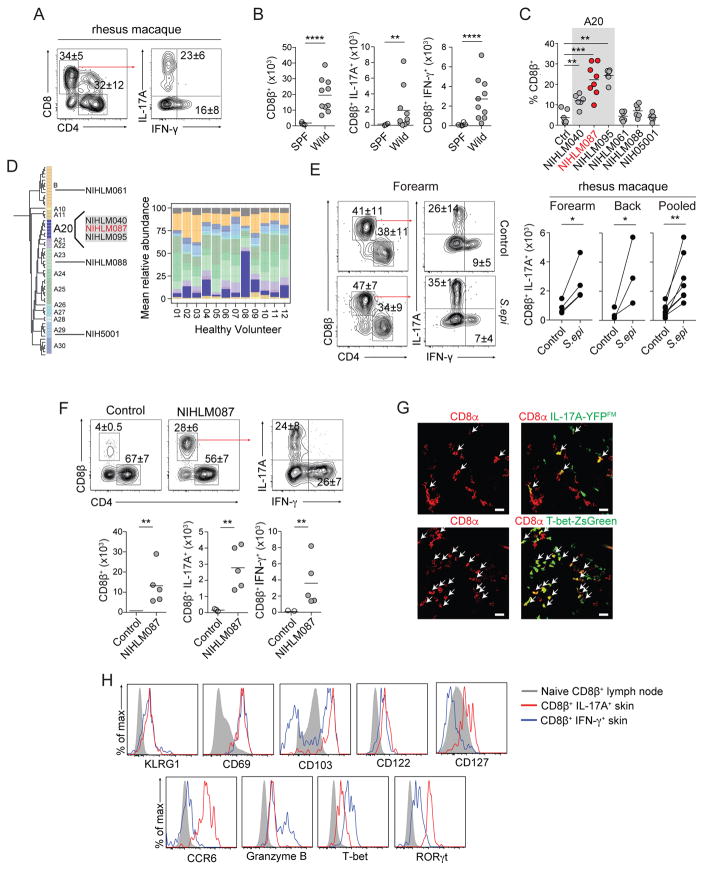
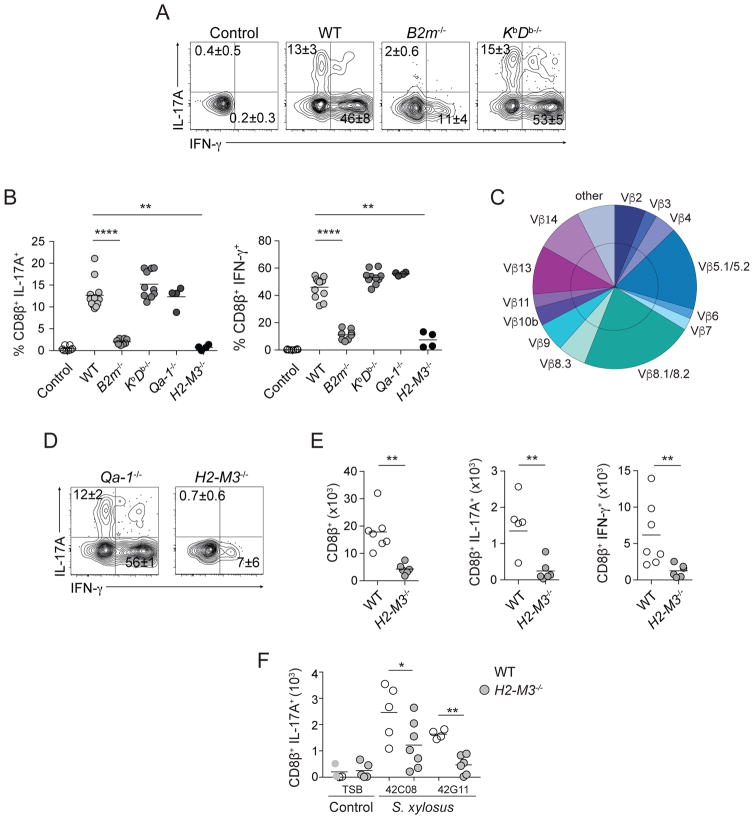
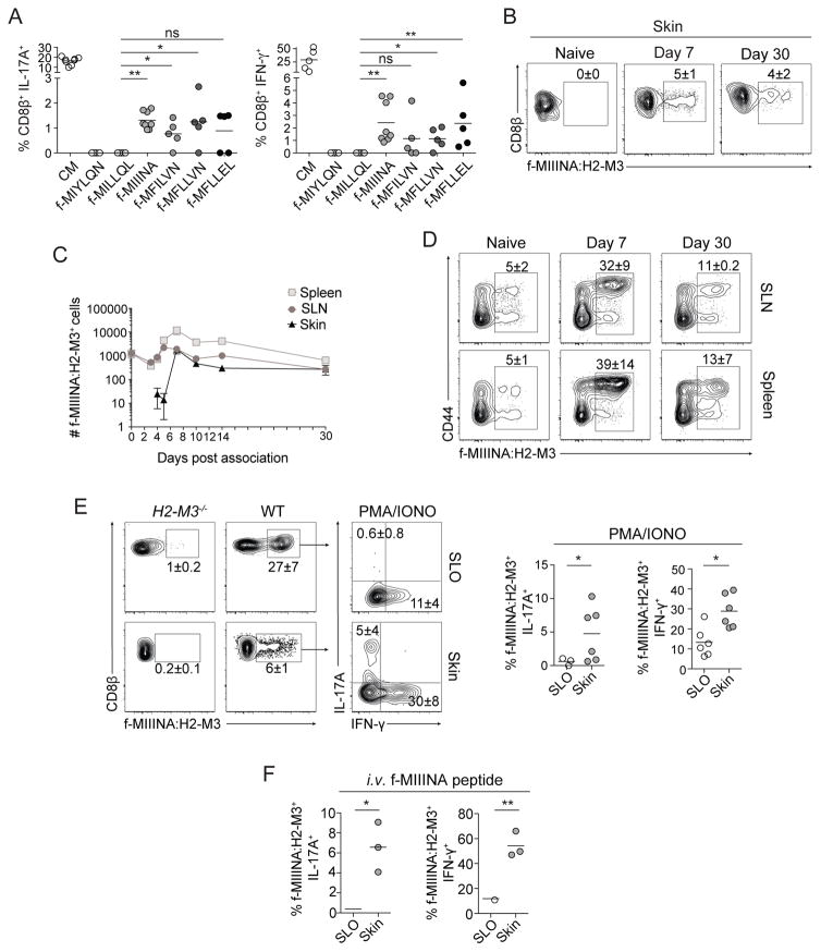
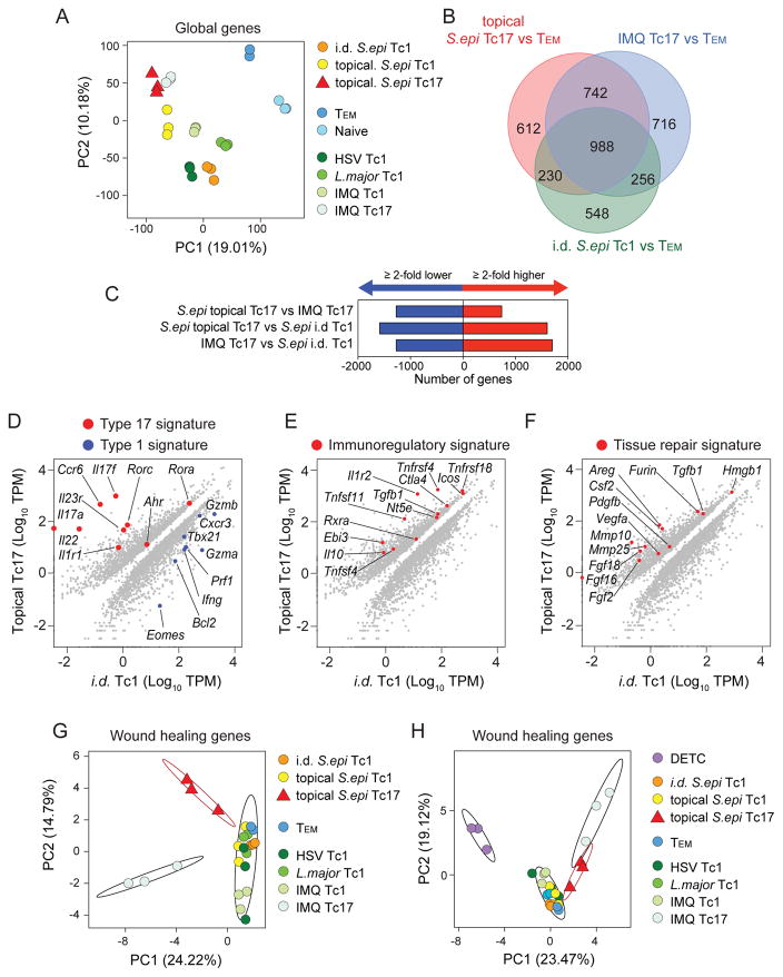
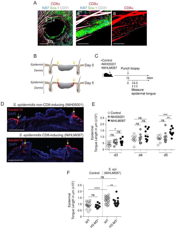
Mammalian barrier surfaces are constitutively colonized by numerous microorganisms. We explored how the microbiota was sensed by the immune system and the defining properties of such responses. Here, we show that a skin commensal can induce T cell responses in a manner that is restricted to non-classical MHC class I molecules. These responses are uncoupled from inflammation and highly distinct from pathogen-induced cells. Commensal-specific T cells express a defined gene signature that is characterized by expression of effector genes together with immunoregulatory and tissue-repair signatures. As such, non-classical MHCI-restricted commensal-specific immune responses not only promoted protection to pathogens, but also accelerated skin wound closure. Thus, the microbiota can induce a highly physiological and pleiotropic form of adaptive immunity that couples antimicrobial function with tissue repair. Our work also reveals that non-classical MHC class I molecules, an evolutionarily ancient arm of the immune system, can promote homeostatic immunity to the microbiota.

Keywords: H2-M3; MHCIb; Staphylococcus epidermidis; microbiota; non-classical MHC class I; skin immunity; tissue repair.


Conflict of interest statement

DECLARATION OF INTERESTS

The authors declare no competing financial interests.

Figures

Figure 1
Figure 1. Tc17 cells are enriched in the skin of non-human primates and mice
A) Frequencies of IL-17A+ or IFN-γ+ CD8+ T cells isolated from Rhesus macaque. All cells were isolated from glabella. Left plot is gated on live CD45+ CD3ε+ T cells. B) Numbers of total, IL-17A+, or IFN-γ+ CD8β+ T cells in naïve SPF or wild-caught mice. All cells were isolated from ear pinnae. Plots are gated on live CD45+ TCRβ+ CD8β+ T cells. Each dot represents an individual mouse. C) Frequencies of CD8β+ T cells in control mice or mice topically associated with the indicated S. epidermidis isolate. S. epidermidis isolate highlighted in red (NIHLM087) is the strain used for all remaining experiments unless specified otherwise. All cells were isolated from ear pinnae. Plots are gated on live CD45+ TCRβ+ CD8β+ T cells. Each dot represents an individual mouse. D) Phylogenetic tree of S. epidermidis clades and relative abundance plot of S. epidermidis clades across healthy volunteers. Individual bacterial clades are differentiated by color. E) Frequencies and numbers of IL-17A+ or IFN-γ+ CD8+ T cells in unassociated (control) or two weeks post S. epidermidis (NIHLM087) association (contralateral side) isolated from Rhesus macaques. All cells were isolated from dorsal forearm or upper back. Left plot is gated on live CD45+ CD3ε+ T cells. Paired points denote same animal. F) Frequencies and numbers of total, IL-17A+, or IFN-γ+ CD8β+ T cells in naïve SPF mice (control) or two weeks post S. epidermidis (NIHLM087) association. All cells were isolated from ear pinnae. Plots are gated on live CD45+ TCRβ+ CD8β+ T cells. Each dot in graph represents an individual mouse. G) Representative confocal imaging volume projected along the z-axis of epidermal skin from mice 14 days post S. epidermidis (NIHLM087) association. Arrows show CD8α+IL-17A-Cre+, Rosa26-YFP+ (IL-17A-YFPFM=fate map) (top), CD8α+ T-bet-ZsGreen+ (bottom) cells. Scale bars, 20μm. H) Expression of: KLRG1, CD69, CD103, CD122, CD127, CCR6, Granzyme B, T-bet, or RORγt by CD8β+ T cells in ear pinnae of mice, two weeks post S. epidermidis association. Plots are gated on live CD45+ TCRβ+ CD8β+ T cells. Solid Gray=CD44low CD8β+ T cells in skin draining lymph nodes. Blue line=IFN-γ+ CD8β+ T cells in ear pinnae. Red line=IL-17A+ CD8β+ T cells in ear pinnae. (A–C, E, G) cells were stimulated with PMA and Ionomycin. (B–C, F) Student’s t test was used to measure significance. **P<0.005, *** P<0.0005, **** P<0.0001. (E) Paired student’s t test was used to measure significance. * P<0.05, **P<0.005 Data are representative of 2–3 independent experiments.
Figure 2
Figure 2. S. epidermidis-specific CD8+ T cells are restricted to the MHCIb molecule H2-M3
A,B,D) In vitro recall of commensal induced CD8+ T cells from skin with heat-killed S. epidermidis loaded splenic DCs. Frequencies of IFN-γ or IL-17A-producing CD8β+ T cells after overnight culture. Splenic dendritic cells were enriched from wild-type (WT), β 2m−/−, KbDb−/−, Qa-1−/−, or H2-M3−/− mice. Control=unloaded WT splenic DCs. C) Frequencies of Vβ chain usage among total CD8β+ T cells from skin. E) Numbers of total, IFN- γ, or IL-17A-producing CD8β+ T cells in S. epidermidis associated WT or H2-M3−/− mice. Cells were stimulated with PMA and Ionomycin. F) Numbers of IL-17A+ CD8β+ T cells in WT or H2-M3−/− mice topically associated with TSB (control) or with the indicated S. xylosus isolate. All cells were isolated from ear pinnae. Plots are gated on live CD45+ TCRβ+ CD8β+ T cells. Each dot represents an individual mouse. All cells were isolated from ear pinnae of S. epidermidis or S. xylosus associated mice 2 weeks after application. Plots are gated on live CD45+ TCRβ+ CD8β+ T cells. Each dot represents an individual mouse. Student’s t test was used to measure significance. *P<0.05, **P<0.005, *** P<0.0005. Data are representative of 2–4 independent experiments.
Figure 3
Figure 3. H2-M3 restricted CD8+ T cells are specific to S. epidermidis derived fMet peptide ligands
A) In vitro recall of S. epidermidis-induced CD8+ T cells from FACsorted skin with S. epidermidis conditioned medium (CM) or fMet peptide loaded splenic DCs. Frequencies of IFN-γ or IL-17A-producing CD8β+ T cells after overnight culture. Gated on live CD45+ TCRβ+ CD8β+ T cells. Splenic dendritic cells were enriched from wild-type mice. B–F) Frequencies and numbers of f-MIIINA:H2-M3 tetramer positive cells or tetramer positive IFN-γ or IL-17A-producing CD8β+ T cells in (B,C,E,F) ear pinnae, (C–D) magnetic bead enriched spleen or skin draining lymph nodes (SLN), or (E–F) magnetic bead enriched combined spleen and skin draining lymph nodes (SLO) of (B–F) intact WT (SPF) or (E) H2-M3−/− mice. B–D) cells were isolated from naïve mice or mice after indicated time-point following first S. epidermidis skin application. C) Data are represented as mean ± SEM. E) Cells were stimulated with PMA and Ionomycin. F) Mice were injected with 100μg f-MIIINA peptide intravenously and tissues harvested after three hours. E–F) All cells were isolated from S. epidermidis associated mice 2 weeks after application. B–F) Plots are gated on live CD45+ Lineage (MHCII, CD11b, γδTCR) CD90.2+ CD3ε+ CD8β+ T cells. Student’s t test was used to measure significance. * P<0.05, ** P<0.005. Data are representative of 2–4 independent experiments.
Figure 4
Figure 4. Commensal-specific Tc17 cells are a distinct population and enriched in transcripts associated with immune regulation and tissue repair
A) Principle component analysis of global gene expression from FACsorted populations identified in (Figure S4A). B) Venn diagram from RNA-seq analysis of genes differentially expressed in pairwise comparisons between topical S. epidermidis-elicited Tc17, imiquimod (IMQ)-elicited Tc17, and intradermal (i.d.) S. epidermidis-elicited Tc1 cells in skin relative to skin draining lymph node Tem cells in naïve mice. Numbers of genes with greater >2-fold increase relative to Tem are shown. C) Numbers of genes with a >2-fold increase (red) or decrease (blue) in expression levels between the indicated populations. D–F) Diagonal plots showing differentially expressed genes (>2-fold, Padj<0.05) comparing topical S. epidermidis-elicited Tc17 to i.d. S. epidermidis-elicited Tc1 cells in skin. G–H) Principle component analysis of curated wound healing gene expression from FACsorted populations identified in (Figure S4A). Ellipses denote 95% confidence intervals of the mean of selected cell populations: topical S. epidermidis-elicited Tc17, imiquimod (IMQ)-elicited Tc17, and all others.
Figure 5
Figure 5. Commensal-specific CD8+ T cells accelerate wound healing
A) Representative imaging of whole mount ear pinnae at 3 days after 2.5mm ear punch biopsy from mice (15 days post initial S. epidermidis association). Whole mounts were stained for CD8+ T cells (CD8α), keratinocytes (Sca-1), blood vessels (CD31), cycling cells (Ki67) and imaged by confocal microscopy. Scale bars: 150μm, 1000μm, and 1000μm, respectively. Hair is autofluorescent in CD8α and CD31 channels. B) Schematic depicting progress of re-epithelialization over time after wounding. Yellow arrows denote wound edge. Red indicates advancing epidermal tongue of basal keratinocytes that stains brightly for Keratin 14. C) Mice were topically associated with growth medium alone (control), non-CD8+ T cell inducing S. epidermidis isolate (NIH05001), or CD8+ T cell inducing S. epidermidis isolate (NIHLM087) followed by a backskin punch biopsy twelve days later. Epidermal keratinocyte tongue length was measured at days 3,4, and 5 post wounding. D) Immunofluorescence images of backskin wounds at day 5 after punch biopsy. Sections are immunolabeled for basal epidermal keratinocytes (K14) and co-stained with DAPI. Scale bars, 1000μm. Yellow arrows denote wound edge. E) Quantification of the length of the tongue of epidermal keratinocytes that migrate in from the wound edges during re-epithelialization of the wound bed in WT mice. Mice were topically skin associated with growth medium (control), a non-CD8+ T cell inducing S. epidermidis (NIH5001) strain, or a CD8+ T cell inducing S. epidermidis (NIHLM087) strain. Tongue lengths were measured at 3–5 days after backskin punch biopsy. F) Quantification of the length of the tongue of epidermal keratinocytes that migrate in from the wound edges during re-epithelialization of the wound bed in WT or H2-M3−/− mice. Mice were topically skin associated with growth medium (control) or NIHLM087. Tongue lengths were measured at 5 days after backskin punch biopsy. Student’s t test was used to measure significance. * P<0.05, ** P<0.005, *** P<0.0005, **** P<0.0001. Data are representative of 2–4 independent experiments.

Comment in

Similar articles

Cited by

References

    1. Adams EJ, Luoma AM. The adaptable major histocompatibility complex (MHC) fold: structure and function of nonclassical and MHC class I-like molecules. Annu Rev Immunol. 2013;31:529–561. - PubMed
    1. An D, Oh SF, Olszak T, Neves JF, Avci FY, Erturk-Hasdemir D, Lu X, Zeissig S, Blumberg RS, Kasper DL. Sphingolipids from a symbiotic microbe regulate homeostasis of host intestinal natural killer T cells. Cell. 2014;156:123–133. - PMC - PubMed
    1. Anders S, Huber W. Differential expression analysis for sequence count data. Genome Biol. 2010;11:R106. - PMC - PubMed
    1. Belkaid Y, Hand TW. Role of the microbiota in immunity and inflammation. Cell. 2014;157:121–141. - PMC - PubMed
    1. Belkaid Y, Segre JA. Dialogue between skin microbiota and immunity. Science. 2014;346:954–959. - PubMed

Publication types

Substances