. 2018 Mar 15;25(3):309-317.e4.
doi: 10.1016/j.chembiol.2017.12.012. Epub 2018 Jan 18.

Lipids Shape the Electron Acceptor-Binding Site of the Peripheral Membrane Protein Dihydroorotate Dehydrogenase

Affiliations

Lipids Shape the Electron Acceptor-Binding Site of the Peripheral Membrane Protein Dihydroorotate Dehydrogenase

Joana Costeira-Paulo et al. Cell Chem Biol. .

Abstract

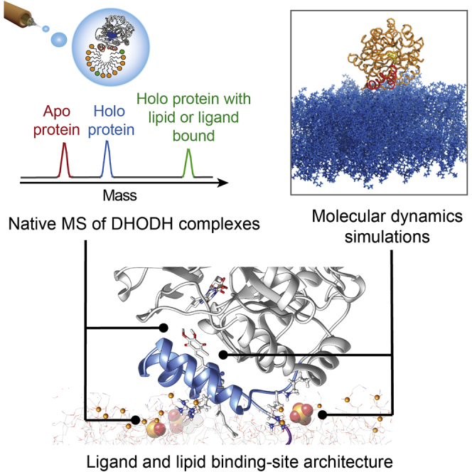
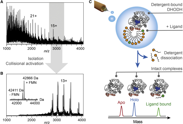
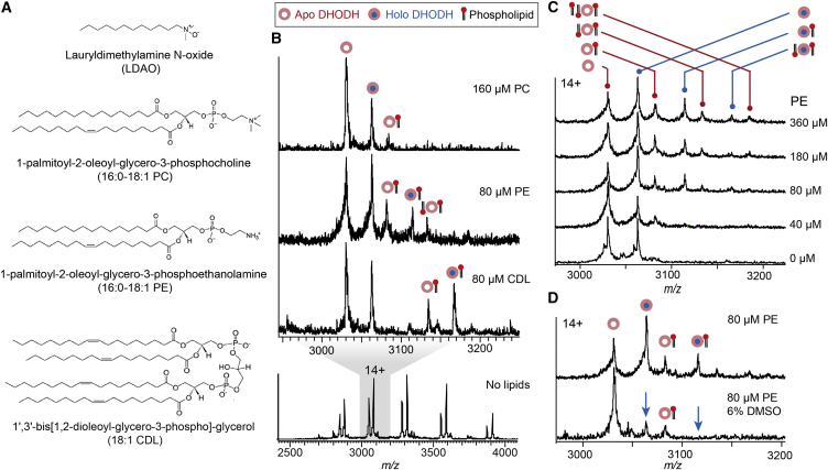
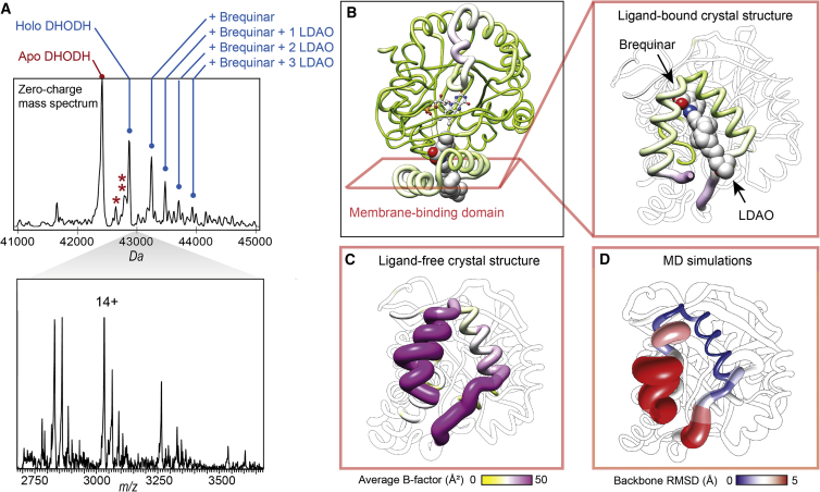
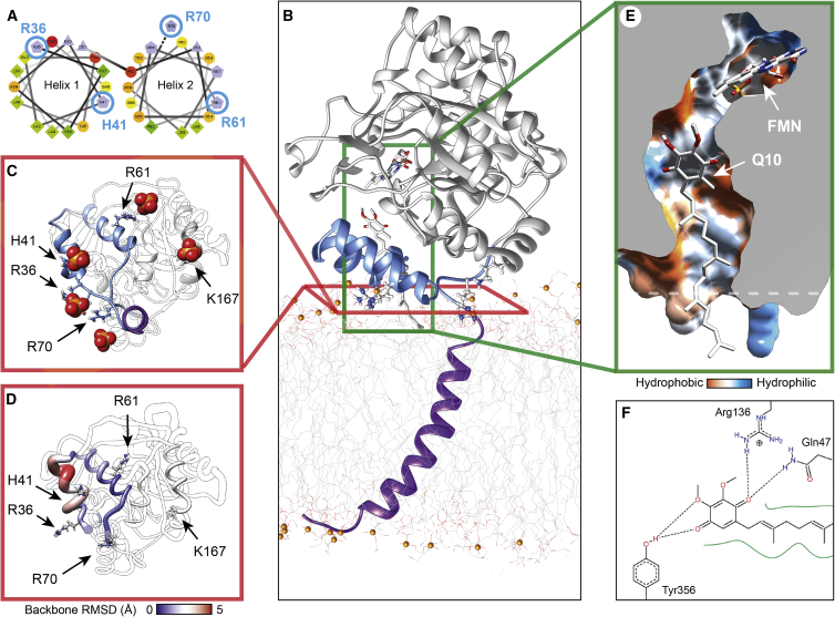
The interactions between proteins and biological membranes are important for drug development, but remain notoriously refractory to structural investigation. We combine non-denaturing mass spectrometry (MS) with molecular dynamics (MD) simulations to unravel the connections among co-factor, lipid, and inhibitor binding in the peripheral membrane protein dihydroorotate dehydrogenase (DHODH), a key anticancer target. Interrogation of intact DHODH complexes by MS reveals that phospholipids bind via their charged head groups at a limited number of sites, while binding of the inhibitor brequinar involves simultaneous association with detergent molecules. MD simulations show that lipids support flexible segments in the membrane-binding domain and position the inhibitor and electron acceptor-binding site away from the membrane surface, similar to the electron acceptor-binding site in respiratory chain complex I. By complementing MS with MD simulations, we demonstrate how a peripheral membrane protein uses lipids to modulate its structure in a similar manner as integral membrane proteins.

Keywords: membrane protein folding; molecular dynamics simulations; non-denaturing mass spectrometry; protein-lipid interactions.


Figures

None
Graphical abstract
Figure 1
Figure 1
Non-denaturing MS of DHODH Complexes (A) The nESI-MS spectrum of the full m/z range shows peaks corresponding to apo-DHODH with a broad charge state distribution. (B) Collisional activation and isolation of the region between m/z 2,700 and 3,300 (shown in gray in A) reveals two series of well-resolved protein signals corresponding to apo-DHODH as well as DHODH bound to the FMN co-factor. The deconvolved zero-charge spectrum is shown as an insert. (C) The MS strategy for integral membrane proteins facilitates the analysis of intact DHODH complexes. Release of the desolvated protein from detergent by collisional activation preserves interactions with the FMN co-factor as well as exogenous ligands. See also Figure S1.
Figure 2
Figure 2
DHODH Exhibits Limited Binding of Mitochondrial Phospholipids (A) Structures of the detergent LDAO and the three major phospholipid species in the human mitochondrial membrane. (B) nESI-MS shows that binding of PC, PE, and CDL, can be preserved in the gas phase. CDL exhibits the most stable association with DHODH, while PC interactions are not well retained. (C) Mass spectra of DHODH in the presence of increasing amounts of PE reveal a maximum number of lipid adducts for both apo- and holo-DHODH. Above a 20-fold excess (180 μM) of PE over DHODH, a maximum of three lipids are bound per protein. (D) The DHODH-lipid complexes are less sensitive to DMSO-induced dissociation than the DHODH-FMN complexes (indicated by blue arrows). Spectra recorded without and with DMSO are normalized to the 14+ charge state of holo- and apo-DHODH, respectively. See also Figure S2.
Figure 3
Figure 3
Effects of Ligand Interactions on the Flexible Membrane-Binding Domain (A) nESI-MS of DHODH in the presence of brequinar shows a series of adduct peaks for holo-DHODH corresponding to one bound brequinar molecule, as well as one brequinar and one, two, or three LDAO molecules. The deconvolved zero-charge spectrum is shown above the corresponding mass spectrum. One and two asterisks indicate peaks corresponding to apo-DHODH with LDAO and brequinar, respectively. (B) The crystal structure of human DHODH with a brequinar analogue (PDB: 1D3G) shows binding of the inhibitor and an LDAO molecule in the hydrophobic channel leading to the FMN co-factor. (C) Comparison with ligand-free DHODH (PDB: 2PRM) and in complex with brequinar and LDAO (PDB: 1D3G) rendered according to the average B factor show that binding of brequinar and LDAO significantly reduces the conformational flexibility of the membrane-binding domain. (D) MD simulations of ligand-free truncated DHODH in solution confirm the conformational flexibility of the membrane-binding domain. The protein is rendered according to the RMSDs between the equilibrated starting structure and the end structure after 200 ns all-atom MD simulations. See also Figure S3.
Figure 4
Figure 4
Membrane Interactions Shape the Q10 Binding Site (A) The N-terminal segment of truncated DHODH contains two amphipathic helices. Hydrophobic residues are shown in green, basic residues in contact with the membrane are labeled in blue. (B) All-atom MD of full-length DHODH on a PE bilayer shows association of charged residues with the lipid head groups, partially lifting the membrane-binding domain (blue) off the membrane surface. Here, a partial Q10 molecule has been modeled into the binding site. (C) The close clustering of the charged residues limits the number of simultaneously bound PE molecules. Phosphate head groups within 3 Å of the protein surface after 200 ns simulations are rendered as spheres and basic residues as sticks. (D) Membrane association reduces conformational fluctuations in the membrane-binding domain. The protein is rendered according to the RMSDs between the equilibrated starting structure and the end structure after 200 ns. (E) In the membrane-bound protein, the Q10 binding site forms a narrow reaction chamber that extends away from the membrane and is sealed by the hydrophobic tail (shown as sticks). The dashed line indicates the membrane surface. (F) In this model, Q10 is coordinated by the strictly conserved residues R136 and Y356. See also Figure S4.

References

    1. Abraham M.J., Murtola T., Schulz R., Páll S., Smith J.C., Hess B., Lindahl E. GROMACS: high performance molecular simulations through multi-level parallelism from laptops to supercomputers. SoftwareX. 2015;1–2:19–25.
    1. Allison T.M., Reading E., Liko I., Baldwin A.J., Laganowsky A., Robinson C.V. Quantifying the stabilizing effects of protein–ligand interactions in the gas phase. Nat. Commun. 2015;6:8551. - PMC - PubMed
    1. Angerer H., Nasiri H.R., Niedergesäß V., Kerscher S., Schwalbe H., Brandt U. Tracing the tail of ubiquinone in mitochondrial complex I. Biochim. Biophys. Acta. 2012;1817:1776–1784. - PubMed
    1. Baradaran R., Berrisford J.M., Minhas G.S., Sazanov L.A. Crystal structure of the entire respiratory complex I. Nature. 2013;494:443–448. - PMC - PubMed
    1. Barrera N.P., Di Bartolo N., Booth P.J., Robinson C.V. Micelles protect membrane complexes from solution to vacuum. Science. 2008;321:243–246. - PubMed

Publication types

MeSH terms