

Effector T Cells in Multiple Sclerosis
- PMID: 29358315
- PMCID: PMC5880159
- DOI: 10.1101/cshperspect.a029025
Effector T Cells in Multiple Sclerosis
Abstract
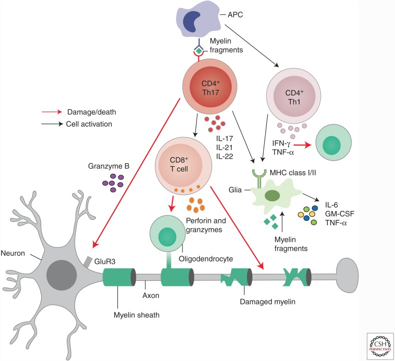
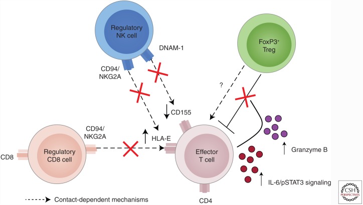
Multiple sclerosis (MS) has long been considered a CD4 T-cell disease, primarily because of the findings that the strongest genetic risk for MS is the major histocompatibility complex (MHC) class II locus, and that T cells play a central role in directing the immune response. The importance that the T helper (Th)1 cytokine, interferon γ (IFN-γ), and the Th17 cytokine, interleukin (IL)-17, play in MS pathogenesis is indicated by recent clinical trial data by the enhanced presence of Th1/Th17 cells in central nervous system (CNS) tissue, cerebrospinal fluid (CSF), and blood, and by research on animal models of MS, such as experimental autoimmune encephalomyelitis (EAE). Although the majority of research on MS pathogenesis has centered on the role of effector CD4 T cells, accumulating data suggests that CD8 T cells may play a significant role in the human disease. In fact, in contrast to most animal models, the primary T cell found in the CNS in patients with MS, is the CD8 T cell. As patient-derived effector T cells are also resistant to mechanisms of dominant tolerance such as that induced by interaction with regulatory T cells (Tregs), their reduced response to regulation may also contribute to the unchecked effector T-cell activity in patients with MS. These concepts will be discussed below.
Copyright © 2018 Cold Spring Harbor Laboratory Press; all rights reserved.
Figures
References
-
- Allegretta M, Nicklas JA, Sriram S, Albertini RJ. 1990. T cells responsive to myelin basic protein in patients with multiple sclerosis. Science 247: 718–721. - PubMed
-
- Aloisi F, Ria F, Adorini L. 2000. Regulation of T-cell responses by CNS antigen-presenting cells: Different roles for microglia and astrocytes. Immunol Today 21: 141–147. - PubMed
-
- Annibali V, Ristori G, Angelini DF, Serafini B, Mechelli R, Cannoni S, Romano S, Paolillo A, Abderrahim H, Diamantini A, et al. 2011. CD161highCD8+ T cells bear pathogenetic potential in multiple sclerosis. Brain 134: 542–554. - PubMed
Publication types
MeSH terms
LinkOut - more resources
Full Text Sources
Other Literature Sources
Medical
Research Materials