Review
. 2018 Mar 1;8(3):a028936.
doi: 10.1101/cshperspect.a028936.

Multiple Sclerosis Pathology

Affiliations
Review

Multiple Sclerosis Pathology

Hans Lassmann. Cold Spring Harb Perspect Med. .

Abstract

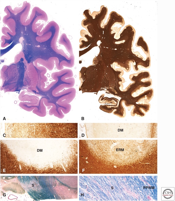
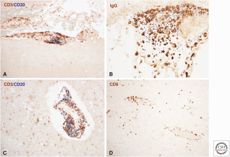
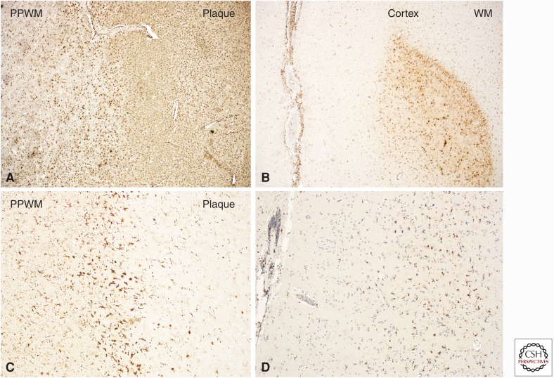
Multiple sclerosis (MS) is a chronic inflammatory demyelinating disease of the central nervous system (CNS), which gives rise to focal lesions in the gray and white matter and to diffuse neurodegeneration in the entire brain. In this review, the spectrum of MS lesions and their relation to the inflammatory process is described. Pathology suggests that inflammation drives tissue injury at all stages of the disease. Focal inflammatory infiltrates in the meninges and the perivascular spaces appear to produce soluble factors, which induce demyelination or neurodegeneration either directly or indirectly through microglia activation. The nature of these soluble factors, which are responsible for demyelinating activity in sera and cerebrospinal fluid of the patients, is currently undefined. Demyelination and neurodegeneration is finally accomplished by oxidative injury and mitochondrial damage leading to a state of "virtual hypoxia."


Figures

Figure 1.
Figure 1.
Pathological changes in the brain of a patient with secondary progressive multiple sclerosis (MS). Large confluent focal demyelinated lesions are present in the white matter (A). In addition, there is extensive subpial cortical demyelination, which can only be seen when sensitive immunocytochemistry for myelin proteins (e.g., proteolipid protein) is used (B). In contrast to the normal pattern of myelin in the cerebral cortex, as shown in C, there is complete loss of myelin in subpial lesions (D). Demyelinated plaques in the white matter may appear as inactive demyelinated lesions ([DMs] in E), as early remyelinated lesions with a low density of thin myelin sheaths only visible by immunocytochemistry for myelin proteins (ERM in F) or as remyelinated shadow plaques (G and H).
Figure 2.
Figure 2.
Inflammation in the multiple sclerosis (MS) brain. Meningeal inflammatory infiltrates are composed of CD3+ T cells, CD20+ B cells (A), and immunoglobulin-positive plasma cells (B). The inflammatory infiltrates appear as aggregates with separated domains for T cells, B cells, and plasma cells (A and B). No lymphocytes are present in the underlying cortical tissue (A). In white matter lesions, profound inflammatory infiltrates are seen in the perivascular space, which contain T cells and B cells (C). In addition, there is a diffuse infiltration of T cells, but not of B cells in the lesioned tissue (C). The diffuse infiltrates in the lesions nearly exclusively consist of CD8+ T lymphocytes (D).
Figure 3.
Figure 3.
Macrophages and microglia in active multiple sclerosis (MS) lesions. Classical active white matter lesions are densely infiltrated by macrophages and show massive microglia activation with expression of the phagocytosis-associated marker CD68 at the lesion margin and in the adjacent periplaque white matter (PPWM) (A). Active cortical lesions are associated with inflammation in the meninges, whereas the zone of active demyelination is infiltrated by activated microglia with some intermingled macrophages (B). The cortical and white matter areas are labeled as Cortex and WM, respectively. Slowly expanding lesions in the white matter are characterized by a dense rim of activated microglia with some intermingled macrophages at the lesion edge, stained with the microglia marker Iba1. In the lesion center, there is a very low microglia density (C). Similarly, slowly expanding lesions are also seen in the cortex, associated with meningeal inflammation and the presence of activated Iba1-positive microglia at the zone of active myelin injury. As in chronic white matter lesions also in the cortex, the demyelinated zones are nearly completely devoid of microglia (D).

References

    1. Albert M, Antel J, Brück W, Stadelmann C. 2007. Extensive cortical remyelination in patients with chronic multiple sclerosis. Brain Pathol 17: 129–138. - PMC - PubMed
    1. Androdias G, Reynolds R, Chanal M, Ritleng C, Confavreux C, Nataf S. 2010. Meningeal T cells associate with diffuse axonal loss in multiple sclerosis spinal cords. Ann Neurol 68: 465–476. - PubMed
    1. Babbe H, Roers A, Waisman A, Lassmann H, Goebels N, Hohlfeld R, Friese M, Schröder R, Deckert M, Schmidt S, et al. 2000. Clonal expansion of CD8+ T cells dominate the T cell infiltrate in active multiple sclerosis lesions as shown by micromanipulation and single cell polymerase chain reaction. J Exp Med 192: 393–404. - PMC - PubMed
    1. Babinski J. 1885. Recherches sur ĺanatomie pathologique de la sclerose en plaque et etude comparative des diverses varietes de la scleroses de la moelle [Research on the anatomic pathology of multiple sclerosis and a comparative study on different forms of scaring of the spinal cord]. Arch Physiol (Paris) 5–6: 186–207.
    1. Balashov KE, Rottman JB, Weiner HL, Hancock WW. 1999. CCR5+ and CXCR3+ T cells are increased in multiple sclerosis and their ligands MIP-1α and IP-10 are expressed in demyelinating brain lesions. Proc Natl Acad Sci 96: 6873–6878. - PMC - PubMed

MeSH terms