. 2018 Mar 9;293(10):3793-3805.
doi: 10.1074/jbc.RA117.000696. Epub 2018 Jan 22.

The CD300e molecule in mice is an immune-activating receptor

Affiliations

The CD300e molecule in mice is an immune-activating receptor

Masamichi Isobe et al. J Biol Chem. .

Abstract

CD300 molecules (CD300s) belong to paired activating and inhibitory receptor families, which mediate immune responses. Human CD300e (hCD300e) is expressed in monocytes and myeloid dendritic cells and transmits an immune-activating signal by interacting with DNAX-activating protein 12 (DAP12). However, the CD300e ortholog in mice (mCD300e) is poorly characterized. Here, we found that mCD300e is also an immune-activating receptor. We found that mCD300e engagement triggers cytokine production in mCD300e-transduced bone marrow-derived mast cells (BMMCs). Loss of DAP12 and another signaling protein, FcRγ, did not affect surface expression of transduced mCD300e, but abrogated mCD300e-mediated cytokine production in the BMMCs. Co-immunoprecipitation experiments revealed that mCD300e physically interacts with both FcRγ and DAP12, suggesting that mCD300e delivers an activating signal via these two proteins. Binding and reporter assays with the mCD300e extracellular domain identified sphingomyelin as a ligand of both mCD300e and hCD300e. Notably, the binding of sphingomyelin to mCD300e stimulated cytokine production in the transduced BMMCs in an FcRγ- and DAP12-dependent manner. Flow cytometric analysis with an mCD300e-specific Ab disclosed that mCD300e expression is highly restricted to CD115+Ly-6Clow/int peripheral blood monocytes, corresponding to CD14dim/+CD16+ human nonclassical and intermediate monocytes. Loss of FcRγ or DAP12 lowered the surface expression of endogenous mCD300e in the CD115+Ly-6Clow/int monocytes. Stimulation with sphingomyelin failed to activate the CD115+Ly-6Clow/int mouse monocytes, but induced hCD300e-mediated cytokine production in the CD14dimCD16+ human monocytes. Taken together, these observations indicate that mCD300e recognizes sphingomyelin and thereby regulates nonclassical and intermediate monocyte functions through FcRγ and DAP12.

Keywords: cell signaling; cell surface receptor; cellular immune response; immunology; monocyte.


Conflict of interest statement

The authors declare that they have no conflicts of interest with the contents of this article

Figures
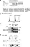
Figure 1.
Figure 1.
mCD300e is an N-glycosylated surface receptor. A, the phylogenetic tree of mouse LMIR3 (CLM-1/CD300f), LMIR4 (CLM-5), LMIR5 (CLM-7/CD300b), LMIR6 (CLM-2/CD300e), and LMIR7 (CLM-3) is shown on the basis of homology with the immunoglobulin-like domain (upper panel). The percentage of amino acid sequence identity of the immunoglobulin-like domain is indicated. Alignment of amino acid sequences for mCD300e is shown (lower panel). The putative signal sequence is shown in lowercase. An immunoglobulin-like domain is boxed. The transmembrane domain is underlined. The potential N-linked glycosylation site is shaded. The positively charged amino acid residue lysine within the transmembrane domain is shown in bold. B, Ba/F3 cells were transduced with FLAG-tagged mCD300e or mock. The transfectants were stained with mouse anti-FLAG Ab or mouse IgG1 Ab followed by PE-conjugated anti-mouse IgG goat F(ab′)2 Ab. C, lysates of Ba/F3 cells expressing FLAG-tagged mCD300e, mCD300e-N84Q, or mock were pretreated with or without N-glycosidase F before immunoprecipitation with mouse anti-FLAG Ab and subsequently immunoblotted with rabbit anti-FLAG Ab. D, 293T cells were transiently co-transfected with FLAG-tagged mCD300e construct or mock together with Myc-tagged FcRγ or DAP12 construct or mock. Immunoprecipitates of lysates of these transfectants with mouse anti-FLAG Ab were probed with anti-Myc Ab or rabbit anti-FLAG Ab. B–D, a representative of three independent experiments is shown. N-Gly, IB, or IP indicates N-glycosidase F, immunoblot, or immunoprecipitation, respectively.
Figure 2.
Figure 2.
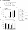
mCD300e delivers an activating signal in BMMC transfectants in both FcRγ- and DAP12-dependent manners. A, BMMCs transduced with FLAG-tagged mCD300e or mock were stained with FITC-conjugated anti-FcϵRIα Ab and PE-conjugated anti-c-Kit Ab (left panel) or with mouse anti-FLAG Ab or mouse IgG1 Ab followed by PE-conjugated anti-mouse IgG goat F(ab′)2 Ab (right panel). B, BMMCs transduced with FLAG-tagged mCD300e or mock were stimulated with plate-coated anti-FLAG Ab or mouse IgG1 as control or with 100 nm PMA for 12 h. IL-6 released into the culture supernatants were measured by ELISA. C, FLAG-tagged mCD300e-transduced BMMCs from WT, FcRγ−/−, DAP12−/−, or FcRγ−/−DAP12−/− mice were stained with FITC-conjugated anti-FcϵRIα Ab and PE-conjugated anti-c-Kit Ab (left panel) or with mouse anti-FLAG Ab or mouse IgG1 Ab followed by PE-conjugated anti-mouse goat IgG F(ab′)2 Ab (right panel). D, FLAG-tagged mCD300e-transduced BMMCs from WT, FcRγ−/−, DAP12−/−, or FcRγ−/−DAP12−/− mice were stimulated with plate-coated anti-FLAG Ab or mouse IgG1 Ab as control or with 100 nm PMA for 12 h. IL-6 released into the culture supernatants were measured by ELISA. A and C, a representative of three independent experiments is shown. B and D, all data points correspond to the mean ± S.D. of three independent experiments. Statistically significant differences are shown. *, p < 0.05 or **, p < 0.01 (Student's t test).
Figure 3.
Figure 3.
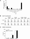
The generation of specific antibody against mCD300e. A, relative expression levels of mCD300e in indicated tissues were estimated by real-time PCR. The amount of expression was indicated relative to that in BM. B, Ba/F3 cells were transduced with FLAG-tagged mouse LMIR1 (CD300a), LMIR2, LMIR3 (CD300f), LMIR4, LMIR5 (CD300b), LMIR6 (CD300e), LMIR7, or mock. The transfectants were stained either with mouse anti-FLAG Ab or mouse IgG1 Ab followed by PE-conjugated anti-mouse IgG goat F(ab′)2 Ab (upper panel) or with biotinylated anti-mCD300e Ab or Armenian hamster IgG Ab followed by PE-conjugated streptavidin (lower panel). A and B, data are representative of three independent experiments. C, BMMCs transduced with FLAG-tagged mCD300e or mock were stimulated with plate-coated anti-mCD300e Ab or Armenia hamster IgG Ab as control or with 100 ng/ml of PMA for 12 h. IL-6 released into the culture supernatants were measured by ELISA. All data points correspond to the mean ± S.D. of three independent experiments. Statistically significant differences are shown. *, p < 0.05 (Student's t test).
Figure 4.
Figure 4.
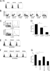
mCD300e is highly expressed in CD115+Ly-6Clow/int monocytes. A, mouse PB cells were stained with FITC-conjugated anti-CD3, CD19, or Ly-6G Ab and with biotinylated anti-mCD300e Ab or Armenian hamster IgG Ab followed by PE-conjugated streptavidin. Expression of mCD300e in CD3+, CD19+, or Ly-6G+ cells was shown. B, mouse PB cells were stained with FITC-conjugated CD11b, CD11c, F4/80, Ly-6C, CD80, CD86, or MHC class II (MHC-II) Ab and with biotinylated anti-mCD300e Ab or Armenian hamster IgG Ab followed by PE-conjugated streptavidin. C, mouse PB cells were stained with FITC-conjugated anti-Ly-6C Ab and PE-conjugated anti-CD115 Ab (upper left panel) or FITC-conjugated anti-Ly-6C Ab and biotinylated anti-mCD300e Ab or Armenian hamster IgG Ab followed by PE-conjugated streptavidin (lower left panel). Histograms showed expression of Ly-6C in CD115+ PB cells (upper right panel) or in mCD300e+ PB cells (lower right panel). D, mean fluorescent intensity (MFI) of mCD300e in CD115+Ly-6Clow, CD115+Ly-6Cint, or CD115+Ly-6Chigh PB monocytes was measured by flow cytometry. E, BM, spleen, or thymus cells were stained with FITC-conjugated anti-CD80 Ab and biotinylated anti-mCD300e Ab or Armenian hamster IgG Ab followed by PE-conjugated streptavidin. F, BMMCs, BMmDCs, BMpDCs, or BMMφ were stained with biotinylated anti-mCD300e Ab or Armenian hamster IgG Ab followed by PE-conjugated streptavidin. G, MFI of mCD300e in CD11c+ PB monocytes from WT, FcRγ−/−, DAP12−/−, or FcRγ−/−DAP12−/− mice were measured by flow cytometry. A–C, E, and F, data are representative of three independent experiments. D and G, all data points correspond to the mean ± S.D. of four independent experiments. Statistically significant differences are shown. *, p < 0.05 or **, p < 0.01 (Student's t test).
Figure 5.
Figure 5.
Sphingomyelin is a candidate ligand for mCD300e. A, mCD300e–Fc or Fc was incubated on plates coated with the indicated lipids. mCD300e–Fc or Fc bound to the plates was quantified by ELISA. All of the data points correspond to the mean ± S.D. of three independent experiments. Statistically significant differences are shown. **, p < 0.01 (Student's t test). PS, PC, PE, SM, or SPC indicates phosphatidylserine, phosphatidylcholine, phosphatidylethanolamine, sphingomyelin, or sphingosylphosphocholine. B, flow cytometry of GFP expression of mCD300e–2B4-GFP cells or 2B4-GFP cells that were incubated for 24 h on plates coated with indicated lipids or with anti-FLAG Ab or anti-mCD300e Ab. C, mCD300e–2B4-GFP cells were incubated for 24 h on plates coated with sphingomyelin in the presence of 20 μg/ml of either a soluble anti-mCD300e Ab or Armenia hamster IgG Ab as control. B and C, data are representative of three independent experiments.
Figure 6.
Figure 6.
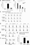
A possible role of the sphingomyelin–CD300e interaction. A and B, IL-6 released into the culture supernatants were measured by ELISA. All data points correspond to the mean ± S.D. of four independent experiments. Statistically significant differences are shown. *, p < 0.05 or **, p < 0.01 (Student's t test). A, BMMCs transduced with FLAG-tagged mCD300e were stimulated with plate-coated sphingomyelin or vehicle in the presence of 20 μg/ml of either soluble anti-mCD300e Ab or Armenia hamster IgG Ab as a control. B, FLAG-tagged mCD300e-transduced BMMCs from WT, FcRγ−/−, DAP12−/−, or FcRγ−/−DAP12−/− mice were stimulated with plate-coated sphingomyelin or vehicle. C, Ba/F3 cells were transduced with FLAG-tagged human CD300a, CD300b, CD300c, CD300e, CD300f, or mock. The transfectants were stained either with mouse anti-FLAG Ab or mouse IgG1 Ab followed by PE-conjugated anti-mouse IgG goat F(ab′)2 Ab (upper panel) or with anti-hCD300e Ab (233804), anti-hCD300e Ab (233812), or rat IgG2a Ab followed by PE-conjugated anti-rat IgG goat F(ab′)2 Ab (middle and lower panels). D, flow cytometry of GFP expression of hCD300e–2B4-GFP cells or 2B4-GFP cells that were incubated for 24 h on plates coated with indicated lipids or with anti-FLAG Ab or anti-hCD300e Ab (233804). E, flow cytometry of GFP expression of hCD300e–2B4-GFP cells that were incubated for 24 h on plates coated with sphingomyelin in the presence of 20 μg/ml of either soluble anti-hCD300e Ab (233812) or rat IgG2a Ab as a control. F, human PB cells were stained with APC-conjugated anti-CD14 Ab and FITC-conjugated anti-CD16 Ab and with anti-hCD300e Ab (233812) or rat IgG2a Ab followed by PE-conjugated anti-rat IgG goat F(ab′)2 Ab. Expression of hCD300e in CD14+CD16, CD14+CD16+, or CD14dimCD16+ PB cells was shown. C–F, data are representative of three independent experiments. G, CD14dimCD16+ cells sorted from human PB were stimulated with plate-coated sphingomyelin or vehicle in the presence of 20 μg/ml of either a soluble anti-hCD300e Ab (233812) or rat IgG2a Ab as a control. The levels of IL-6 released into the culture supernatants were measured by ELISA. All data points correspond to the mean ± S.D. of three independent experiments. Statistically significant differences are shown. *, p < 0.05 or **, p < 0.01 (Student's t test).

References

    1. Colonna M. (2003) TREMs in the immune system and beyond. Nat. Rev. Immunol. 3, 445–453 10.1038/nri1106 - DOI - PubMed
    1. Ravetch J. V., and Lanier L. L. (2000) Immune inhibitory receptors. Science 290, 84–89 10.1126/science.290.5489.84 - DOI - PubMed
    1. Hardison S. E., and Brown G. D. (2012) C-type lectin receptors orchestrate antifungal immunity. Nat. Immunol. 13, 817–822 - PMC - PubMed
    1. Chung D. H., Humphrey M. B., Nakamura M. C., Ginzinger D. G., Seaman W. E., and Daws M. R. (2003) CMRF-35-like molecule-1, a novel mouse myeloid receptor, can inhibit osteoclast formation. J. Immunol. 171, 6541–6548 10.4049/jimmunol.171.12.6541 - DOI - PubMed
    1. Yotsumoto K., Okoshi Y., Shibuya K., Yamazaki S., Tahara-Hanaoka S., Honda S., Osawa M., Kuroiwa A., Matsuda Y., Tenen D. G., Iwama A., Nakauchi H., and Shibuya A. (2003) Paired activating and inhibitory immunoglobulin-like receptors, MAIR-I and MAIR-II, regulate mast cell and macrophage activation. J. Exp. Med. 198, 223–233 10.1084/jem.20021825 - DOI - PMC - PubMed

Publication types

MeSH terms