. 2018 Feb 6;115(6):1204-1209.
doi: 10.1073/pnas.1717282115. Epub 2018 Jan 22.

Aberration-corrected cryoimmersion light microscopy

Affiliations

Aberration-corrected cryoimmersion light microscopy

Raffaele Faoro et al. Proc Natl Acad Sci U S A. .

Abstract

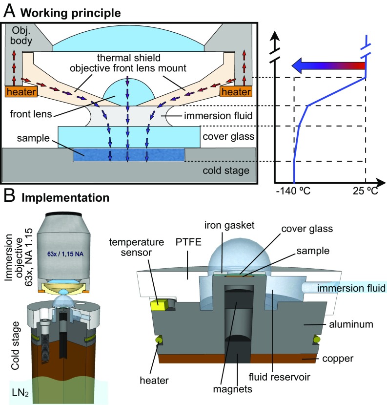
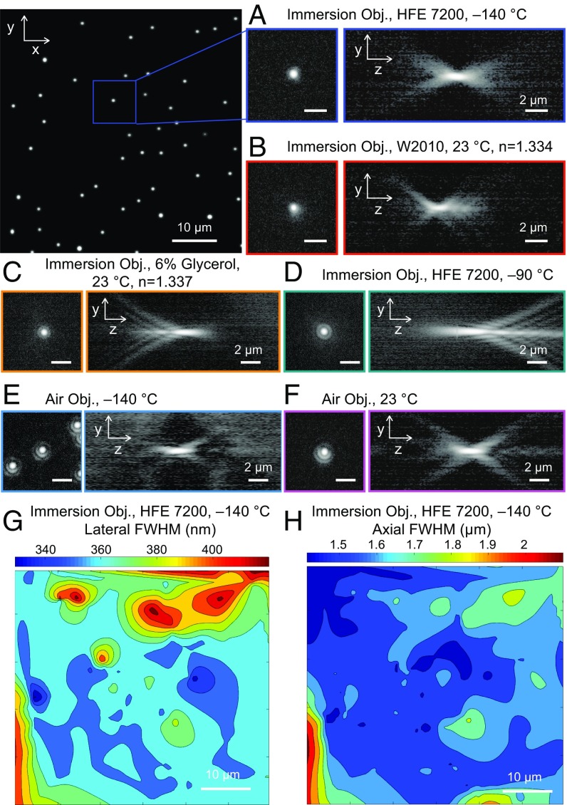
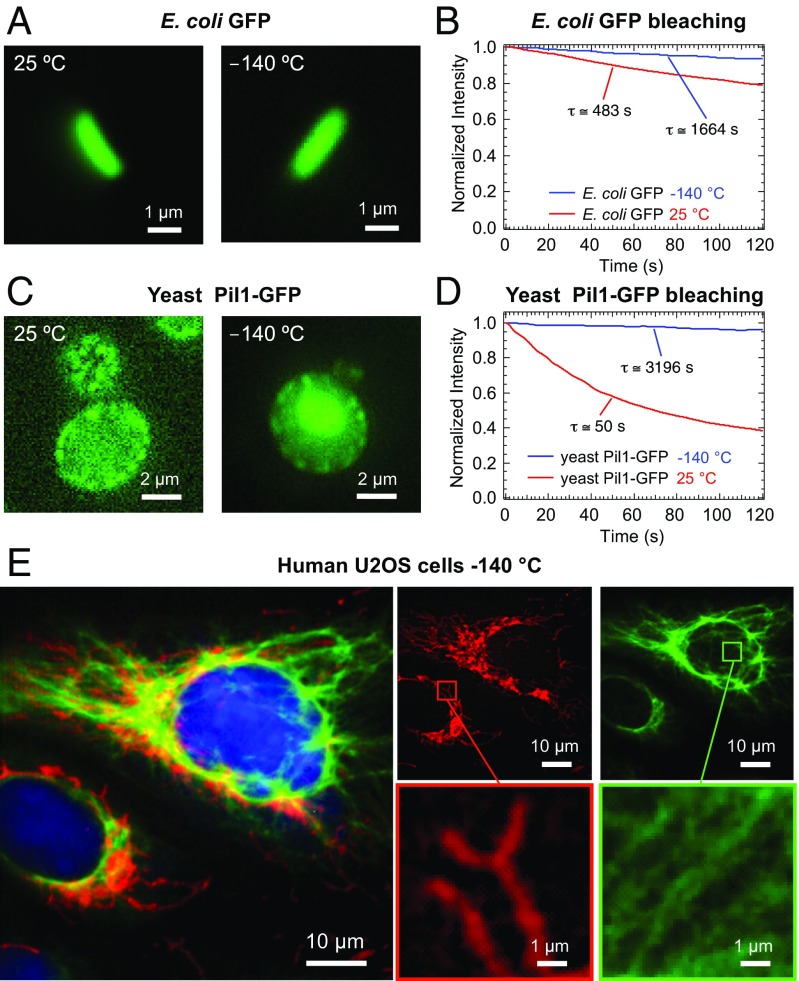
Cryogenic fluorescent light microscopy of flash-frozen cells stands out by artifact-free fixation and very little photobleaching of the fluorophores used. To attain the highest level of resolution, aberration-free immersion objectives with accurately matched immersion media are required, but both do not exist for imaging below the glass-transition temperature of water. Here, we resolve this challenge by combining a cryoimmersion medium, HFE-7200, which matches the refractive index of room-temperature water, with a technological concept in which the body of the objective and the front lens are not in thermal equilibrium. We implemented this concept by replacing the metallic front-lens mount of a standard bioimaging water immersion objective with an insulating ceramic mount heated around its perimeter. In this way, the objective metal housing can be maintained at room temperature, while creating a thermally shielded cold microenvironment around the sample and front lens. To demonstrate the range of potential applications, we show that our method can provide superior contrast in Escherichia coli and yeast cells expressing fluorescent proteins and resolve submicrometer structures in multicolor immunolabeled human bone osteosarcoma epithelial (U2OS) cells at [Formula: see text]C.

Keywords: cryo-light microscopy; cryofixation; cryofluorescence microscopy; fluorescence imaging; high-NA immersion objective.


Conflict of interest statement

The authors declare no conflict of interest.

Figures

Fig. 1.
Fig. 1.
(A) Schematic cross-section of an immersion objective with the front lens mount made of Macor operating as a thermal shield. The heat flows across the sample, cover glass, immersion fluid, front lens, and ceramic mount. The low thermal expansion coefficient of the machinable ceramic allows for reducing the thermal stresses. The high thermal resistance of Macor allows confining most of the temperature gradient in the front lens mount, as shown in the diagram. Note that due to the high thermal conductivity of the metallic cold stage and the intimate contact with the sample, the stage temperature closely reflects the temperature of the sample. (B) The cold stage used for the implementation of cryoimmersion microscopy was designed to keep both sample and cryoimmersion fluid at a stable temperature by combining heating and temperature sensing. The stage is composed of two parts, a bottom copper bar, which is in direct contact with liquid nitrogen, and a top anodized aluminum post. The sample is placed on top of the aluminum post and is in direct contact with the metal below a coverslip. The coverslip and sample are secured magnetically to the post by using an iron gasket and a magnet glued inside the aluminum post. The immersion fluid is supplied to the reservoir through a channel in the polytetrafluoroethylene (PTFE) cap. The opening in the PTFE cap allows forming a cold drop around the sample.
Fig. 2.
Fig. 2.
(A) Lateral and axial PSF measured at 140°C with the cryoimmersion objective (Obj.) and the immersion fluid HFE-7200. Fitting the PSF to a two-dimensional (2D) Gaussian (Fig. S2) allows us to determine lateral and axial resolution. The intensity is normalized and shown on a logarithmic scale. (B) Lateral and axial PSF measured at room temperature with the cryoimmersion objective and the commercial immersion fluid W2010. (C) Lateral and axial PSF measured at room temperature with the cryoimmersion objective and 6% glycerol (n=1.337) as immersion fluid. The refractive index mismatch produces positive spherical aberrations. (D) Lateral and axial PSF measured at −90 °C with the cryoimmersion objective and the immersion fluid HFE-7200. The refractive index of the immersion fluid at −90 °C is smaller than the refractive index of water at room temperature, producing negative spherical aberrations. (E) Lateral and axial PSF measured at 140°C with the 0.75-NA air objective. (F) Lateral and axial PSF measured at room temperature with the air objective. (All scale bars in AF are 2 μm.) (G) A 2D heat map of the measured lateral FWHM at −140 °C. (H) A 2D heat map of the measured axial FWHM at −140 °C.
Fig. 3.
Fig. 3.
Imaging of cryofixed biological specimens with the prototyped cryoimmersion objective. (A) Room-temperature (Left) and cryogenic (Right) widefield fluorescence images of Escherichia coli expressing GFP. (B) Photobleaching curves for GFP expressed in E. coli. The red curve shows decay at room temperature, and blue curve shows decay at 140°C. In cryoconditions, the GFP bleaching is suppressed 3.5 times. (C) Room-temperature (Left) and cryogenic (Right) widefield fluorescence images of yeast cells expressing GFP-tagged Pil1. (D) Photobleaching curves for GFP expressed in yeast cells. The red curve shows decay at room temperature, and the blue curve shows decay at 140°C. In cryoconditions, the GFP bleaching is suppressed 64 times. (E) Three-color widefield cryofluorescence image of plunge frozen U2OS cells labeled with Alexa Fluor 488 (vimentin cytoskeleton), Alexa Fluor 594 (Tom20 mitochondrial protein), and DAPI (cell nuclei).

References

    1. Kaufmann R, Hagen C, Grünewald K. Fluorescence cryo-microscopy: Current challenges and prospects. Curr Opin Chem Biol. 2014;20:86–91. - PMC - PubMed
    1. Loussert Fonta C, Humbel BM. Correlative microscopy. Arch Biochem Biophys. 2015;581:98–110. - PubMed
    1. Schwartz CL, Sarbash VI, Ataullakhanov FI, Mcintosh JR, Nicastro D. Cryo-fluorescence microscopy facilitates correlations between light and cryo-electron microscopy and reduces the rate of photobleaching. J Microsc. 2007;227:98–109. - PubMed
    1. Kaufmann R, et al. Super-resolution microscopy using standard fluorescent proteins in intact cells under cryo-conditions. Nano Lett. 2014;14:4171–4175. - PMC - PubMed
    1. Moerner WE, Orrit M. Illuminating single molecules in condensed matter. Science. 1999;283:1670–1676. - PubMed

Publication types

Substances