. 2018 Jan 22;9(1):319.
doi: 10.1038/s41467-017-02749-w.

Culex pipiens crossing type diversity is governed by an amplified and polymorphic operon of Wolbachia

Affiliations

Culex pipiens crossing type diversity is governed by an amplified and polymorphic operon of Wolbachia

Manon Bonneau et al. Nat Commun. .

Erratum in

Abstract

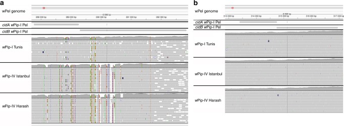
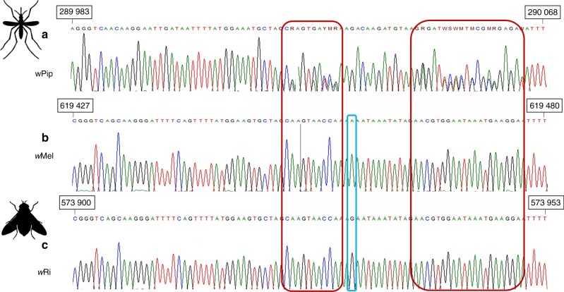
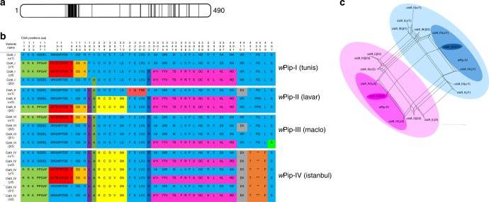
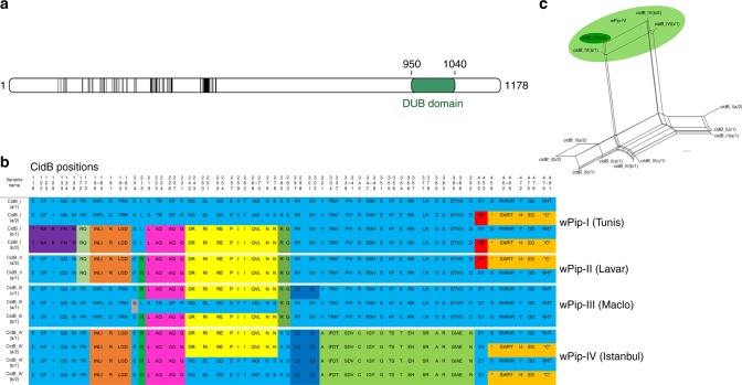
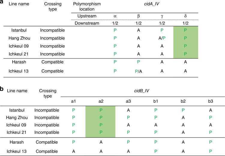
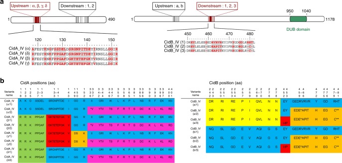
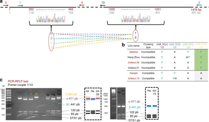
Culex pipiens mosquitoes are infected with Wolbachia (wPip) that cause an important diversity of cytoplasmic incompatibilities (CIs). Functional transgenic studies have implicated the cidA-cidB operon from wPip and its homolog in wMel in CI between infected Drosophila males and uninfected females. However, the genetic basis of the CI diversity induced by different Wolbachia strains was unknown. We show here that the remarkable diversity of CI in the C. pipiens complex is due to the presence, in all tested wPip genomes, of several copies of the cidA-cidB operon, which undergoes diversification through recombination events. In 183 isofemale lines of C. pipiens collected worldwide, specific variations of the cidA-cidB gene repertoires are found to match crossing types. The diversification of cidA-cidB is consistent with the hypothesis of a toxin-antitoxin system in which the gene cidB co-diversifies with the gene cidA, particularly in putative domains of reciprocal interactions.


Conflict of interest statement

The authors declare no competing financial interests.

Figures

Fig. 1
Fig. 1
The cidA/cidB operon displays polymorphism within and between different wPip strains, whereas no polymorphism is observed for the cinA/cinB operon. Mapping onto the reference genome wPip_Pel of the Illumina reads from the Tunis line infected with wPipI, and from the Harash and Istanbul lines infected with wPipIV. Colored residues are different from those in the reference wPip_Pel sequence. The pattern above the dotted lines represents the number of Illumina reads that have mapped for each position. The red box on the wPip_Pel reference genome allows the IGV user to locate the zone that is visualized with more details on the bottom panels. a Mapping of the cidA/cidB operon reads onto the wPip_Pel reference genome. Polymorphism between wPip groups was detected (differences between Harash and Istanbul infected with wPipIV, Tunis, and wPip_Pel infected with wPipI). Polymorphism was also detected within Wolbachia groups (variations observed between Tunis and wPip_Pel or between Harash and Istanbul). The operon was also found to be polymorphic within the same isofemale line of C. pipiens. b Mapping of the cinA/cinB operon reads onto the wPip_Pel reference genome. No polymorphism was detected between reads from the three isofemale lines and the reference line
Fig. 2
Fig. 2
Sanger sequencing electrophoregrams of homologous regions of a cidB in wPip, b cifB in wMel, c and cifB in wRi. The numbers in the black boxes indicate the positions of these regions in the wMel, wRi, and wPip_Pel genomes. The blue rectangle highlights the only polymorphism between wRi and wMel. Unlike the Wolbachia strains of Drosophila, wPip strains consistently give mixed signals (showed in red rectangles), suggesting the presence of at least two different sequences of this gene in DNA samples
Fig. 3
Fig. 3
The repertoire of CidA variants in the different wPip groups. a Localization of polymorphic zones within CidA proteins. Schematic representation of the CidA protein with polymorphic zones between CidA variants of the four wPip groups highlighted in black. b The polymorphism between CidA variants is distributed as blocks of variable amino acids. Protein sequences alignment of the CidA variants found in the four Wolbachia strains Tunis, Lavar, Maclo, and Istanbul. The first sequence is used as a reference to determine the polymorphic region. For more clarity, only polymorphic positions of the alignment are represented and amino-acid positions are not contiguous. When more than two contiguous amino acids were variable the “-” symbol was used between the first and the last variable position of the zone. Colors show polymorphic blocks of residues present in variants regardless of their phylogenetic wPip group (I–IV). However, no variant (i.e., complete CidA sequence) is common to wPip strains from different groups. c cidA variants result from block recombination. Each edge (or set of parallel edges) corresponds to a split in the data set and has a length equal to the weight of the split. Incompatible splits produced by recombination are represented by boxes in the network. Most of the cidA variants are connected by multiple pathways resulting from block recombination between them. The largest circles highlight the two groups of cidA variants sharing the same amino-acid sequence from position 336 to 401 (b). Intermediate darker circles highlight cidA variants observed in the wPipIV group. The smallest bold circles highlight the two versions (1 and 2) of the cidA_IV(δ) variant matching the “incompatible” crossing type of mosquito lines infected with wPipIV (see the text)
Fig. 4
Fig. 4
The repertoire of CidB variants in the different wPip groups. a Localization of polymorphic zones within CidB proteins. Schematic representation of the CidB protein with polymorphic zones between CidB variants of the four wPip groups highlighted in black. The deubiquitylating protease domain (DUB) present in CidB, represented in green, displayed no polymorphism between wPip strains. b The polymorphism between CidB variants is distributed as blocks of variable amino acids. Protein sequences alignment of the CidB variants found in the four Wolbachia strains Tunis, Lavar, Maclo, and Istanbul. The first sequence is used as reference to determine the polymorphic region. For more clarity, only polymorphic positions of the alignment are represented and amino-acid positions are not contiguous. When more than two contiguous amino acids were variable the “-” symbol was used between the first and the last variable position of the zone. Colors show polymorphic blocks of residues present in variants regardless of their phylogenetic wPip group (I–IV). However, no full variant (i.e., complete CidB sequence) is common to wPip strains from different groups. c cidB variants result from block recombination. Each edge (or set of parallel edges) corresponds to a split in the data set and has length equal to the weight of the split. Incompatible splits produced by recombination are represented by boxes in the network. Most of the cidB variants are connected by multiple pathways resulting from block recombination between them. The light green circle highlights cidB_IV variants, which cluster together due to their similar amino-acid sequences from position 321 to position 392 (b). The cidB_IV(a/2) variant matching the “incompatible” crossing type of mosquito lines infected with wPipIV (see the text) is highlighted by the darker green circle
Fig. 5
Fig. 5
cidA_IV(δ) and cidB_IV(a/2) are present specifically in lines infected with wPipIV strains displaying the “incompatible” crossing type. The “incompatible” or “compatible” crossing type of the C. pipiens lines infected with wPipIV strains were determined by crossing males with females infected with wPipI, wPipII, or wPipIII. A “compatible” type corresponds to a line in which all males are compatible when crossed with all tested females, and an “incompatible” type corresponds to a line in which males are incompatible when crossed with all tested females. The column of the two variants cidA IV(δ) and cidB_IV(a/2) associated with the “incompatible” crossing type were shaded in green. a Distribution of cidA variants (P for present, A for absent) in six wPipIV strains. Each column corresponds to the specific upstream polymorphic region named α, β, γ, or δ. The downstream polymorphism is represented by either the sequence 1 or the sequence 2 (1/2). For a given upstream sequence P/A means that the strain exhibits the sequence 1 but not the sequence 2. On the opposite A/P means that the strain exhibits the sequence 2 but not the sequence 1. b Distribution of cidB variants (P for present, A for absent) in six wPipIV strains
Fig. 6
Fig. 6
The putative domains of interaction between the CidA_IV and CidB_IV proteins. a Localization of polymorphic zones within CidA_IV and CidB_IV proteins. Secondary structure prediction and fold-recognition analysis predicted that the CidA protein could almost exclusively display protein/protein interaction repeated motifs as ankyrin- (or HEAT-like repeats). Similar domains of ~30 amino acids (expected size for ankyrin domains) are also present in CidB. The deubiquitylating protease domain (DUB) present in CidB, represented in green, displayed no polymorphism between wPipIV strains as already observed between wPip strains from different groups. The polymorphism of the wPipIV CidA and CidB variants associated with crossing type (“compatible” vs. “incompatible”, see Fig. 5 for definition) is highlighted in red. The corresponding variations in protein sequences are reported below the schematic representation of the full-length proteins, with the variable residues highlighted in red. Other sequence variations not predicted to be involved in CI variations are shown in black. b CidA_IV and CidB_IV variants sequences detected in all sequenced wPip strains from group IV. CidA_IV variants display two regions of polymorphism resulting from recombination revealed by block colors: the upstream one from 118 to 152 aa and the downstream one from 336 to 401 aa. Four possible sequences (noted α, β, γ, or δ) were found in the upstream polymorphic region followed by one of the two sequences in the downstream region (noted 1 or 2). Only the upstream polymorphic region of CidA is associated with crossing type variations. CidB_IV variants display two regions of polymorphism resulting from recombination: the upstream one from 242 to 278 aa and the second one from 450 to 481 aa. Two possible sequences (noted a and b) were found in the upstream polymorphic region followed by one of the three possible sequences in the downstream polymorphic region (noted 1, 2, and 3). Only the downstream region is associated with crossing type variations
Fig. 7
Fig. 7
cidA_IV gene polymorphic regions and PCR-RFLP tests for specific variants. a Schematic representation of the architecture of cidA_IV polymorphism. The black line represents the cidA_IV sequence with a focus on the two polymorphic regions. Non-polymorphic regions were shortened and represented as a dashed line. Numbers in red under the line represent nucleotide positions, and the numbers in blue indicate amino-acid positions. The overlapping oligonucleotides used for PCR amplification are represented by arrows numbered as in Supplementary Table 2. Both polymorphic regions were studied, but only the upstream matched the compatibility profile; its four different sequences (α, β γ, and δ) were followed by one of the two possible sequences (1 or 2) in the downstream polymorphic region. A different color code was used for α (purple), β (light blue), γ (light green), and δ (yellow) sequences in the upstream part of the cidA gene. b The repertoire of cidA_IV variants is different in “compatible” and “incompatible” lines. cidA_IV(δ) is present only in lines with “incompatible” crossing type. The names of the C. pipiens lines used to set-up the PCR-RFLP (c) test are highlighted in red. c PCR-RFLP tests for distinguishing between cidA_IV variants on the basis of the upstream polymorphic region. A 778 bp fragment was amplified with primers 1/13. Double digestion with ApoI and Hpy188I distinguished between cidA_IV(α) (six fragments: 471; 122; 57; 53; 51; and 24 bp), cidA_IV(β) and cidA_IV(γ) (six fragments: 441; 122; 83; 57; 51; and 24 bp), and cidA_IV(δ) (five fragments: 524; 122; 57; 51; and 24 bp). Panel to the left of the electrophoresis gel: PCR-RFLP on DNA from clones; right panel: PCR-RFLP on DNA from the Istanbul, Harash, Ichkeul 13, and Ichkeul 09 lines. On the right of the gel, a schematic representation of the PCR-RFLP profiles of these lines. Digestion bands that are specific of the variants are represented with the color code established in a. Bands that are not used to discriminate between the variants are represented in black on the bottom of the schematic gel
Fig. 8
Fig. 8
cidB_IV gene polymorphic regions and RFLP tests for specific variants. a Schematic representation of the polymorphism architecture of cidB_IV. The black line represents the cidB_IV sequence with a focus on the polymorphic region. Non-polymorphic regions were shortened and represented as a dashed line. Numbers in red represent nucleotide positions and those in blue indicate amino-acid positions. The overlapping oligonucleotides used for amplification are represented by arrows numbered as in Supplementary Table 2. Both polymorphic regions are indicated but only the downstream (on the left, base pairs 1347–1428, amino acids 450–481) matches the compatibility profiles. The two sequences of the upstream polymorphic zone (a and b) are followed by one of the three possible sequences (1, 2, and 3) in the downstream polymorphic region. A different color code was used for 1 (orange), 2 (blue), and 3 (green) different sequences in the downstream part of the cidB gene. b The cidB_IV variant repertoire differs between “compatible” and “incompatible” lines. cidB_IV(a/2) is present only in lines with “incompatible” crossing type. The names of the C. pipiens lines used to set up the PCR-RFLP (c) test are highlighted in red. c PCR-RFLP tests for distinguishing between cidB_IV variants on the basis of the second polymorphic region. A 1267–1276 bp fragment was amplified with primers 7/8. Double digestion with BanI and TaqaI distinguished between cidB_IV(1) (three fragments: 892; 239; and 145 bp), cidB_IV(2) (two fragments: 1028 and 239 bp), and cidB(3) (three fragments: 861; 239; and 167 bp). Panel to the left of the electrophoresis gel: PCR-RFLP on DNA from clones; right panel: PCR-RFLP on DNA from lines. The panel on the right shows schematic representations of the PCR-RFLP profiles for these lines. Digestion bands that are specific of the variants are represented with the color code established in the a. Bands that are not used in to discriminate between the variants are represented in black on the bottom of the schematic gel

References

    1. Yen JH, Barr AR. New hypothesis of the cause of cytoplasmic incompatibility in Culex pipiens L. Nature. 1971;232:657–658. doi: 10.1038/232657a0. - DOI - PubMed
    1. Poinsot D, Charlat S, Merçot H. On the mechanism of Wolbachia-induced cytoplasmic incompatibility: confronting the models with the facts. BioEssays. 2003;25:259–265. doi: 10.1002/bies.10234. - DOI - PubMed
    1. Hurst LD. The evolution of cytoplasmic incompatibility or when spite can be successful. J. Theor. Biol. 1991;148:269–277. doi: 10.1016/S0022-5193(05)80344-3. - DOI - PubMed
    1. Werren JH. Biology of Wolbachia. Annu. Rev. Entomol. 1997;42:587–609. doi: 10.1146/annurev.ento.42.1.587. - DOI - PubMed
    1. Foster J, et al. The Wolbachia genome of Brugia malayi: endosymbiont evolution within a human pathogenic nematode. PLoS Biol. 2005;3:0599–0614. doi: 10.1371/journal.pbio.0030121. - DOI - PMC - PubMed

Publication types

MeSH terms

Substances