. 2018 Jan 22;8(1):1305.
doi: 10.1038/s41598-018-19289-y.

Ex Vivo Tracer Efficacy in Optical Imaging of Staphylococcus Aureus Nuclease Activity

Affiliations

Ex Vivo Tracer Efficacy in Optical Imaging of Staphylococcus Aureus Nuclease Activity

Colin W K Rosman et al. Sci Rep. .

Abstract

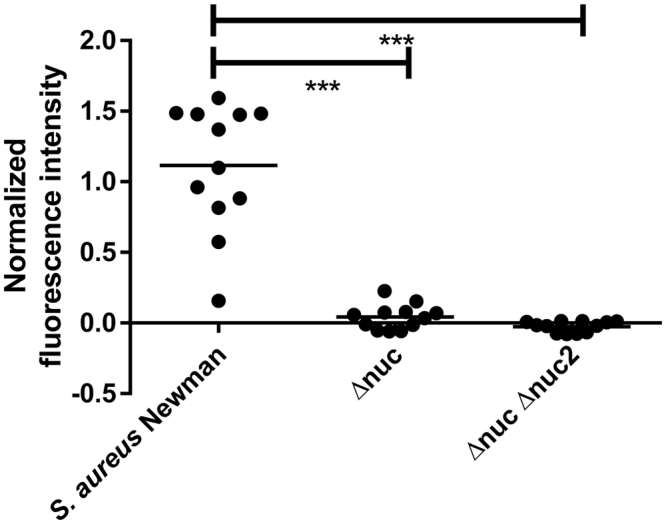
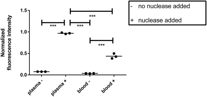
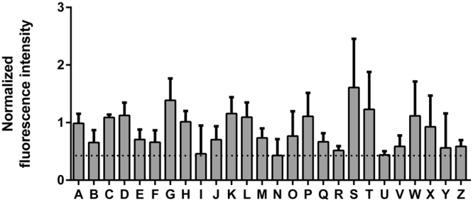
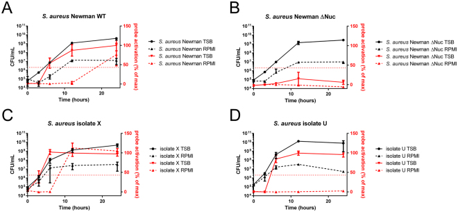
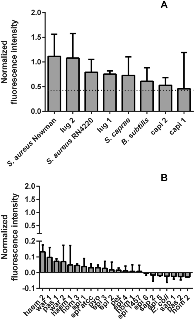
The key to effective treatment of bacterial infections is a swift and reliable diagnosis. Current clinical standards of bacterial diagnosis are slow and laborious. There are several anatomical imaging modalities that can detect inflammation, but none can distinguish between bacterial and sterile inflammation. Novel tracers such as smart activatable fluorescent probes represent a promising development that allow fast and specific testing without the use of ionizing radiation. Previously, a smart activatable probe was developed that is a substrate for the micrococcal nuclease as produced by Staphylococcus aureus. In the present study, the function of this probe was validated. Practical applicability in terms of sensitivity was assessed by incubation of the probe with 26 clinical S. aureus isolates, and probe specificity was verified by incubation with 30 clinical isolates and laboratory strains of various bacterial pathogens. The results show that the nuclease-specific probe was activated by all tested S. aureus isolates and laboratory strains with a threshold of ~106-107 cells/mL. The probe was also activated by certain opportunistic staphylococci. We therefore propose that the studied nuclease probe represents a significant step forward to address the need for a rapid, practical, and precise method to detect infections caused by S. aureus.


Conflict of interest statement

The authors declare that they have no competing interests.

Figures

Figure 1
Figure 1
P2&3 TT-probe activation by S. aureus Newman (Newman), S. aureus Newman Δnuc (Δnuc) and S. aureus Newman Δnuc, Δnuc2 (Δnuc, Δnuc2). The horizontal line represents the mean. ***Indicates P < 0.0001.
Figure 2
Figure 2
Normalized fluorescence intensity after probe incubation in human blood and plasma. Horizontal line represents the mean. indicated no nuclease was added. +indicates nuclease was added. **Indicates P < 0.001, ***indicates P < 0.0001.
Figure 3
Figure 3
Normalized fluorescence intensity after incubation of clinical S. aureus isolates. A/Z indicate the isolates that were incubated. The dotted horizontal line represents the positive cutoff. Bars indicate mean fluorescence. Capped lines indicate standard deviation.
Figure 4
Figure 4
Activation of the P2&3 TT-probe by S. aureus grown to different stages in TSB and RPMI. Left Y-axis and lines in black indicate CFU per mL, right Y-axis and lines in red indicate probe activation as a percentage of the maximum activation. All experiments were done in triplicate, and the plots present the average numbers of the measurements. Capped lines indicate standard deviation. Strains included were: A, S. aureus Newman wild-type; B, nuclease deficient S. aureus mutant (Δnuc); C, S. aureus clinical isolate X; and D, S. aureus clinical isolate U.
Figure 5
Figure 5
P2&3 TT-probe activiation by different bacterial isolates. (A) Normalized fluorescense intensity upon P2&3 TT-probe activation by of all strains that tested positive. B. subtilis was grown in LB and the respective measurements were normalized with LB controls. All other strains were grown in TSB. (B) normalized fluorescence intensity of strains that tested negative with the P2&3 TT-probe. E. coli was grown in LB and the respective measurements were normalized with LB controls. All other strains were grown in TSB.

Similar articles

Cited by

References

    1. European Centre for Disease Prevention and Control. Annual epidemiological report 2014. Antimicrobial resistance and healthcare-associated infections. Available at: http://ecdc.europa.eu/en/publications/Publications/antimicrobial-resista... (Accessed 25 May 2016) (2015).
    1. Carnicer-Pont D, et al. Risk factors for hospital-acquired methicillin-resistant Staphylococcus aureus bacteraemia: a case-control study. Epidemiol. Infect. 2006;134:1167–1173. doi: 10.1017/S0950268806006327. - DOI - PMC - PubMed
    1. Carek PJ, Dickerson LM, Sack JL. Diagnosis and management of osteomyelitis. Am. Fam. Physician. 2011;65:1751. - PubMed
    1. Clerc O, Prod’hom G, Greub G, Zanetti G, Senn L. Adult native septic arthritis: a review of 10 years of experience and lessons for empirical antibiotic therapy. J. Antimicrob. Chemother. 2011;66:1168–1173. doi: 10.1093/jac/dkr047. - DOI - PubMed
    1. Agarwal V, Chauhan S, Gupta RK. Pyomyositis. Neuroimaging Clin. North. Am. 2002;21:975–983. doi: 10.1016/j.nic.2011.07.011. - DOI - PubMed

MeSH terms

LinkOut - more resources