. 2018 May;25(5):966-982.
doi: 10.1038/s41418-017-0036-9. Epub 2018 Jan 22.

TLR3 Mediates Repair and Regeneration of Damaged Neonatal Heart through Glycolysis Dependent YAP1 Regulated miR-152 Expression

Affiliations

TLR3 Mediates Repair and Regeneration of Damaged Neonatal Heart through Glycolysis Dependent YAP1 Regulated miR-152 Expression

Xiaohui Wang et al. Cell Death Differ. 2018 May.

Abstract

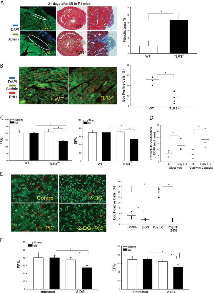
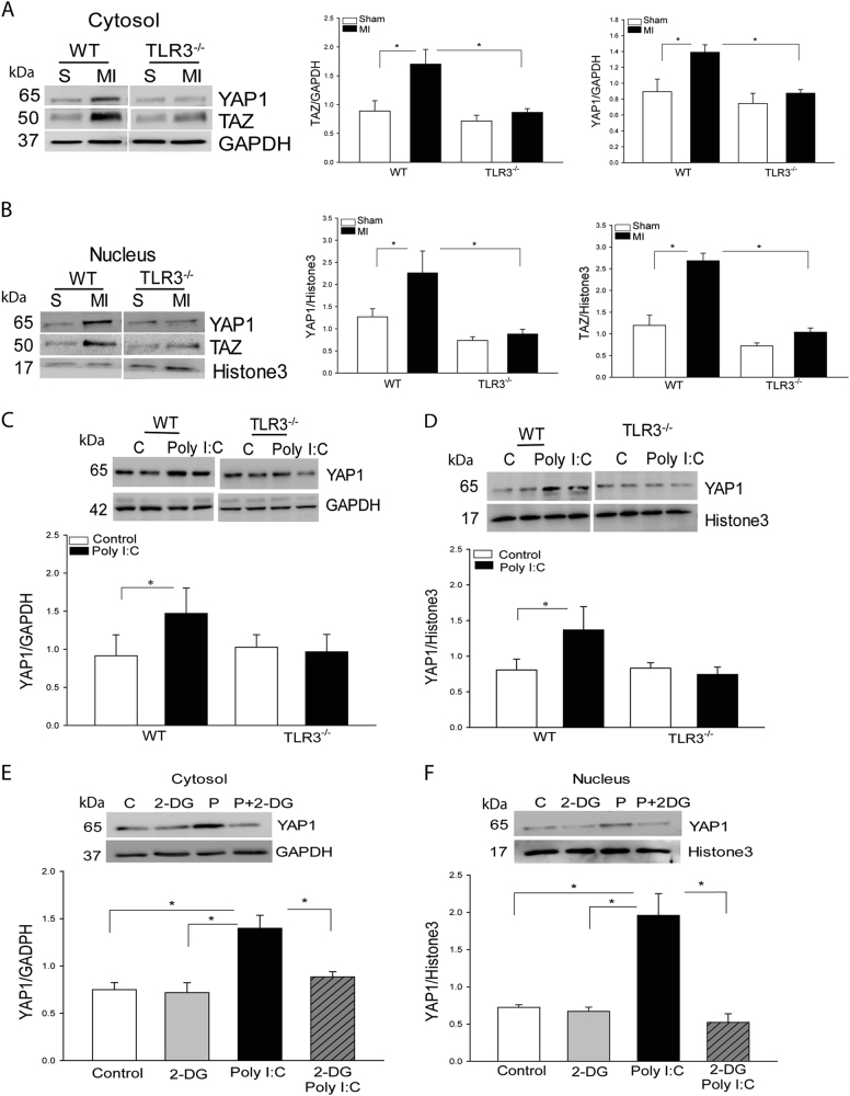
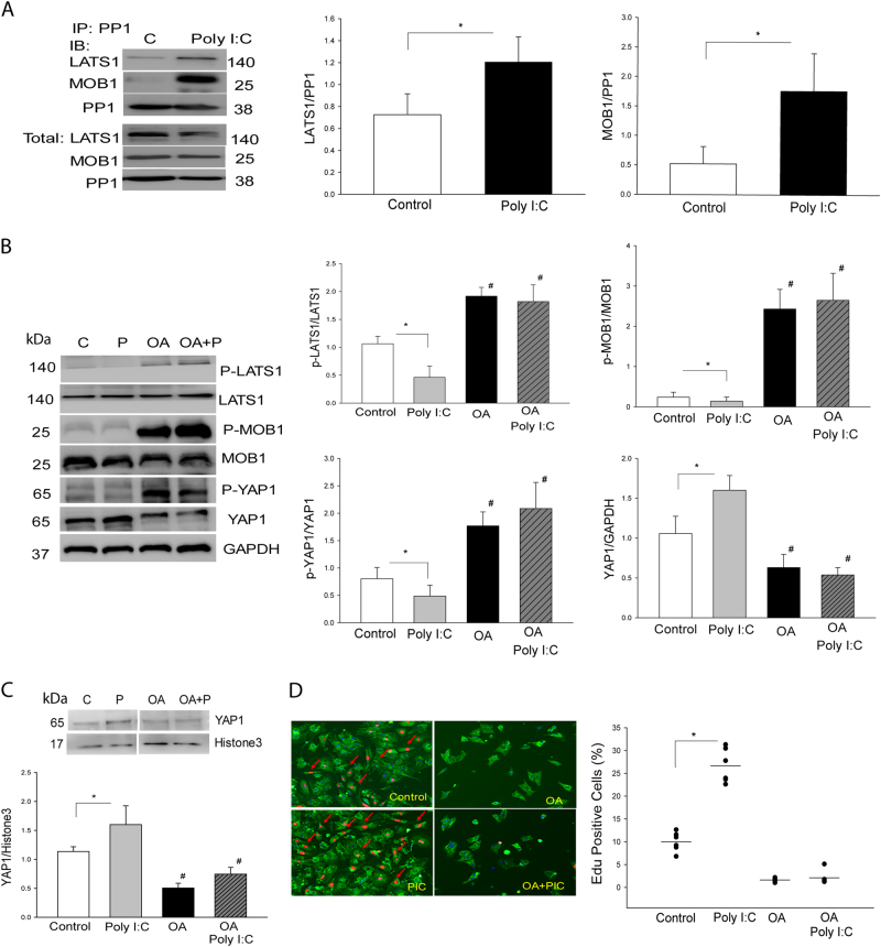
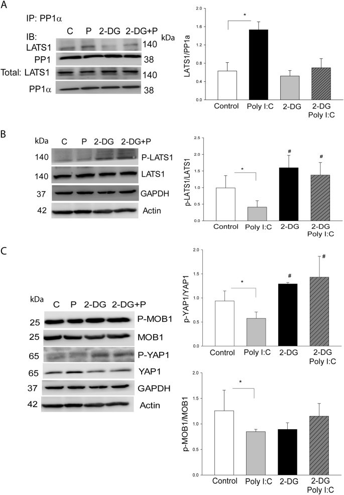
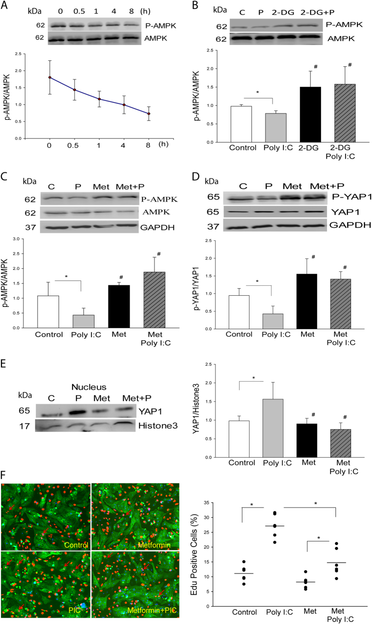
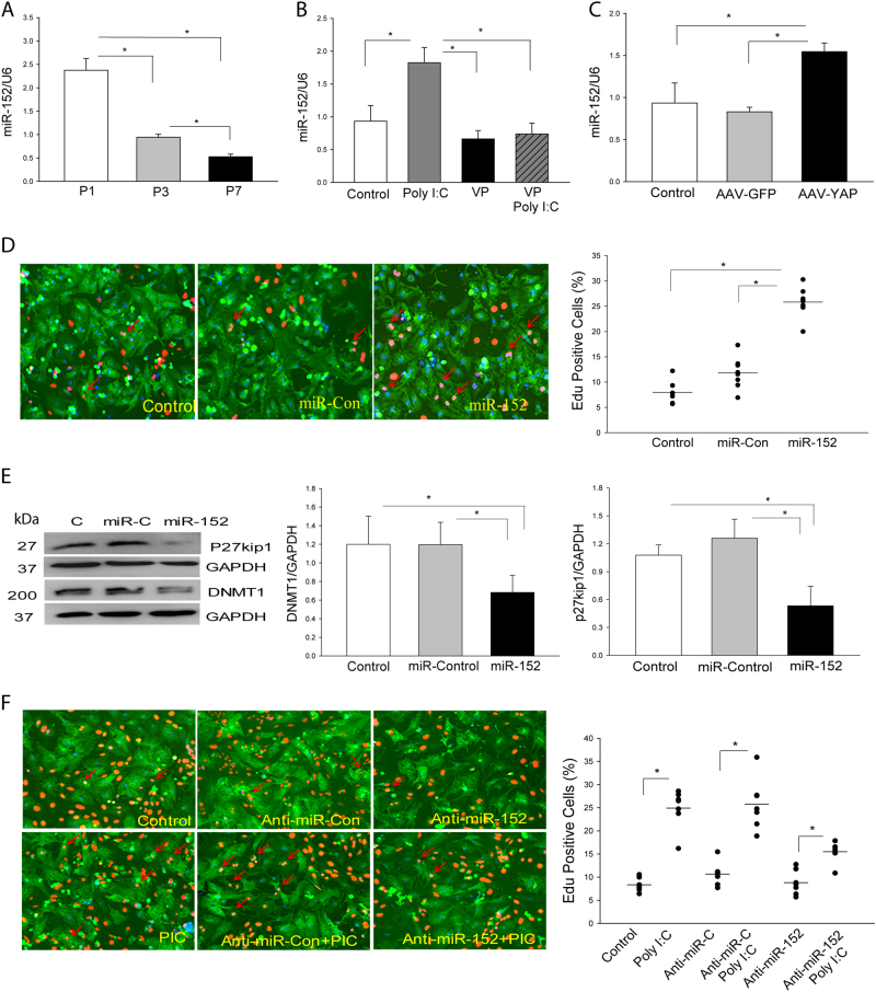
The present study investigated whether TLR3 is required for neonatal heart repair and regeneration following myocardial infarction (MI). TLR3 deficient neonatal mice exhibited impaired cardiac functional recovery and a larger infarct size, while wild type neonatal mice showed cardiac functional recovery and small infarct size after MI. The data suggest that TLR3 is essential for the regeneration and repair of damaged neonatal myocardium. In vitro treatment of neonatal cardiomyocytes with a TLR3 ligand, Poly (I:C), significantly enhances glycolytic metabolism, YAP1 activation and proliferation of cardiomyocytes which were prevented by a glycolysis inhibitor, 2-deoxyglucose (2-DG). Administration of 2-DG to neonatal mice abolished cardiac functional recovery and YAP activation after MI, suggesting that TLR3-mediated regeneration and repair of the damaged neonatal myocardium is through glycolytic-dependent YAP1 activation. Inhibition of YAP1 activation abolished Poly (I:C) induced proliferation of neonatal cardiomyocytes. Interestingly, activation of YAP1 increases the expression of miR-152 which represses the expression of cell cycle inhibitory proteins, P27kip1 and DNMT1, leading to cardiomyocyte proliferation. We conclude that TLR3 is required for neonatal heart regeneration and repair after MI. The mechanisms involve glycolytic-dependent YAP1 activation, resulting in miR-152 expression which targets DNMT1/p27kip1.


Conflict of interest statement

The authors declare that there are no competing financial interests.

Figures

Fig. 1
Fig. 1. TLR3 deficiency impaired regeneration and repair of damaged hearts following myocardial infarction
1 day old (P1) neonatal wild type (WT) and TLR3 deficient (TLR3-/-) mice were subjected to myocardial infarction (MI). Hearts were harvested 21 days after MI. a Heart tissue sections were stained with hematoxylin and eosin (H&E) or Masson-trichrome dye after MI. Fibrous area was quantified by image analysis in Masson-trichrome staining. b TLR3 deficiency reduced neonatal cardiomyocyte proliferation. EdU incorporation into the nuclei of cardiomyocytes from TLR3-/- neonatal mice was significantly reduced after MI. c TLR3-/- impaired cardiac functional recovery. 1 day old (P1) neonatal wild type (WT) and TLR3 deficient mice were subjected to myocardial infarction (MI) and cardiac function was measured by echocardiography. d The TLR3 ligand, Poly (I:C) increased glycolysis and glycolytic capacity in isolated neonatal cardiomyocytes. e Inhibition of glycolysis by 2-Deoxy-D-glucose (2-DG) attenuated Poly (I:C) induced increases in EdU incorporation into the nuclei of neonatal cardiomyocytes e and cardiac functional recovery of P1 WT neonatal mice after MI f. n = 4–8/group. *p < 0.05 compared with indicated groups
Fig. 2
Fig. 2. Myocardial infarction induced increases in YAP1/TAZ expression and nuclear translocation in WT neonatal hearts, but not in TLR3 deficient neonatal mice.
1 day old (P1) neonatal wild type (WT) and TLR3 deficient (TLR3-/-) mice were subjected to (MI). a, b Cytosolic and nuclear proteins were isolated from the neonatal hearts for analysis of YAP and TAZ levels in the cytosol a and the nuclei b. c, d The TLR3 ligand, Poly I:C increased the level of YAP1 in the cytosol c and promotes YAP1 nuclear translocation d in WT neonatal cardiomyocytes, but not in TLR3-/- neonatal cardiomyocytes. e, f Inhibition of glycolysis by 2-DG prevents Poly I:C-induced increases in YAP1 expression in cytosol e and nuclear translocation f. n = 3–6/group. *p < 0.05 compared with indicated groups
Fig. 3
Fig. 3. YAP1 activation is required for TLR3-mediated neonatal cardiomyocyte proliferation.
a The levels of YAP/TAZ in the myocardium are greater in P1 and P3 neonatal mice and are gradually decreased in P7, P14 and P21 neonatal mice. b, c Increased YAP levels in neonatal cardiomyocytes by transfection of AAV virus expressing activated YAP1 increased YAP1 levels (B) and promoted neonatal cardiomyocyte proliferation c. d-f Inhibition of YAP1 expression by specific siRNA for YAP1 or YAP inhibitor, verteporfin (VP) markedly suppressed Poly I:C (PIC)-induced neonatal cardiomyocyte proliferation d, YAP1 expression in cytosol e, f and nuclear translocation g. n = 3–8/group. *p < 0.05 compared with indicated groups. #p < 0.05 compared with the control group
Fig. 4
Fig. 4. The TLR3 ligand, Poly (I:C) reduced LATS1, MOB1, and YAP1 phosphorylation in neonatal cardiomyocytes
Neonatal cardiomyocytes were treated with Poly (I:C) for different time points. Total RNA and cytosolic proteins were isolated for PCR assay of mRNA and Western blot analysis of LATS, MOB1 and YAP.a Poly (I:C) treatment had no effect on YAP1 or TAZ mRNA expression. b, c Poly (I:C) treatment significantly reduced the phosphorylation of LATS1 b, MOB1 and YAP1 c. n = 3–5/group
Fig. 5
Fig. 5. PP1a is involved in Poly (I:C) induced LATS1 and MOB1 dephosphorylation, YAP1 activation, and neonatal cardiomyocyte proliferation
a Poly (I:C) treatment induced an interaction of PP1 with LATS1 and MOB1 in neonatal cardiomyocytes. Neonatal cardiomyocytes were treated with Poly (I:C) and cellular proteins were isolated for the immunoprecipitation with specific anti-PP1 antibody. The immunoprecipitates were subjected to immunoblot with anti-LATS1 and anti-MOB1, respectively. b PP1 inhibitor Okadaic acid (OA) treatment increased phosphorylation of LATS, MBO1, and YAP1 and attenuated Poly (I:C) induced decreases in the phosphorylation levels of LATS1, MOB1 and YAP1 in the neonatal cardiomyocytes. c, d PP1 inhibitor OA treatment prevented Poly (I:C) induced YAP1 nuclear translocation c and neonatal cardiomyocyte proliferation d. n = 3–8/group. *p < 0.05 compared with indicated groups. #p < 0.05 compared with the control group
Fig. 6
Fig. 6. Glycolysis induced by Poly (I:C) mediates PP1a dependent LATS1 and YAP1 dephosphorylation.
a Treatment of neonatal cardiomyocytes with the  glycolysis inhibitor, 2-DG, attenuated Poly I:C-induced interaction of PP1 with LATS1. b, c Inhibition of glycolysis with 2-DG increased the levels of phosphorylated LATS b and YAP1 c in neonatal cardiomyocytes. c 2-DG treatment did not alter Poly (I:C) decreased MOB1 phosphorylation. n = 3–5/group. *p < 0.05 compared with indicated groups. #p < 0.05 compared with the control group
Fig. 7
Fig. 7. AMPK is involved in TLR3 ligand, Poly (I:C)-induced YAP1 activation and neonatal cardiomyocyte proliferation.
a Treatment of neonatal cardiomyocytes with Poly (I:C) significantly decreased the levels of phosphorylated AMPK in neonatal cardiomyocytes. b Inhibition of glycolysis by 2-DG increased AMPK phosphorylation and abolished Poly (I:C) induced decreases in the levels of dephosphorylated AMPK. c, d Treatment of neonatal cardiomyocytes with an  AMPK activator, metformin, prevents Poly (I:C) induced dephosphorylation of AMPK c and YAP1 d. e, f Treatment of neonatal cardiomyocytes with the AMPK activator metformin prevented Poly (I:C) induced YAP1 nuclear translocation e and neonatal cardiomyocyte proliferation f. n = 3–6/group. *p < 0.05 compared with indicated groups. #p < 0.05 compared with the control group
Fig. 8
Fig. 8. MicroRNA-152 (miR-152) is involved in TLR3-mediated YAP1 activation and neonatal cardiomyocyte proliferation.
a The levels of miR-152 expression are greater in the myocardium of P1 neonatal mice and gradually decrease in P3 and P7 neonatal hearts. b, c YAP1 is required for miR-152 expression in neonatal cardiomyocytes. b Treatment of neonatal cardiomyocytes with YAP inhibitor, VP abolished Poly (I:C) induced increases in miR-152 expression. c Transfection of neonatal cardiomyocytes with AAV virus carrying activated YAP1 increased miR-152 expression. d, e Transfection of neonatal cardiomyocytes with miR-152 mimics promotes neonatal cardiomyocyte proliferation d and represses p27kip1 and DNMT1 expression e. f Treatment of neonatal cardiomyocytes with anti-miR-152 mimics attenuates Poly (I:C) induced neonatal cardiomyocyte proliferation. n = 3–6/group. *p < 0.05 compared with indicated groups. #p < 0.05 compared with the control group
Fig. 9
Fig. 9. Illustration of TLR3-mediated YAP1 activation and miR-152 expression in cardiomyocyte proliferation
Activation of TLR3 increased glycolysis, resulting in inactivation of LATS and AMPK by reduced phosphorylation. Inactivated LATS and AMPK lead to activation of YAP1 which, as a co-transcriptional factor regulated miR-152 expression. miR-152 suppressed p27kip1 and DNMT1 expression, promoting cell proliferation

Similar articles

Cited by

References

    1. Go AS, Mozaffarian D, Roger VL, Benjamin EJ, Berry JD, Blaha MJ, et al. Heart disease and stroke statistics--2014 update: a report from the American Heart Association. Circulation. 2014;129:e28–e292. doi: 10.1161/01.cir.0000441139.02102.80. - DOI - PMC - PubMed
    1. Porrello ER, Mahmoud AI, Simpson E, Hill JA, Richardson JA, Olson EN, et al. Transient regenerative potential of the neonatal mouse heart. Science. 2011;331:1078–80. doi: 10.1126/science.1200708. - DOI - PMC - PubMed
    1. Poss KD, Wilson LG, Keating MT. Heart regeneration in zebrafish. Science. 2002;298:2188–90. doi: 10.1126/science.1077857. - DOI - PubMed
    1. Bryant DM, O’Meara CC, Ho NN, Gannon J, Cai L, Lee RT. A systematic analysis of neonatal mouse heart regeneration after apical resection. J Mol Cell Cardiol 2014;79:315–18. - PMC - PubMed
    1. Harris DA, Das AM. Control of mitochondrial ATP synthesis in the heart. Biochem J. 1991;280(Pt 3):561–73. doi: 10.1042/bj2800561. - DOI - PMC - PubMed

Publication types

MeSH terms