. 2018 Feb;10(2):170-176.
doi: 10.1038/nchem.2889. Epub 2017 Nov 20.

Structure-based inhibitors of tau aggregation

Affiliations

Structure-based inhibitors of tau aggregation

P M Seidler et al. Nat Chem. 2018 Feb.

Abstract

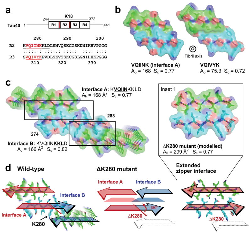
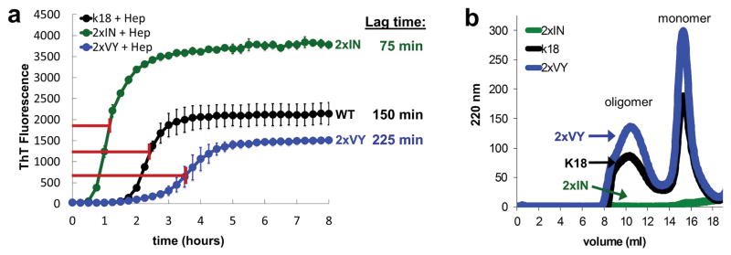
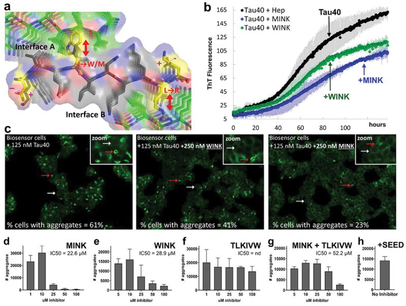
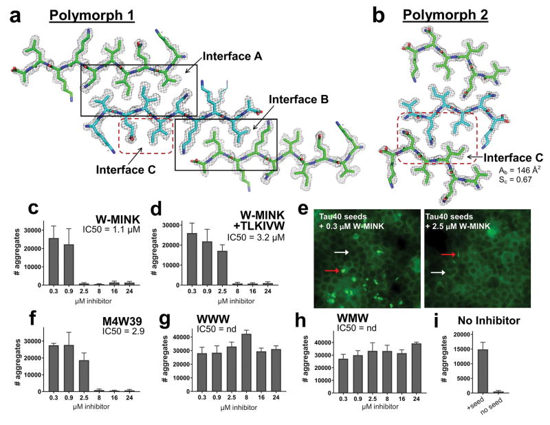
Aggregated tau protein is associated with over 20 neurological disorders, which include Alzheimer's disease. Previous work has shown that tau's sequence segments VQIINK and VQIVYK drive its aggregation, but inhibitors based on the structure of the VQIVYK segment only partially inhibit full-length tau aggregation and are ineffective at inhibiting seeding by full-length fibrils. Here we show that the VQIINK segment is the more powerful driver of tau aggregation. Two structures of this segment determined by the cryo-electron microscopy method micro-electron diffraction explain its dominant influence on tau aggregation. Of practical significance, the structures lead to the design of inhibitors that not only inhibit tau aggregation but also inhibit the ability of exogenous full-length tau fibrils to seed intracellular tau in HEK293 biosensor cells into amyloid. We also raise the possibility that the two VQIINK structures represent amyloid polymorphs of tau that may account for a subset of prion-like strains of tau.


Figures

Figure 1
Figure 1
Atomic structures of amyloid fibrils formed by segments of Tau, viewed down the fibril axes. (a) Schematic of full-length Tau showing the positions of VQIINK and VQIVYK (colored red) in the microtubule binding domain which contains four repeats (R1–4) together termed K18. Shown below is a sequence alignment of Repeats 2 and 3 (R2 and R3) from human Tau with the VQIINK and VQIVYK segments underlined. (b) Comparison of buried surface area Ab and shape complementarity Sc for the VQIINK (interface A; this paper) and VQIVYK (Sawaya et al. 2007; PBD 2ON9) steric zippers. (c) The two steric zipper interfaces, A and B, in the ten residue KVQIINKKLD crystal, shown as stick models with superimposed van der Waals atomic radii. The two interfaces have similar buried areas and shape complementarities. Numbering in C corresponds to the N- and C- termini for the β-sheet colored in cyan. (d) Arrangement of interfaces A and B in the ten residue wild-type KVQIINKKLD structure (left) and the predicted arrangement in the ΔK280 mutant (center and right). Trapezoids in the center diagram represent steric zipper forming residues that are predicted to line the interface between the mated β-strands. The colored arrows show the directions of β-strands forming the steric zippers. Oxygen atoms are red; nitrogen atoms are blue, and mainchain atoms are green for Chains A and C, and cyan for Chain B. In the wild-type structure, interfaces A (red) and B (blue) are formed on opposite faces of the VQIINK β-sheet. Deletion of residue K280 is predicted to reverse the orientation of C-terminal residues by 180º about the β-strand axes (center) merging steric zipper interfaces A and B into a single extended steric zipper interface with greater Ab and Sc as calculated from the ΔK280 model (right and Inset 1).
Figure 2
Figure 2
Time dependence of fibrillization and oligomerization for wild-type K18 construct and 2xIN and 2xVY K18 mutant constructs. (a) Averaged ThT fluorescence curves of wild-type K18 construct and engineered 2xIN and 2xVY K18 constructs at 50 μM in the presence of heparin with shaking at 700 rpm 37 ºC. Lag times determined from the half-maximum values of the curves shown are given for the respective constructs on the right. Error bars show the standard deviation of triplicate ThT measurements. (b) Analysis of oligomers measured by S200 size exclusion chromatography. Peaks corresponding to the soluble oligomer species are marked with an arrow.
Figure 3
Figure 3
Structure-based design of Phase 1 inhibitors of VQIINK aggregation. (a) Logic of inhibitor design. The inhibitors (Table 1) bind on the tips of VQIINK fibrils and introduce steric clashes that disrupt interfaces A and B. Based on the structures, the inhibitor sequences were designed as follows. Either a tryptophan (inhibitor WINK) or methionine (inhibitor MINK) were incorporated to block interface A. Both inhibitors contain an arginine to disrupt interface B. The ends of the inhibitor were charge reversed to promote electrostatic attraction of the inhibitor peptide to the fibril sequence. (b) The effects of designed inhibitors on the formation of fibrils of 25 μM full-length Tau (Tau40) plus a two-fold molar excess of MINK or WINK inhibitor peptide with shaking at 700 rpm at 37 ºC. Error bars show the standard deviation of triplicate ThT measurements from two independent experiments. (c) The effects of designed inhibitors on the transfer of fibrils of Tau40 into HEK293 biosensor stably expressing a full-length (4R1N P301S) YFP fusion. The cells were seeded with 125 nM Tau40 fiber (final concentration); the seeds were grown for 120 hours in the presence or absence of a two-fold excess of WINK or MINK. Representative cells containing aggregates are marked by red arrows, and cells without by white arrows. Percentages of cells with aggregates were calculated by dividing the number of aggregates in the field of view by the number of cells. Inset box shows a zoomed image from within the presented field of view. (d–h) 4R1N Tau-YFP biosensor cells seeded with pre-capped Tau40 fibers treated with MINK, WINK, TLKIVW, or MINK + TLKIVW inhibitor peptides at indicated concentrations. Bar graphs show the average number of aggregates at the indicated inhibitor concentrations, and error bars show the standard deviation of triplicate measurements. IC50’s were calculated from the dose-response curves shown. Panels B, C and D suggest that MINK is a more effective inhibitor than WINK.
Figure 4
Figure 4
The inhibition of Tau aggregation by Phase 2 designed inhibitors. (a and b) Comparison of the 10-residue (Polymorph 1) and 6-residue (Polymorph 2) VQIINK MicroED structures, viewed down the fibril axes. 2fo-fc electron density maps are contoured to 1.0 σ, and an alternate conformation modelled at 50% occupancy rendered in line format is shown for Lys281 in polymorph 1. (a) The 10-residue segment forms a face-to-face Class 1 zipper, whereas (b) the 6-residue VQIINK segment forms a face-to-back Class 4 zipper. (c–i) Seeding by Tau40 fibers pre-capped with Phase 2 VQIINK inhibitors (described in Table 1) designed to block interfaces A, B and C, measured in 4R1N Tau-YFP biosensor cells. Bar graphs show the average number of aggregates at the indicated inhibitor concentrations, and error bars show the standard deviation of triplicate measurements. Fluorescence images in E show seeded cells treated with 0.3 or 2.5 μM W-MINK inhibitor. Representative cells containing aggregates are marked by red arrows, and cells without by white arrows. Panel I shows quantified levels of aggregation in unseeded 4R1N Tau biosensor cells, and cells seeded by untreated Tau40 fibrils.

References

    1. Margittai M, Langen R. Side Chain-dependent Stacking Modulates Tau Filament Structure. Journal of Biological Chemistry. 2006;281:37820–37827. doi: 10.1074/jbc.M605336200. - DOI - PubMed
    1. von Bergen M, et al. Assembly of τ protein into Alzheimer paired helical filaments depends on a local sequence motif (306VQIVYK311) forming β structure. Proceedings of the National Academy of Sciences. 2000;97:5129–5134. doi: 10.1073/pnas.97.10.5129. - DOI - PMC - PubMed
    1. Goedert M, Eisenberg DS, Crowther RA. Annual Review of Neuroscience. 2017;40 doi: 10.1146/annurev-neuro-072116-031153. - DOI - PubMed
    1. Schwarz AJ, et al. Regional profiles of the candidate tau PET ligand recapitulate key features of Braak histopathological stages. Brain. 2016;139:1539–1550. doi: 10.1093/brain/aww023. - DOI - PubMed
    1. Manczak M, Reddy PH. Abnormal interaction of oligomeric amyloid-β with phosphorylated tau: implications to synaptic dysfunction and neuronal damage. Journal of Alzheimer’s disease: JAD. 2013;36:285–295. doi: 10.3233/JAD-130275. - DOI - PMC - PubMed

Publication types

MeSH terms