. 2018 Mar;28(3):307-322.
doi: 10.1038/cr.2018.14. Epub 2018 Jan 23.

Structure of the human activated spliceosome in three conformational states

Affiliations

Structure of the human activated spliceosome in three conformational states

Xiaofeng Zhang et al. Cell Res. 2018 Mar.

Abstract

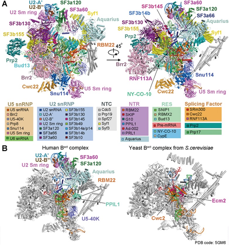
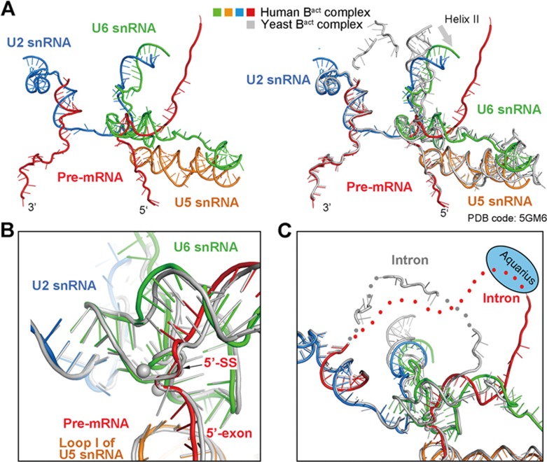
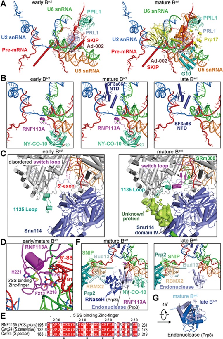
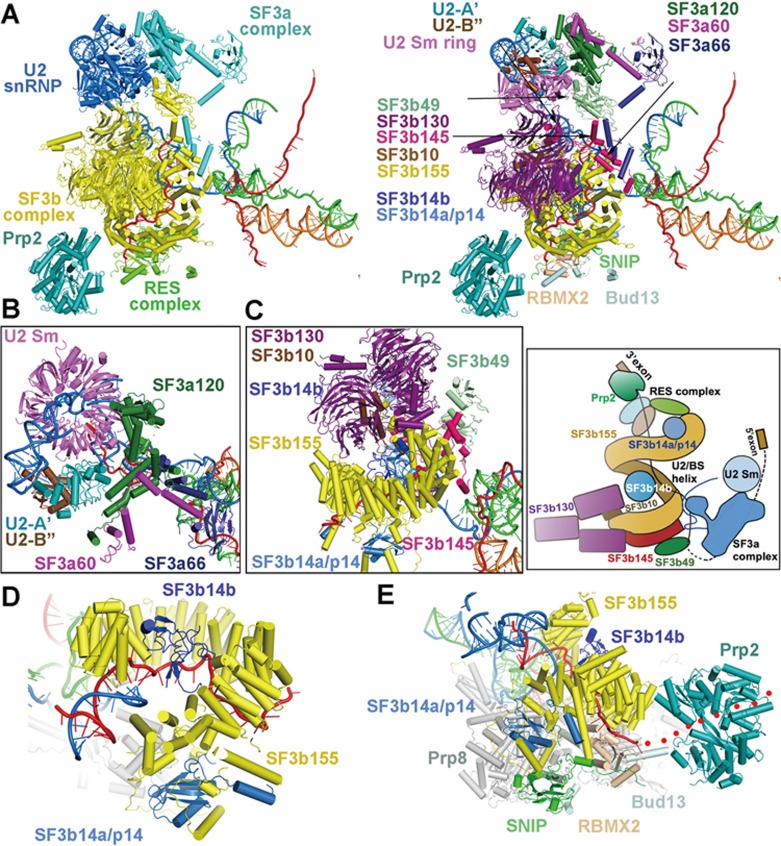
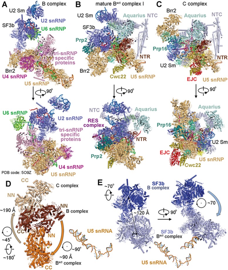
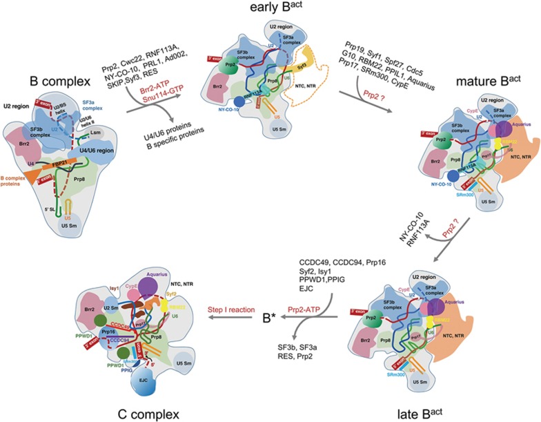
During each cycle of pre-mRNA splicing, the pre-catalytic spliceosome (B complex) is converted into the activated spliceosome (Bact complex), which has a well-formed active site but cannot proceed to the branching reaction. Here, we present the cryo-EM structure of the human Bact complex in three distinct conformational states. The EM map allows atomic modeling of nearly all protein components of the U2 small nuclear ribonucleoprotein (snRNP), including three of the SF3a complex and seven of the SF3b complex. The structure of the human Bact complex contains 52 proteins, U2, U5, and U6 small nuclear RNA (snRNA), and a pre-mRNA. Three distinct conformations have been captured, representing the early, mature, and late states of the human Bact complex. These complexes differ in the orientation of the Switch loop of Prp8, the splicing factors RNF113A and NY-CO-10, and most components of the NineTeen complex (NTC) and the NTC-related complex. Analysis of these three complexes and comparison with the B and C complexes reveal an ordered flux of components in the B-to-Bact and the Bact-to-B* transitions, which ultimately prime the active site for the branching reaction.


Figures

Figure 1
Figure 1
Cryo-EM structure of the human activated spliceosome (the Bact complex). (A) Two views of the human mature Bact complex. The protein and RNA components are color-coded and tabulated below the images. The structure of the mature Bact complex shown here includes 52 proteins, three snRNAs, and one pre-mRNA, with a combined molecular mass of about 1.8 MDa. U2, U5, and U6 snRNAs are colored marine, orange, and green, respectively. Pre-mRNA is colored red. This coloring scheme is preserved throughout this manuscript. (B) Structural comparison between the human and yeast Bact complexes,. For protein components, only those that are unique in either spliceosome are colored. All shared protein components are shown grey. All structural images were created using PyMol.
Figure 2
Figure 2
The RNA elements and the splicing active site of the human mature Bact complex. (A) Structure of the RNA elements in the core of the human mature Bact complex. The color-coded RNA elements of the human Bact complex are shown in the left panel, and their superposition with those of the S. cerevisiae Bact complex is displayed in the right panel. All yeast RNA elements are colored grey. The helix II of the U2/U6 duplex in the human Bact complex is bent relative to that in the yeast complex. (B) Structural overlay of the active site RNA elements between the human and S. cerevisiae Bact complexes. (C) The U6/intron duplex in the human Bact complex is considerably longer than that in the S. cerevisiae Bact complex.
Figure 3
Figure 3
The early, mature, and late Bact complexes represent three different conformational states of the human spliceosome. (A) Structural comparison between the early Bact complex (left panel) and the mature Bact complex (right panel). Shown here are only the RNA elements and the protein components that undergo dynamic changes in the core of the spliceosome in the transition of the early to mature Bact complex. Compared to that of the early Bact complex, the core of the mature Bact complex contains four additional proteins: G10, Prp17, RBM22, and the N-terminal domain (NTD) of the SF3a component SF3a66. (B) Close-up views on the early (left panel), mature (middle panel), and late (right panel) Bact complexes. Notably, the splicing factors RNF113A (Cwc24 in S. cerevisiae) and NY-CO-10 (Cwc27 in S. cerevisiae) are loaded in the early and the mature, but not the late, Bact complexes. On the other hand, SF3s66 is loaded in the mature and late Bact complexes, but not the early Bact complex. (C) A close-up comparison of the Switch loop regions between the early and mature Bact complexes. The Switch loop of Prp8 is stabilized by an extended sequence (named 1135-loop) and the splicing factor SRm300 (Cwc21 in S. cerevisiae) in the mature Bact complex (right panel). SRm300 is absent and the 1135-loop is shifted away in the early Bact complex (left panel); consequently the Switch loop is flexible and remains unidentified. (D) A close-up view on the recognition of the guanine nucleotide (G1) at the 5-end of the 5′-splice site (5′SS) by the splicing factor RNF113A. The stacking of the guanine base against the aromatic rings of Phe213 and Phe219 of RNF113A is reminiscent of that in the yeast Bact complex. (E) Sequence alignment between the human RNF113A and its yeast orthologues Cwc24 (S. cerevisiae) and Cwf24 (S. pombe). The three key residues involved in recognition of G1 of the 5′SS (Phe213, Lys218, and Phe219) are highly conserved. (F) Close-up views on the role of the endonuclease-like domain and the RNaseH-like domain of Prp8 in the mature and late Bact complexes. In the mature Bact complex (left panel), both domains of Prp8 appear to stabilize the binding of RNF113A and NY-CO-10 in the spliceosome. The RNaseH-like domain also binds Bud13 of the RES complex. In the late Bact complex (right panel), RNF113A and NY-CO-10 have been dissociated, leading to the dislocation of the RNaseH-like domain and Bud13. (G) Superposition of the endonuclease-like domain of Prp8 between the mature and late Bact complexes. The core machineries of the two complexes are aligned.
Figure 4
Figure 4
Structures of the SF3a and SF3b complexes. (A) Structure of the SF3a and SF3b complexes in the context of key surrounding components. In the left panel, the SF3a and SF3b complexes are colored cyan and yellow, respectively. The U2 snRNA, the RES complex, and Prp2 are colored blue, green, and teal, respectively. The RNA elements are displayed for orientation. In the right panel, the individual components of the SF3a and SF3b complexes are color-coded and labeled. The SF3a complex consists of three proteins: SF3a60, SF3a66, and SF3a120. The SF3b complex comprises seven proteins SF3b10, SF3b14a/p14, SF3b14b, SF3b49, SF3b130, SF3b145, and SF3b155. (B) A close-up view on the SF3a complex and its interactions with the U2 snRNP subcomplex involving U2 Sm ring. (C) A close-up view on the SF3b complex and the interactions among its constituents. The structure is shown in the left panel and the cartoon representation is displayed in the right panel. (D) A close-up view on the components SF3b14b and SF3b14a/p14 of the SF3b complex. (E) A close-up view on the 3′-end sequences of the pre-mRNA and nearby protein components. The 3′-end sequences of the pre-mRNA are bound by RBMX2 of the RES complex. The dotted lines leading to the RNA-binding groove of Prp2 indicate the path of the RNA sequences downstream of the last ordered nucleotide in the structure.
Figure 5
Figure 5
Structural comparison among the B complex, the Bact complex, and the C complex. (A) Structure of the human B complex. Two perpendicular views are shown. The tri-snRNP-specific proteins, and U2, U4, U5, and U6 snRNPs are colored pink, blue, orange, magenta, and green, respectively. (B) Structure of the human mature Bact complex. Two perpendicular views are shown, and these two views are identical to those in C of the human C complex. Ribonucleoprotein remodeling from the B to the Bact complex is the most dramatic in the splicing cycle, involving dissociation of the U4 snRNP and tri-snRNP-specific proteins and recruitment of the NTC and NTR components along with several splicing factors and the ATPase/helicase Prp2. (C) Structure of the human C complex. Compared to the Bact complex, the SF3a/SF3b complexes along with Prp2 and the splicing factor RNF113A have been dissociated, and the exon junction complex (EJC) along with the step I factors CCDC49/CCDC94 and the ATPase/helicase Prp16 have been recruited. (D) Movement of the ATPase/helicase Brr2 in the B-Bact-C transition. The U5 snRNA molecules from the three human spliceosomal complexes are superimposed. (E) Movement of the SF3b complex in the B-to-Bact transition. The U5 snRNA molecules from the human B and Bact complexes are superimposed.
Figure 6
Figure 6
A structure-based model of the ribonucleoprotein remodeling from the B complex to the C complex. The B-to-Bact transition represents the most complex transition in the pre-mRNA splicing cycle. The ATPase/helicase Brr2 drives the formation of the early Bact complex, where the NTC and NTR components are yet to be recruited. In the active site of the early Bact complex, the splicing factor RNF113A and the PPI NY-CO-10 are already loaded but the N-terminal domain (NTD) of SF3a66 along with G10 and Prp17 are yet to be recruited. Next, components of the NTC and NTR, Prp17, along with the NTD of SF3a66, are recruited to form the mature Bact complex.

References

    1. Wahl MC, Will CL, Luhrmann R. The spliceosome: design principles of a dynamic RNP machine. Cell 2009; 136:701–718. - PubMed
    1. Chan SP, Kao DI, Tsai WY, Cheng SC. The Prp19p-associated complex in spliceosome activation. Science 2003; 302:279–282. - PubMed
    1. Ohi MD, Gould KL. Characterization of interactions among the Cef1p-Prp19p-associated splicing complex. RNA 2002; 8:798–815. - PMC - PubMed
    1. Fabrizio P, Dannenberg J, Dube P, et al. The evolutionarily conserved core design of the catalytic activation step of the yeast spliceosome. Mol Cell 2009; 36:593–608. - PubMed
    1. Yan C, Wan R, Bai R, Huang G, Shi Y. Structure of a yeast activated spliceosome at 3.5 A resolution. Science 2016; 353:904–911. - PubMed

MeSH terms

Substances