. 2018 Jan 23;13(1):e0191621.
doi: 10.1371/journal.pone.0191621. eCollection 2018.

Identification and analysis of OsttaDSP, a phosphoglucan phosphatase from Ostreococcus tauri

Affiliations

Identification and analysis of OsttaDSP, a phosphoglucan phosphatase from Ostreococcus tauri

Julieta B Carrillo et al. PLoS One. .

Abstract

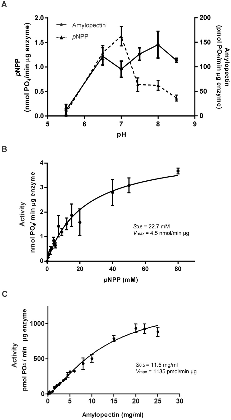
Ostreococcus tauri, the smallest free-living (non-symbiotic) eukaryote yet described, is a unicellular green alga of the Prasinophyceae family. It has a very simple cellular organization and presents a unique starch granule and chloroplast. However, its starch metabolism exhibits a complexity comparable to higher plants, with multiple enzyme forms for each metabolic reaction. Glucan phosphatases, a family of enzymes functionally conserved in animals and plants, are essential for normal starch or glycogen degradation in plants and mammals, respectively. Despite the importance of O. tauri microalgae in evolution, there is no information available concerning the enzymes involved in reversible phosphorylation of starch. Here, we report the molecular cloning and heterologous expression of the gene coding for a dual specific phosphatase from O. tauri (OsttaDSP), homologous to Arabidopsis thaliana LSF2. The recombinant enzyme was purified to electrophoretic homogeneity to characterize its oligomeric and kinetic properties accurately. OsttaDSP is a homodimer of 54.5 kDa that binds and dephosphorylates amylopectin. Also, we also determined that residue C162 is involved in catalysis and possibly also in structural stability of the enzyme. Our results could contribute to better understand the role of glucan phosphatases in the metabolism of starch in green algae.


Conflict of interest statement

Competing Interests: The authors have declared that no competing interests exist.

Figures

Fig 1
Fig 1. Amino acidic sequence alignment and homology modelling of OsttaDSP.
A) Amino Acidic Sequence Alignment of OsttaDSP with A. thaliana LSF2. The yellow box highlight the active site. cTP, chloroplast transit peptide; DSP, dual specific phosphatase and CT, C-terminal. Residues involved in glucan binding in A. thaliana LSF2 are indicated within the aromatic channel (green arrowheads), SBS2 (red arrowheads) and SBS3 (blue arrowheads). Residues in the aromatic channel are conserved between OsttaDSP and LSF2, however, residues in SBS2 and SBS3 are not. B) Structural model of A. thaliana LSF2 (4kyq, left) and the proposed model for OsttaDSP (right). α- helix are indicated in red, β- sheets in yellow and loops in green. N, N-terminal and C, C-terminal. C) Superposition of LSF2 (cyan) and OsttaDSP (red) structures. The active site is shown in yellow. D) Superposition between OsttaDSP model and LSF2 structure showing the residues involved in the active site. OsttaDSP residues are shown in red and LSF2 residues are shown in cyan.
Fig 2
Fig 2. Electrophoretic analysis of OsttaDSP.
A) Coomassie Blue staining of a SDS-PAGE of OsttaDSP. B) SDS-PAGE analyzed by western blotting of OsttaDSP and crude extract of O. tauri cells. a-His, a-His antibody; a-SEX4, a-SEX4 antibody immunopurified for OsttaDSP protein. C) Coomassie Blue staining of a native PAGE of OsttaDSP. Arrowheads (►) indicate pure recombinant OsttaDSP and asterisk (*) indicate putative DSPs in the crude extract of O. tauri cells. For SDS-PAGE 1 μg of OsttaDSP was loaded while 3 μg of protein was used for native PAGE analysis. Numbers indicate the molecular masses of markers in kDa. For SDS-PAGE, Page Ruler Prestained Protein Ladder (range 10–170 kDa) was used (Thermo Fisher Scientific, Waltham, MA USA) and, for native electrophoresis, the markers were Amersham High Molecular Weight Calibration Kit (range 66–669 kDa). MMM, molecular mass markers. OsttaDSP, O.tauri OsttaDSP, O. tauri CE, O. tauri crude extract (20 μg).
Fig 3
Fig 3. Kinetic characterization of OsttaDSP.
A) Effect of pH on the activity of recombinant OsttaDSP. Activity was assayed with pNPP (filled triangles/▲) or amylopectin (filled circles/●) as a substrate using 100 mM Sodium Acetate, 50 mMBis-Tris and 50 mM Tris as a buffer. Buffer pH values were adjusted to the values shown in the graphic and used to assay the enzyme. All data are the means ± SD of 3 independent experiments. B) pNPP saturation plot for OsttaDSP determined at pH 7. C) Amylopectin saturation plot for OsttaDSP determined at pH 7.5.
Fig 4
Fig 4. Characterization of OsttaDSP C162S modified protein.
A) Specific activities of OsttaDSP wild-type (WT) and C162S (C162S) enzymes with pNPP as substrate. Both activities were determined at 100 mM of pNPP. All data are the means ± SD of 3 independent experiments. B) Specific activities of OsttaDSP wild-type (WT) and C162S (C162S) modified enzyme in the presence of amylopectin. Both activities were determined at 10 mg/ml (white bars) or 20 mg/ml (black bars) of amylopectin. All data are the means ± SD of 3 independent experiments. C) Native PAGE analysis of OsttaDSP wild-type (WT) and C162S mutated enzyme (C162S). OsttaDSP WT (3 μg) and OsttaDSP C162S (3 μg) were resolved by native PAGE. After native PAGE, gels were either stained with Coomassie Blue or with the activity gel assay using pNPP. Filled arrowheads (►) indicate purified recombinant enzymes stained with Coomassie Blue and empty arrowhead (>) indicates OsttaDSP WT stained with the pNPP in gel activity assay. The number indicates the molecular mass marker in kDa. MMM, molecular mass marker (Amersham High Molecular Weight Calibration Kit (range 66–669 kDa)). D) Amylopectin kinetics of OsttaDSP wild-type (WT) and OsttaDSP C162S modified protein (C162S). Amylopectin saturation plots for OsttaDSP WT (filled circles (●)) and OsttaDSP C162S variant (filled arrowheads (►)) were performed at pH 7.5. All data are the means ± SD of 3 independent experiments.
Fig 5
Fig 5. Affinity gel electrophoresis (AGE) of OsttaDSPwild-type (WT) and C162S mutated protein (C162S).
OsttaDSP WT (WT, 5 μg) and OsttaDSP C162S (C162S, 5 μg) proteins were run on native PAGE without or with amylopectin (0.3 mg/ml) simultaneously under the same conditions. Gels were then revealed by Coomassie Blue staining. The number indicates the molecular mass marker in kDa. MMM, molecular mass markers (Amersham High Molecular Weight Calibration Kit (range 66–669 kDa)).

Similar articles

References

    1. Tester R, Karkalas J, Qi X. Starch structure and digestibility enzyme-substrate relationship. World’s Poultry Science Journal. 2004;60(02):186–95.
    1. Blennow A, Bay-Smidt AM, Olsen CE, Møller BL. The distribution of covalently bound phosphate in the starch granule in relation to starch crystallinity. International Journal of Biological Macromolecules. 2000;27(3):211–8. - PubMed
    1. Carciofi M, Shaik SS, Jensen SL, Blennow A, Svensson JT, Vincze É, et al. Hyperphosphorylation of cereal starch. Journal of Cereal Science. 2011;54(3):339–46.
    1. Ritte G, Lloyd JR, Eckermann N, Rottmann A, Kossmann J, Steup M. The starch-related R1 protein is an α-glucan, water dikinase. Proceedings of the National Academy of Sciences of the United States of America. 2002;99(10):7166–71. doi: 10.1073/pnas.062053099 - DOI - PMC - PubMed
    1. Ritte G, Heydenreich M, Mahlow S, Haebel S, Kotting O, Steup M. Phosphorylation of C6- and C3-positions of glucosyl residues in starch is catalysed by distinct dikinases. FEBS letters. 2006;580(20):4872–6. Epub 2006/08/18. doi: 10.1016/j.febslet.2006.07.085 . - DOI - PubMed

Publication types

MeSH terms

LinkOut - more resources