. 2018 Apr 1;27(7):1174-1185.
doi: 10.1093/hmg/ddy032.

Temporal requirement of dystroglycan glycosylation during brain development and rescue of severe cortical dysplasia via gene delivery in the fetal stage

Affiliations

Temporal requirement of dystroglycan glycosylation during brain development and rescue of severe cortical dysplasia via gene delivery in the fetal stage

Atsushi Sudo et al. Hum Mol Genet. .

Abstract

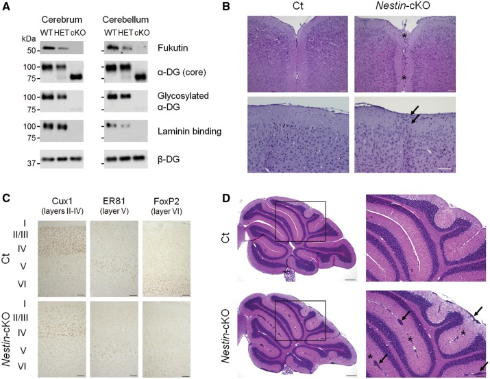
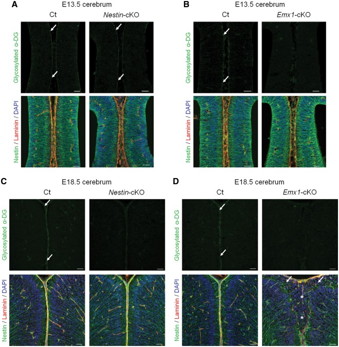
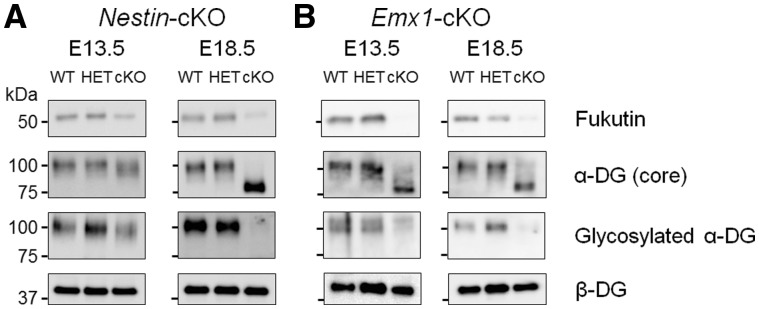
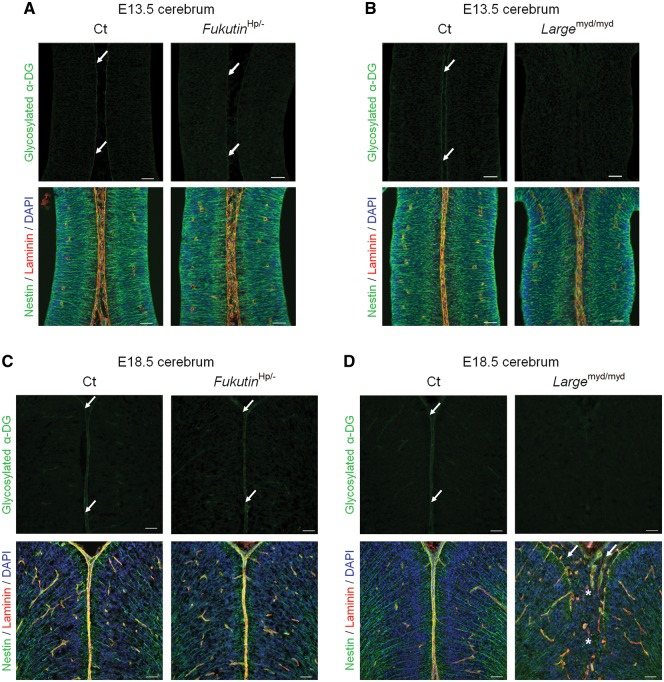
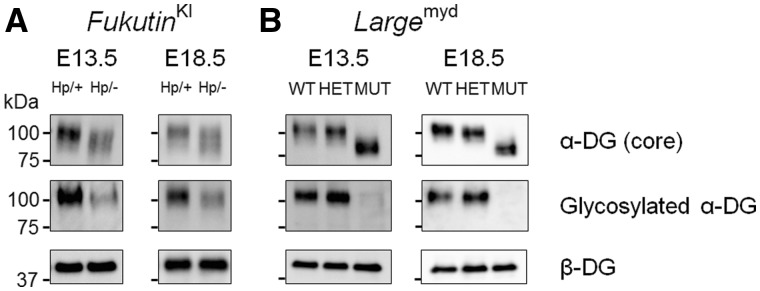
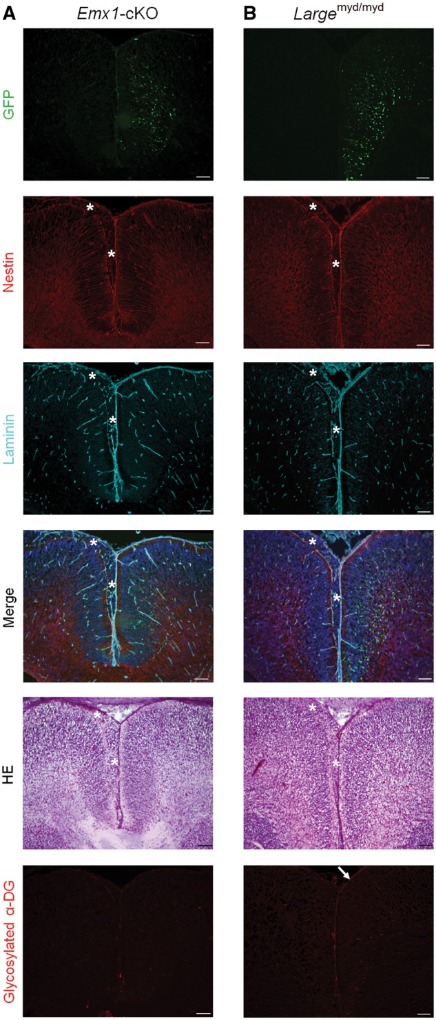
Congenital muscular dystrophies (CMDs) are characterized by progressive weakness and degeneration of skeletal muscle. In several forms of CMD, abnormal glycosylation of α-dystroglycan (α-DG) results in conditions collectively known as dystroglycanopathies, which are associated with central nervous system involvement. We recently demonstrated that fukutin, the gene responsible for Fukuyama congenital muscular dystrophy, encodes the ribitol-phosphate transferase essential for dystroglycan function. Brain pathology in patients with dystroglycanopathy typically includes cobblestone lissencephaly, mental retardation, and refractory epilepsy; however, some patients exhibit average intelligence, with few or almost no structural defects. Currently, there is no effective treatment for dystroglycanopathy, and the mechanisms underlying the generation of this broad clinical spectrum remain unknown. Here, we analysed four distinct mouse models of dystroglycanopathy: two brain-selective fukutin conditional knockout strains (neuronal stem cell-selective Nestin-fukutin-cKO and forebrain-selective Emx1-fukutin-cKO), a FukutinHp strain with the founder retrotransposal insertion in the fukutin gene, and a spontaneous Large-mutant Largemyd strain. These models exhibit variations in the severity of brain pathology, replicating the clinical heterogeneity of dystroglycanopathy. Immunofluorescence analysis of the developing cortex suggested that residual glycosylation of α-DG at embryonic day 13.5 (E13.5), when cortical dysplasia is not yet apparent, may contribute to subsequent phenotypic heterogeneity. Surprisingly, delivery of fukutin or Large into the brains of mice at E12.5 prevented severe brain malformation in Emx1-fukutin-cKO and Largemyd/myd mice, respectively. These findings indicate that spatiotemporal persistence of functionally glycosylated α-DG may be crucial for brain development and modulation of glycosylation during the fetal stage could be a potential therapeutic strategy for dystroglycanopathy.


Figures

Figure 1.
Figure 1.
Generation and characterization of brain-selective Nestin-fukutin-cKO mice. (A) Western blotting analysis of endogenous fukutin expression and α-DG glycosylation in the adult cerebrum and cerebellum. Laminin-binding activity of α-DG was examined by a laminin overlay assay. We used β-DG as a loading control. (B) Mild focal abnormalities were observed in the cerebrum of adult Nestin-fukutin-cKO mice. Hematoxylin and eosin (H&E) staining revealed fusion of the cerebral hemispheres in cKO mice (asterisk). Ectopic cellular infiltration into layer I was observed in cKO mice (arrow). (C) Laminar organization of cerebral cortex. No obvious difference was detected between cKO mice and littermate controls. Cux1 was used as a marker of neurons in layers II-IV of cerebral cortex. ER81 and FoxP2 were used as markers of neurons in layer V or VI, respectively. (D) H&E staining of the cerebellum. Ectopic cells between adjacent cerebellar lobules and at the surface of cerebellar lobules were diffusely distributed in cKO mice (arrow). Cerebellar lobules were fused at many sites (asterisk). Scale bars = (B, C) 100 μm; (D) 300 μm (left column), 100 μm (right column). α-DG, alpha dystroglycan; Ct, control; cKO, conditional knockout.
Figure 2.
Figure 2.
Damage of the basement membrane and disorganization of glial cells underlie cerebral and cerebellar malformations in Nestin-fukutin-cKO mice. (A) (Left column) Immunofluorescence analysis of the developing cortex at E18.5. Radial glia fibers were disorganized and extended into subarachnoid spaces (white arrow) through breaches in the basement membrane (asterisk) in cKO mice. In contrast, they had merged properly in littermate controls. Nestin and laminin were used as markers of radial glia or the basement membrane, respectively. (Right column) H&E staining of serial sections. Ectopic cells were observed at cerebral fissures (black arrow) in cKO mice. (B) Immunofluorescence analysis of the cerebellum at P7. Between adjacent cerebellar lobules, the basement membrane was not detected (asterisk), and many ectopic cells were observed (arrow) in cKO mice. Bergmann glia fibers were disorganized, and reactive gliosis was detected in such regions. In contrast, they had merged properly in littermate controls. GFAP was used as a marker of Bergmann glia. Scale bars = (A, B) 50 μm.
Figure 3.
Figure 3.
Dystroglycan glycosylation and phenotypic correlation during brain development in fukutin-cKO mice. (A, B) Immunofluorescence analysis of the developing cortex at E13.5. (A, upper panel) Nestin-fukutin-cKO mice demonstrated residual glycosylation of α-DG at the glia limitans as in littermate controls (arrow). (B, upper panel) Defective glycosylation of α-DG was observed in Emx1-fukutin-cKO mice. (A and B, lower panel) Both strains exhibited almost normal brain structure regardless of α-DG glycosylation state. (C, D) Immunofluorescence analysis of the developing cortex at E18.5. (C and D, upper panel) Both strains demonstrated defective glycosylation of α-DG in contrast to littermate controls. (C, lower panel) With the exception of focal cortical dysplasia within a limited region, the laminar organization of the cerebral cortex and basement membrane were preserved in Nestin-fukutin-cKO mice. (D, lower panel) Diffuse and severe brain malformations were observed in Emx1-fukutin-cKO mice, in contrast to very mild brain pathology in Nestin-fukutin-cKO mice. Cerebral hemispheres were widely fused (asterisk), and the glia limitans-basement membrane complex was diffusely dissociated in Emx1-fukutin-cKO mice. Many ectopic cells were observed in the subarachnoid space (arrow). Scale bars = (A, B, C, D) 50 μm.
Figure 4.
Figure 4.
Western blotting analysis in the fetal brains of fukutin-cKO mice. (A) Nestin-fukutin-cKO and (B) Emx1-fukutin-cKO strains were analysed at E13.5 and E18.5, respectively. We used β-DG as a loading control.
Figure 5.
Figure 5.
Dystroglycan glycosylation and phenotypic correlation during brain development in other mouse models of dystroglycanopathy. (A, B) Immunofluorescence analysis of the developing cortex at E13.5. (A, upper panel) FukutinHp/- mice demonstrated residual α-DG glycosylation at the glia limitans (arrow). (B, upper panel) α-DG glycosylation was defective in Largemyd/myd mice. (A and B, lower panel) Both strains exhibited almost normal brain structure regardless of α-DG glycosylation state. (C, D) Immunofluorescence analysis of the developing cortex at E18.5. (C, upper panel) Glycosylation of α-DG was observed at the glia limitans in FukutinHp/- mice (arrow). (D, upper panel) Glycosylation of α-DG was consistently defective in Largemyd/myd mice. (C, lower panel) Almost no structural defects were observed in FukutinHp/- mice. (D, lower panel) Diffuse and severe brain malformations were observed in Largemyd/myd mice. Cerebral hemispheres were widely fused (asterisk), and the glia limitans-basement membrane complex was diffusely dissociated in Largemyd/myd mice, similar to findings observed in Emx1-fukutin-cKO mice. Many ectopic cells were observed in the subarachnoid space (arrow). Scale bars = (A, B, C, D) 50 μm.
Figure 6.
Figure 6.
Western blotting analysis in the fetal brains of other mouse models of dystroglycanopathy. (A) FukutinHp and (B) Largemyd strains were analysed at E13.5 and E18.5, respectively. We used β-DG as a loading control.
Figure 7.
Figure 7.
Gene rescue during the middle stage of brain development prevents severe cortical dysplasia. (A) Phenotypic rescue in Emx1-fukutin-cKO mice at E18.5 via delivery of the fukutin gene into the brains of fetal mice at E12.5. Green fluorescent protein (GFP) expression was observed in the plasmid-treated hemisphere (right hemisphere). The glia limitans-basement membrane complex was intact in the right hemisphere. In the left hemisphere, the glia limitans-basement membrane complex was diffusely dissociated, and many ectopic cells were observed in the subarachnoid space (asterisk). (B) Phenotypic rescue in Largemyd/myd mice at E18.5 via delivery of the Large gene into the brains of fetal mice at E12.5. Following gene delivery, severe cortical dysplasia was prevented in Largemyd/myd mice. Scale bars = (A, B) 100 μm.

References

    1. Muntoni F., Voit T. (2004) The congenital muscular dystrophies in 2004: a century of exciting progress. Neuromuscul. Disord., 14, 635–649. - PubMed
    1. Toda T., Kobayashi K., Takeda S., Sasaki J., Kurahashi H., Kano H., Tachikawa M., Wang F., Nagai Y., Taniguchi K.. et al. (2003) Fukuyama-type congenital muscular dystrophy (FCMD) and alpha-dystroglycanopathy. Congenit. Anom. (Kyoto), 43, 97–104. - PubMed
    1. Michele D.E., Campbell K.P. (2003) Dystrophin-glycoprotein complex: post-translational processing and dystroglycan function. J. Biol. Chem., 278, 15457–15460. - PubMed
    1. Ervasti J.M., Campbell K.P. (1993) A role for the dystrophin-glycoprotein complex as a transmembrane linker between laminin and actin. J. Cell Biol., 122, 809–823. - PMC - PubMed
    1. Ibraghimov-Beskrovnaya O., Ervasti J.M., Leveille C.J., Slaughter C.A., Sernett S.W., Campbell K.P. (1992) Primary structure of dystrophin-associated glycoproteins linking dystrophin to the extracellular matrix. Nature, 355, 696–702. - PubMed

Publication types