. 2018 Apr 1;27(7):1136-1149.
doi: 10.1093/hmg/ddy022.

CHEK1 coordinates DNA damage signaling and meiotic progression in the male germline of mice

Affiliations

CHEK1 coordinates DNA damage signaling and meiotic progression in the male germline of mice

Hironori Abe et al. Hum Mol Genet. .

Abstract

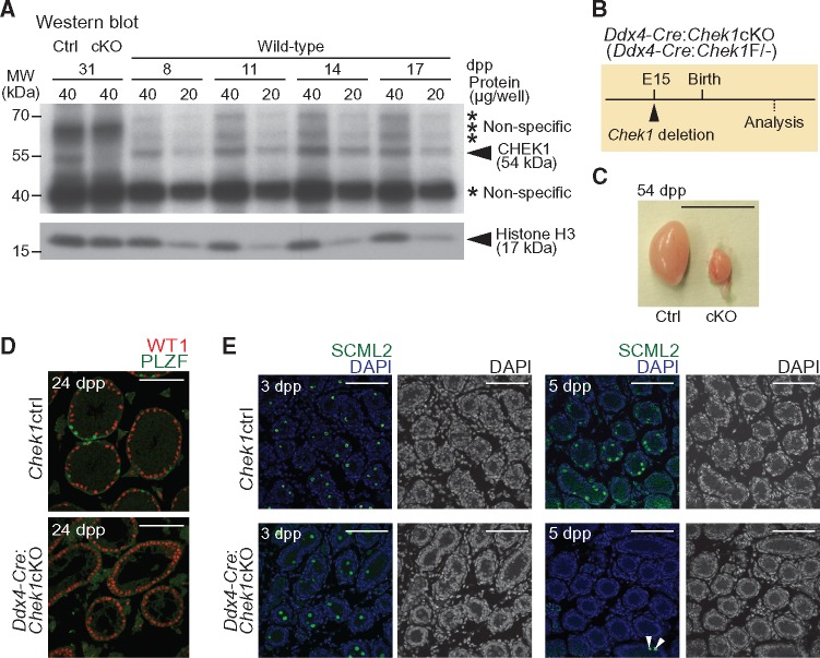
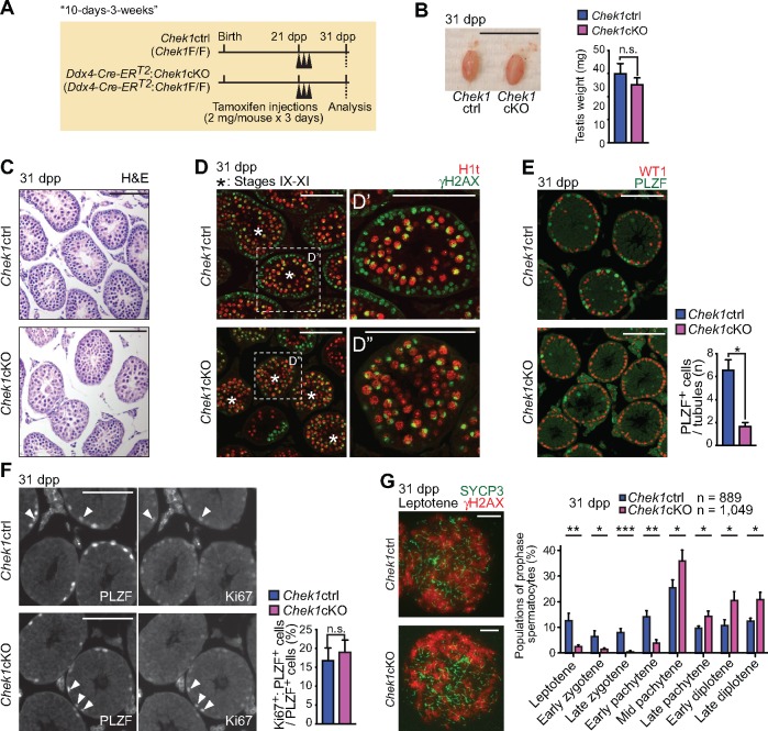
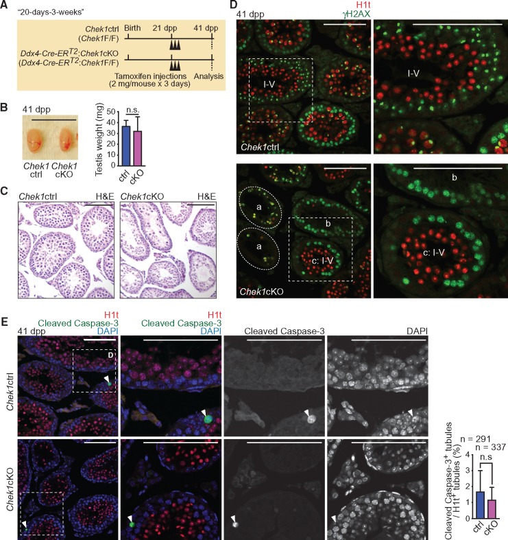
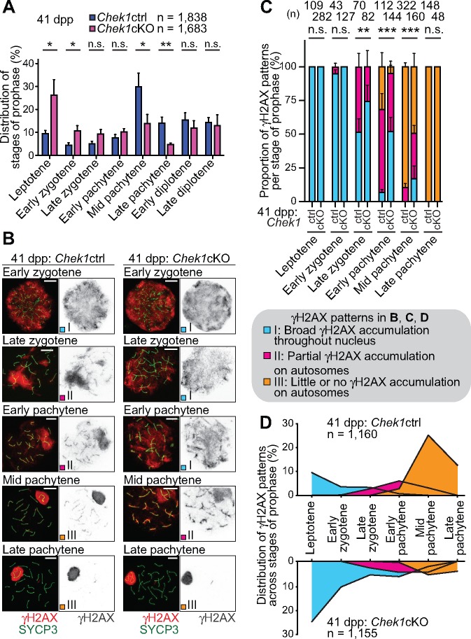
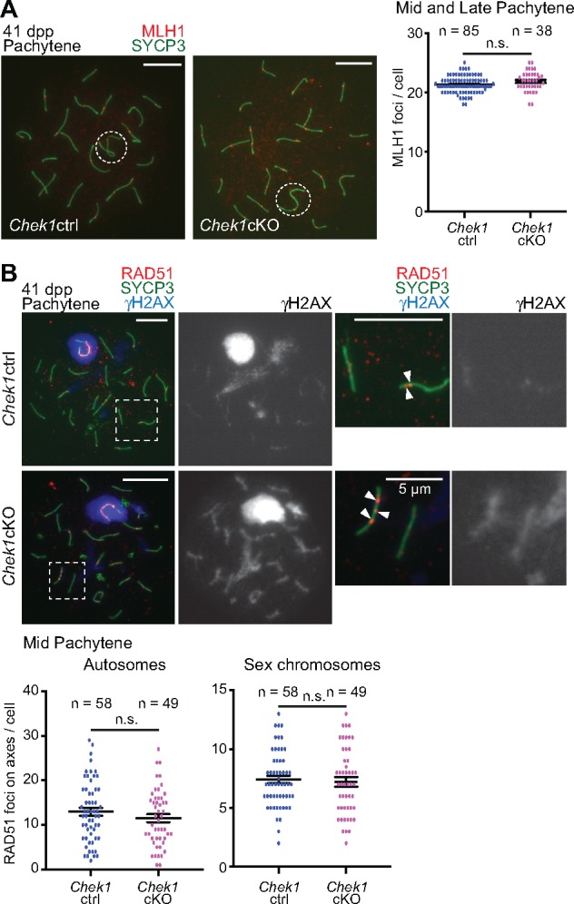
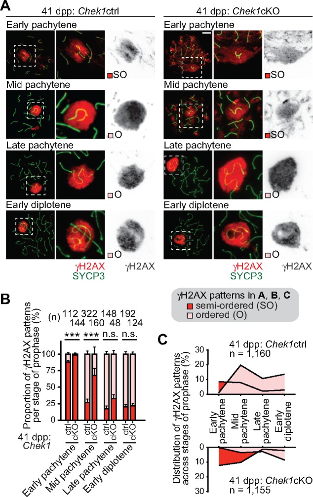
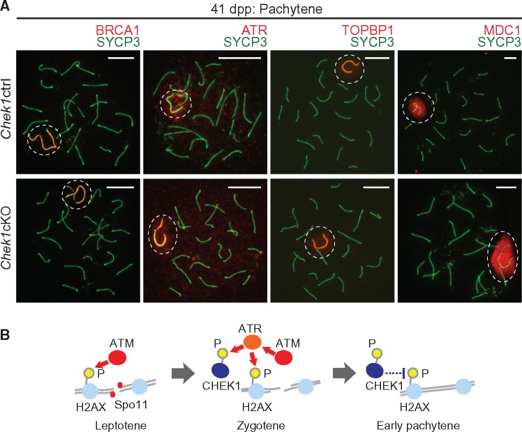
The continuity of life depends on mechanisms in the germline that ensure the integrity of the genome. The DNA damage response/checkpoint kinases ATM and ATR are essential signaling factors in the germline. However, it remains unknown how a downstream transducer, Checkpoint Kinase 1 (CHEK1 or CHK1), mediates signaling in the male germline. Here, we show that CHEK1 has distinct functions in both the mitotic and meiotic phases of the male germline in mice. In the mitotic phase, CHEK1 is required for the resumption of prospermatogonia proliferation after birth and the maintenance of spermatogonia. In the meiotic phase, we uncovered two functions for CHEK1: one is the stage-specific attenuation of DNA damage signaling on autosomes, and the other is coordination of meiotic stage progression. On autosomes, the loss of CHEK1 delays the removal of DNA damage signaling that manifests as phosphorylation of histone variant H2AX at serine 139 (γH2AX). Importantly, CHEK1 does not have a direct function in meiotic sex chromosome inactivation (MSCI), an essential event in male meiosis, in which ATR is a key regulator. Thus, the functions of ATR and CHEK1 are uncoupled in MSCI, in contrast to their roles in DNA damage signaling in somatic cells. Our study reveals stage-specific functions for CHEK1 that ensure the integrity of the male germline.


Figures

Figure 1.
Figure 1.
CHEK1 is required for the proliferation of prospermatogonia. (A) Western blotting of testicular lysate obtained from wild-type mice at 8, 11, 14 and 17 dpp, and from Chek1ctrl and Ddx4-Cre-ERT2: Chek1cKO mice at 31 dpp (tamoxifen injections were at 3 weeks of age, or 21 dpp, and testes were harvested 10 days later). Asterisks indicate the positions of non-specific bands. Anti-CHEK1 antibody (Sigma #C9358) was used. (B) Schematic for Chek1 deletion by Ddx4-Cre. (C) Picture of Chek1ctrl and Chek1cKO testes at 54 dpp. Scale bar: 1 cm. (D) Immunostaining of WT1 and PLZF on testicular sections at 24 dpp. Scale bars: 100 μm. (E) Immunostaining of SCML2 on testicular sections at 3 and 5 dpp. White arrowheads indicate SCML2-positive cells in the Chek1cKO. Nuclei were counterstained with DAPI. Scale bars: 100 μm.
Figure 2.
Figure 2.
CHEK1 is required for the maintenance of spermatogonia. (A) Schematic of the experimental design of the 10-day-3-weeks method. (B) Picture of Chek1ctrl and Chek1cKO testes at 31 dpp. Scale bar: 1 cm. Testis weights are shown as mean ± SEM for three independent pairs of Chek1ctrl and Chek1cKO mice. n.s.: not significant. Significance was determined utilizing an unpaired t-test. (C) Sections stained with H & E at 31 dpp. Scale bars: 100 μm. (D) Immunostaining of H1t and γH2AX on testicular sections at 31 dpp. Asterisks indicate seminiferous tubules at stages IX–XI. Dashed squares are magnified in the right-hand panels. Scale bars: 100 μm. (E) Immunostaining of WT1 and PLZF on testicular sections at 31 dpp. Scale bars: 100 μm. Numbers of PLZF positive cells per seminiferous tubule are shown as mean ± SEM for three independent pairs of Chek1ctrl and Chek1cKO mice. *P < 0.05 (unpaired t-test). (F) Immunostaining of PLZF and Ki-67 on testicular sections at 31 dpp. Arrowheads indicate cells that are double positive for PLZF and Ki-67. Scale bars: 100 μm. Percentages of PLZF-positive cells that are also positive for Ki-67 are shown as the mean ± SEM for three independent pairs Chek1ctrl and Chek1cKO mice. n.s.: not significant. Unpaired t-test. (G) Immunostaining of SYCP3 and γH2AX on meiotic chromosome spreads at 31 dpp. Scale bars: 10 μm. Stage populations during meiotic prophase are shown as the mean ± SEM for five independent pairs of Chek1ctrl and Chek1cKO mice. Total numbers of analyzed nuclei are indicated in the panels. Scale bars: 10 μm. *P < 0.05, **P< 0.01, ***P < 0.001 (unpaired t-test).
Figure 3.
Figure 3.
CHEK1 depletion disrupts the seminiferous cycle independent of apoptosis. (A) Schematic of the experimental design for the 20-day-3-weeks method. (B) Picture of Chek1ctrl and Chek1cKO testes at 41 dpp. Scale bar: 1 cm. Testis weights are shown as the mean ± SEM for three independent pairs of Chek1ctrl and Chek1cKO mice. n.s.: not significant (unpaired t-test). (C) Sections stained with H & E at 41 dpp. Scale bars: 100 μm. (D) Immunostaining of H1t and γH2AX on testicular sections at 41 dpp. A seminiferous tubule at stages I–V was judged by the presence of H1t positive round spermatids. The dashed circle labeled with ‘a’” indicates seminiferous tubules with an absence of germ cells. ‘b’ indicates seminiferous tubules including only leptotene and zygotene spermatocytes. ‘c’ indicates a seminiferous tubule at stages I–V. Dashed squares are magnified in the right-hand panels. Scale bars: 100 μm. (E) Immunostaining of H1t and Cleaved Caspase-3 on testicular sections at 41 dpp. Dashed squares are magnified in the right-hand panels. White arrowheads show Cleaved Caspase-3 positive cells. Nuclei were counterstained with DAPI. Scale bars: 100 μm. Population of H1t-positive seminiferous tubules containing Cleaved Caspase-3 positive cells are shown as the mean ± SEM for three independent pairs of Chek1ctrl and Chek1cKO mice. Total numbers of analyzed seminiferous tubules are indicated in the panels. n.s.: not significant (Chi-squared test).
Figure 4.
Figure 4.
CHEK1 coordinates meiotic progression and the removal of γH2AX from autosomes. (A) Stage populations during prophase are shown as the mean ± SEM for five independent pairs of Chek1ctrl and Chek1cKO mice at 41 dpp. *P < 0.05, **P < 0.01, n.s.: not significant (unpaired t-test). (B) Immunostaining of SYCP3 and γH2AX on meiotic chromosome spreads at 41 dpp. Distribution patterns of γH2AX were classified into three types by criteria described in the figure. Scale bars: 10 μm. (C) Population of each γH2AX pattern in each stage of meiotic prophase for three independent pairs of Chek1ctrl and Chek1cKO mice. **P < 0.01, ***P < 0.001. n.s.: not significant (Chi-squared test). (D) Integrative analyses of spermatocytes classified by the distribution of γH2AX on autosomes in different stages of prophase from three independent pairs of Chek1ctrl and Chek1cKO mice. Total numbers of analyzed nuclei are indicated in the panels.
Figure 5.
Figure 5.
CHEK1 function is independent of meiotic recombination. (A) Immunostaining of SYCP3 with MLH1 on meiotic chromosome spreads at 41 dpp. Dashed circles indicate the XY body. Scale bars: 10 μm. Distributions of data from three independent pairs of Chek1ctrl and Chek1cKO mice are shown as dots. Mean ± SEM. Total numbers of analyzed nuclei are indicated in the panels. n.s.: not significant (unpaired t-test). (B) Immunostaining of SYCP3 with RAD51 and γH2AX on meiotic chromosome spreads at 41 dpp, and the number of RAD51 foci on axes. Dashed squares in left panels are magnified in the two right panels. Arrowheads indicate RAD51 foci. Scale bars: 10 μm unless shown otherwise in the panel. Distributions of data from three independent pairs of Chek1ctrl and Chek1cKO mice are shown as dots. Mean ± SEM. Total numbers of analyzed nuclei are indicated in the panels. n.s.: not significant (unpaired t-test).
Figure 6.
Figure 6.
Loss of Chek1 leads to a delay in the establishment of ATR-dependent γH2AX in MSCI. (A) Immunostaining of SYCP3 and γH2AX on meiotic chromosome spreads. The γH2AX domain gradually becomes confined to sex chromatin; the boundary of the γH2AX domain is initially indeterminate (semiordered: SO) and, later, becomes tightly bounded (ordered: O). Scale bars: 10 μm. (B) Population of each γH2AX pattern in each substage for three independent pairs of Chek1ctrl and Chek1cKO mice. **P < 0.01, ***P < 0.001. n.s.: not significant (Chi-squared test). (C) Integrative analyses of spermatocytes classified by the distribution of γH2AX on the sex chromatin in stages of prophase from three independent pairs of Chek1ctrl and Chek1cKO mice. Total numbers of analyzed nuclei are indicated in the panels. ***P < 0.001. n.s.: not significant (Chi-squared test).
Figure 7.
Figure 7.
CHEK1 is dispensable for the regulation of DDR factors that establish MSCI, and a model of CHEK1 action during meiosis. (A) Immunostaining of SYCP3 with the indicated DDR factors, which are involved in the establishment of MSCI, on meiotic chromosome spreads at 41 dpp. Dashed circles indicate the XY body. Scale bars: 10 μm. (B) Model of action for CHEK1 during meiosis. CHEK1 functions in the context of the ATM-mediated DDR, and phospho-CHEK1 negatively regulates ATM-dependent γH2AX.

References

    1. Drost J.B., Lee W.R. (1995) Biological basis of germline mutation: comparisons of spontaneous germline mutation rates among drosophila, mouse, and human. Environ. Mol. Mutagen., 25, 48–64. - PubMed
    1. Lynch M. (2010) Rate, molecular spectrum, and consequences of human mutation. Proc. Natl. Acad. Sci. U. S. A., 107, 961–968. - PMC - PubMed
    1. Gunes S., Al-Sadaan M., Agarwal A. (2015) Spermatogenesis, DNA damage and DNA repair mechanisms in male infertility. Reprod. Biomed. Online, 31, 309–319. - PubMed
    1. Ciccia A., Elledge S.J. (2010) The DNA damage response: making it safe to play with knives. Mol. Cell, 40, 179–204. - PMC - PubMed
    1. Polo S.E., Jackson S.P. (2011) Dynamics of DNA damage response proteins at DNA breaks: a focus on protein modifications. Genes. Dev., 25, 409–433. - PMC - PubMed

Publication types

Substances