

Structural divergence creates new functional features in alphavirus genomes
- PMID: 29361131
- PMCID: PMC6283419
- DOI: 10.1093/nar/gky012
Structural divergence creates new functional features in alphavirus genomes
Abstract
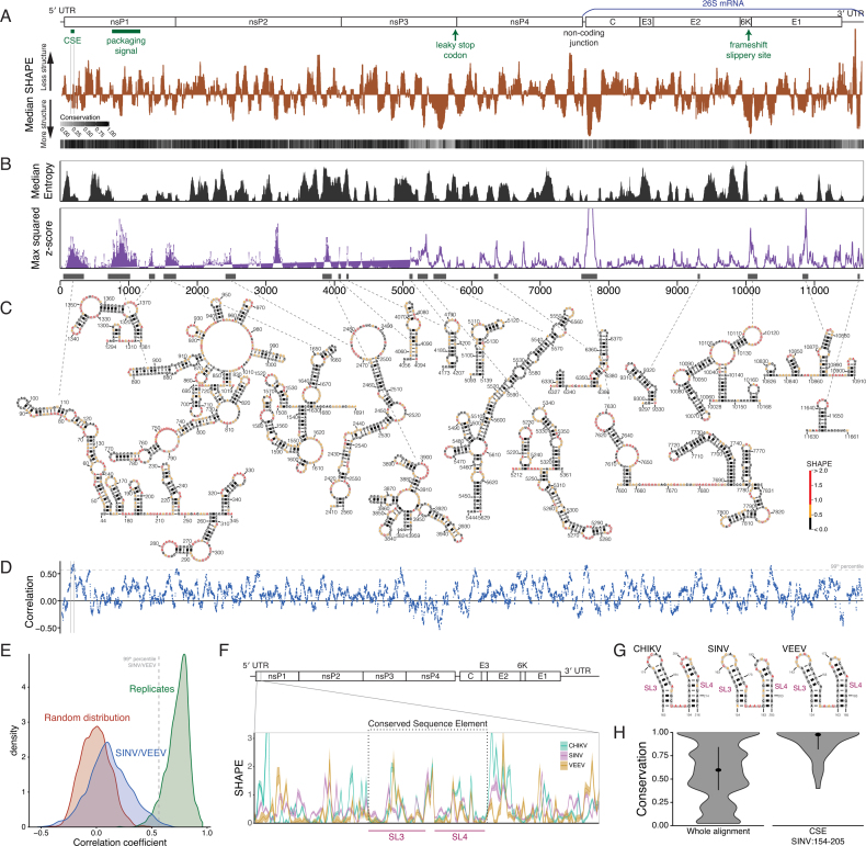
Alphaviruses are mosquito-borne pathogens that cause human diseases ranging from debilitating arthritis to lethal encephalitis. Studies with Sindbis virus (SINV), which causes fever, rash, and arthralgia in humans, and Venezuelan equine encephalitis virus (VEEV), which causes encephalitis, have identified RNA structural elements that play key roles in replication and pathogenesis. However, a complete genomic structural profile has not been established for these viruses. We used the structural probing technique SHAPE-MaP to identify structured elements within the SINV and VEEV genomes. Our SHAPE-directed structural models recapitulate known RNA structures, while also identifying novel structural elements, including a new functional element in the nsP1 region of SINV whose disruption causes a defect in infectivity. Although RNA structural elements are important for multiple aspects of alphavirus biology, we found the majority of RNA structures were not conserved between SINV and VEEV. Our data suggest that alphavirus RNA genomes are highly divergent structurally despite similar genomic architecture and sequence conservation; still, RNA structural elements are critical to the viral life cycle. These findings reframe traditional assumptions about RNA structure and evolution: rather than structures being conserved, alphaviruses frequently evolve new structures that may shape interactions with host immune systems or co-evolve with viral proteins.
Figures

Similar articles
-
The Old World and New World alphaviruses use different virus-specific proteins for induction of transcriptional shutoff.J Virol. 2007 Mar;81(5):2472-84. doi: 10.1128/JVI.02073-06. Epub 2006 Nov 15. J Virol. 2007. PMID: 17108023 Free PMC article.
-
Recombinant sindbis/Venezuelan equine encephalitis virus is highly attenuated and immunogenic.J Virol. 2003 Sep;77(17):9278-86. doi: 10.1128/jvi.77.17.9278-9286.2003. J Virol. 2003. PMID: 12915543 Free PMC article.
-
Ablation of Programmed -1 Ribosomal Frameshifting in Venezuelan Equine Encephalitis Virus Results in Attenuated Neuropathogenicity.J Virol. 2017 Jan 18;91(3):e01766-16. doi: 10.1128/JVI.01766-16. Print 2017 Feb 1. J Virol. 2017. PMID: 27852852 Free PMC article.
-
Current Understanding of the Molecular Basis of Venezuelan Equine Encephalitis Virus Pathogenesis and Vaccine Development.Viruses. 2019 Feb 18;11(2):164. doi: 10.3390/v11020164. Viruses. 2019. PMID: 30781656 Free PMC article. Review.
-
Venezuelan Equine Encephalitis Virus Capsid-The Clever Caper.Viruses. 2017 Sep 29;9(10):279. doi: 10.3390/v9100279. Viruses. 2017. PMID: 28961161 Free PMC article. Review.
Cited by
-
Conserved structural RNA domains in regions coding for cleavage site motifs in hemagglutinin genes of influenza viruses.Virus Evol. 2019 Aug 21;5(2):vez034. doi: 10.1093/ve/vez034. eCollection 2019 Jul. Virus Evol. 2019. PMID: 31456885 Free PMC article.
-
Anomalous Reverse Transcription through Chemical Modifications in Polyadenosine Stretches.Biochemistry. 2020 Jun 16;59(23):2154-2170. doi: 10.1021/acs.biochem.0c00020. Epub 2020 Jun 1. Biochemistry. 2020. PMID: 32407625 Free PMC article.
-
The life cycle of the alphaviruses: From an antiviral perspective.Antiviral Res. 2023 Jan;209:105476. doi: 10.1016/j.antiviral.2022.105476. Epub 2022 Nov 25. Antiviral Res. 2023. PMID: 36436722 Free PMC article.
-
Challenges and approaches to predicting RNA with multiple functional structures.RNA. 2018 Dec;24(12):1615-1624. doi: 10.1261/rna.067827.118. Epub 2018 Aug 24. RNA. 2018. PMID: 30143552 Free PMC article. Review.
-
RNA structures within Venezuelan equine encephalitis virus E1 alter macrophage replication fitness and contribute to viral emergence.bioRxiv [Preprint]. 2024 Apr 9:2024.04.09.588743. doi: 10.1101/2024.04.09.588743. bioRxiv. 2024. Update in: PLoS Pathog. 2024 Sep 27;20(9):e1012179. doi: 10.1371/journal.ppat.1012179. PMID: 38645187 Free PMC article. Updated. Preprint.
References
Publication types
MeSH terms
Substances
Grants and funding
LinkOut - more resources
Full Text Sources
Other Literature Sources
