. 2018 Feb 27;12(2):1544-1563.
doi: 10.1021/acsnano.7b08122. Epub 2018 Jan 26.

Bespoke Pretargeted Nanoradioimmunotherapy for the Treatment of Non-Hodgkin Lymphoma

Affiliations

Bespoke Pretargeted Nanoradioimmunotherapy for the Treatment of Non-Hodgkin Lymphoma

Kin Man Au et al. ACS Nano. .

Erratum in

Abstract

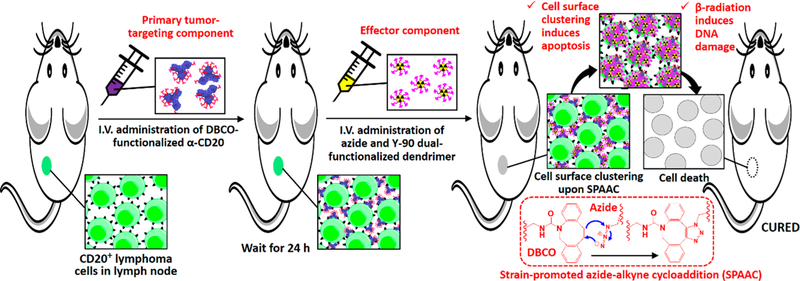
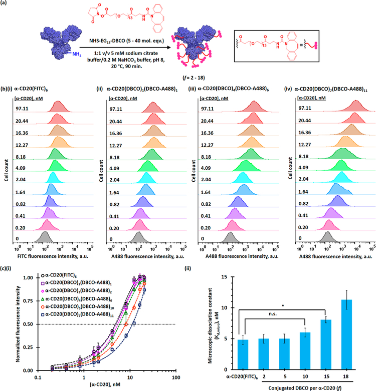
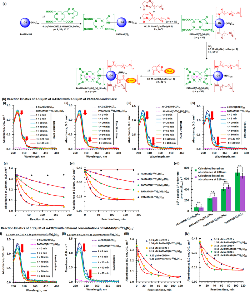
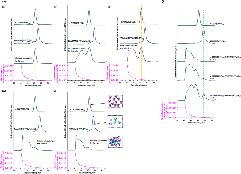
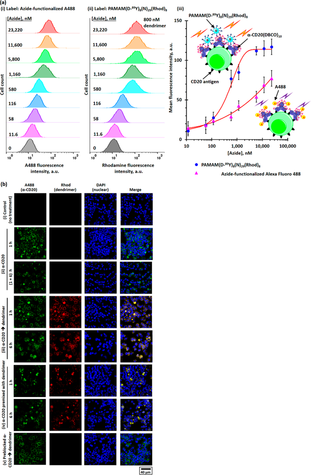
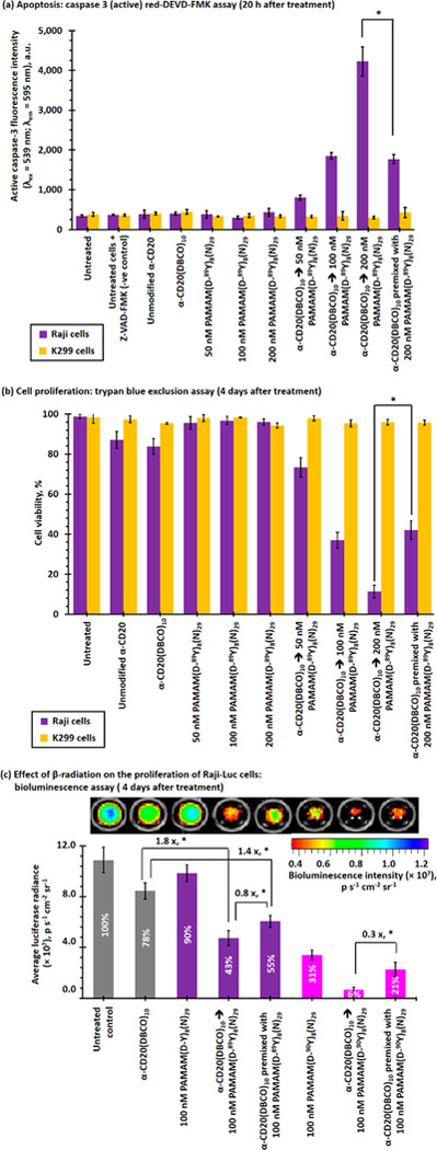
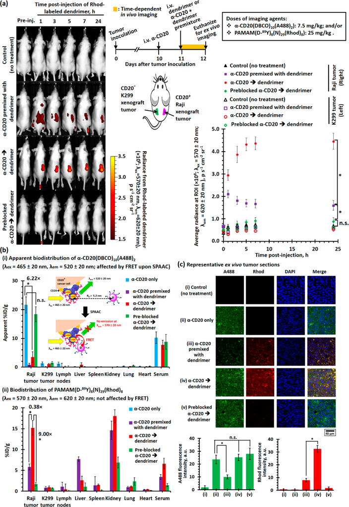
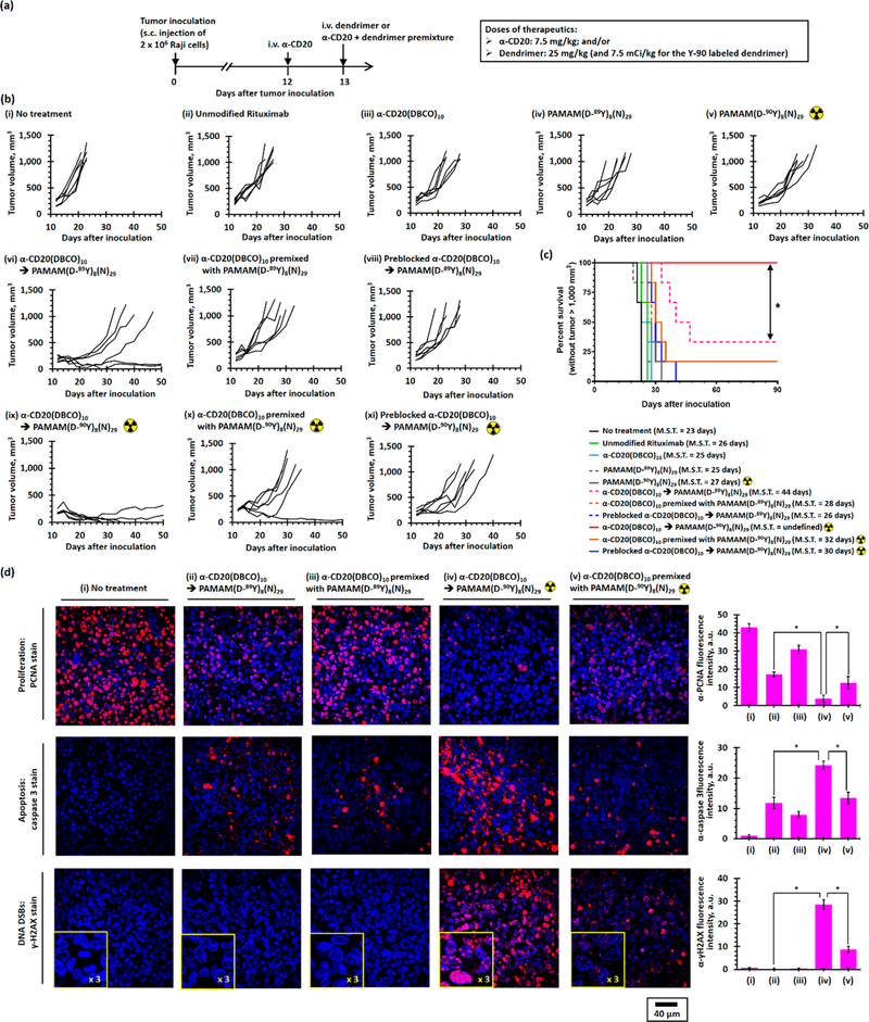
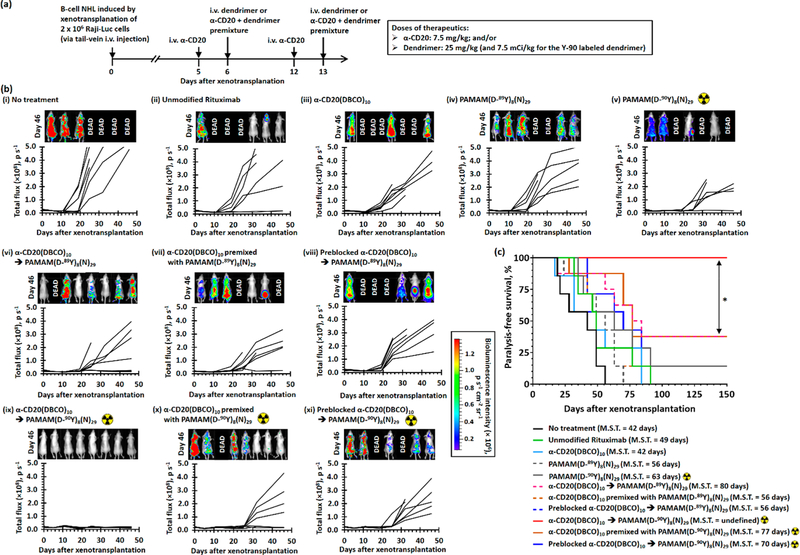
Non-Hodgkin lymphoma (NHL) is one of the most common types of hematologic malignancies. Pretargeted radioimmunotherapy (PRIT), the sequential administration of a bispecific antibody-based primary tumor-targeting component followed by a radionucleotide-labeled treatment effector, has been developed to improve the treatment efficacy and to reduce the side effects of conventional RIT. Despite the preclinical success of PRIT, clinical trials revealed that the immunogenicity of the bispecific antibody as well as the presence of competing endogenous effector molecules often compromised the treatment. One strategy to improve PRIT is to utilize bio-orthogonal ligation reactions to minimize immunogenicity and improve targeting. Herein, we report a translatable pretargeted nanoradioimmunotherapy strategy for the treatment of NHL. This pretargeting system is composed of a dibenzylcyclooctyne (DBCO)-functionalized anti-CD20 antibody (α-CD20) tumor-targeting component and an azide- and yttrium-90-(90Y) dual-functionalized dendrimer. The physicochemical properties of both pretargeting components have been extensively studied. We demonstrated that an optimized dual-functionalized dendrimer can undergo rapid strain-promoted azide-alkyne cycloaddition with the DBCO-functionalized α-CD20 at the physiological conditions. The treatment effector in our pretargeting system can not only selectively deliver radionucleotides to the target tumor cells but also increase the complement-dependent cytotoxicity of α-CD20 and thus enhance the antitumor effects, as justified by comprehensive in vitro and in vivo studies in mouse NHL xenograft and disseminated models.

Keywords: antibody; bioorthogonal ligation reaction; dendrimer; non-Hodgkin lymphoma; pretargeted radioimmunotherapy.


Conflict of interest statement

The authors declare no competing financial interest.

Figures

Figure 1.
Figure 1.
Scheme illustrating the two-step pretargeted radioimmunotherapy strategy for the treatment of non-Hodgkin lymphoma.
Figure 2.
Figure 2.
Functionalization and characterization of rituximab (α-CD20). (a) Functionalization of rituximab for pretargeting. The target degrees of functionalization were 5, 10, 20, 30, and 40, and the actual degrees of functionalization (f) were 2, 5, 10, 15, and 18, respectively.(b) Representative flow cytometry histograms of CD20-antigen overexpressing Raji cells after incubation with different concentrations of (i) FITC-labeled rituximab, (ii) α-CD20(DBCO)1(A488)1, (iii) α-CD20(DBCO)4(A488)6, and (ii) α-CD20(DBCO)7(A488)11. (c)(i) The plot of normalized mean-fluorescence intensities (MFIs) of Raji cells after staining with different concentrations of α-CD20. (ii) Microscopic dissociation constants (Kd,micro) of FITC-labeled rituximab and different DBCO-functionalized α-CD20 with different degrees of functionalization, as determined by the FACS binding assay in the CD20-overexpressing Raji cell line. The microscopic dissociation constant (Kd,micro) is equivalent to the concentration of antibody required to achieve half-saturated binding.
Figure 3.
Figure 3.
Functionalization and evaluation of azide and yttrium dual-functionalized PAMAM G4 for pretargeted immunotherapy and pretargeted radioimmunotherapy. (a) Functionalization azide and yttrium dual-functionalized PAMAM G4. (b) In vitro evaluation of SPAAC between the different dual-functionalized PAMAM and the selected α-CD20(DBCO)10. (i–iv) Time-dependent UV spectra of 3.13 μM (final concentration) of α-CD20(DBCO)10 recorded before and after incubation with 3.13 μM of dual-functionalized PAMAM at 37 °C for 5 to 180 min. (v, vi) Time-dependent UV absorbance at (v) 280 nm and (vi) 310 nm of α-CD20(DBCO)10 after incubation with an equivalent amount of different dual-functionalized PAMAM. (vii) The plot of the pseudo-first-order rate constants (kobs,280 nm and kobs,310 nm) of SPAAC between different dual-functionalized dendrimers and α-CD20(DBCO)10. (c)(i, ii) Time-dependent UV spectra of 3.13 μM (final concentration) of α-CD20(DBCO)10 recorded before and after incubation with (i) 1.56, and (ii) 6.25 μM of dual-functionalized PAMAM(D-89Y)8(N)29 at 37 °C for 5–180 min. (iii, iv) Time-dependent UV absorbance at (iii) 280 nm and (iv) 310 nm of α-CD20(DBCO)10 after incubation with different molar ratios of PAMAM(D-89Y)8(N)29. The concentration-dependent second-order rate constant was calculated to be about 230 M−1 s−1 (see Figure S22). (NB. * represents p < 0.05; n.s. represents statistically insignificant (i.e., p > 0.05).)
Figure 4.
Figure 4.
Cross-linking efficiencies of different dual-functionalized PAMAM after incubated with α-CD20(DBCO)10 at the physiological conditions. (a) SEC-MALLS analysis of α-CD20(DBCO)10, different dual-functionalized PAMAM, and their 1:1 mixture after being incubated at 37 °C for 30 min. (b) SEC-MALLS analysis of α-CD20(DBCO)10, PAMAM(D-89Y)8(N)29, and their mixtures obtained after incubating with different molar ratios of PAMAM(D-89Y)8(N)29 at 37 °C for 30 min. The SEC-MALLS analysis indicates that PAMAM(D-89Y)8(N)29 prefers to form high molecular-weight nanoclusters with α-CD20(DBCO)10 rather than form well-defined antibody-dendrimer conjugates.
Figure 5.
Figure 5.
In vitro evaluation of the α-CD20(DBCO)10 and PAMAM(D-89Y)8(N)29(Rhod)8-based pretargeted system. (a) Flow cytometry histograms of α-CD20(DBCO)10-pretreated Raji cells after incubation with different concentrations of (i) azide-functionalized A488 and (ii) PAMAM(D-89Y)8(N)29(Rhod)8 containing 11.6–23,220 nM of terminal azide. (iii) The plot of MFIs of α-CD20(DBCO)10-pretreated Raji cells after being incubated with different concentrations of azide-functionalized A488 and PAMAM(D-89Y)8(N)29(Rhod)8, as determined by the flow cytometry method. (b) Representative CLSM images of (i) unstained Raji cells (control); (ii) Raji cells stained with α-CD20(DBCO)10(A488)2 for 1 h, washed, with and without further incubation at 37 °C for another 6 h; (iii) Raji cells pretreated with α-CD20(DBCO)10(A488)2 for 1 h before staining with PAMAM(D-89Y)8(N)29(Rhod)8 for another 1 or 6 h; (iv) Raji cells incubated with the antibody-dendrimer premixture for 1 or 6 h; and (v) Raji cells incubated with preblocked α-CD20(DBCO)10(A488)2 for 1 h before being incubated with PAMAM(D-89Y)8(N)29(Rhod)8 for another 1 h.
Figure 6.
Figure 6.
In vitro toxicities of direct and pretargeted treatments with α-CD20(DBCO)10 and nonradioactive PAMAM-(D-89Y)8(N)29/radioactive PAMAM(D-90Y)8(N)29 in CD20 K299 and CD20+ Raji cell lines. (a) Caspase 3 activities of Raji and K299 cells recorded 20 h after direct or sequential treatment with α-CD20(DBCO)10(A488)2 and/or PAMAM(D-89Y)8(N)29(Rhod)8. High caspase 3 activities indicate early stage apoptosis (i.e., programmed cell death). (b) Proliferation of K299 and Raji cells recorded 4 days after direct or sequential treatment with α-CD20(DBCO)10(A488)2 and/or PAMAM(D-89Y)8(N)29(Rhod)8, as determined by trypan blue exclusion assay. (c) Proliferation of Raji-Luc cells after direct or sequential treatment with α-CD20-(DBCO)10(A488)2 and/or nonradioactive PAMAM-(D-89Y)8(N)29 (Rhod)8/radioactive PAMAM(D-89Y)8(N)29(Rhod)8, as determined by the luciferase-based bioluminescence assay recorded 4 days after the initial treatment. The inserted number in the histograms represent the relative cell viabilities. (NB. * represents p < 0.05; n.s. represents statistically insignificant (i.e., p > 0.05).)
Figure 7.
Figure 7.
Biodistribution of rhodamine-labeled dual-functionalized PAMAM administrated via direct-targeting or pretargeting methods in K299 and Raji dual-exnograft tumor bearing mice. (a)(i) Representative time-dependent in vivo fluorescence images (λex = 570 ± 20 nm, λem = 620 ± 20 nm) of the Raji and K299 dual-xenograft tumor-bearing mice recorded after i.v. administration of α-CD20(DBCO)10(A488)2 (7.5 mg/kg) and PAMAM(D-89Y)8(N)29(Rhod)8 (25 mg/kg). The α-CD20 and dendrimer were either administrated as a premixture at day 11 (after tumor inoculation) or sequentially administrated as α-CD20 (or preblocked α-CD20) at day 10 and dendrimer at day 11 (24 h after the administration of α-CD20). The in vivo imaging study recorded the fluorescence emitted from the systemically administrated rhodamine-labeled dendrimer. The inserts show the treatment schedule and locations of the xenograft tumors. (ii) The plot of time-dependent average radiances at the regions of interest (ROI, i.e., xenograft tumor sites) of different imaging-group mice (n = 5 per group). (b) “Apparent” biodistributions of (i) α-CD20(DBCO)10(A488)2 and (ii) PAMAM(D-89Y)8(N)29(Rhod)8 determined by the ex vivo fluorescence imaging technique. The inserted cartoon shows rhodamine in the dual-functionalized dendrimer quenches the fluorescence emitted from the A488 in α-CD20 upon SPAAC because of FRET. (c) Representative CLSM images and the fluorescence intensities of A488 and rhodamine channels of the ex vivo Raji xenograft tumor sessions collected from different imaging-group mice. The confocal microscope was operated under the compensation mode to allow concurrent observations of fluorescence emitted from A488 and rhodamine. (NB. * denotes p < 0.05, and n.s. denotes insignificant.)
Figure 8.
Figure 8.
In vivo antilymphoma efficacy study in Raji xenograft tumor-bearing mice. (a) Treatment schedule. Xenograft tumor was inoculated by s.c. injection of 2 × 10⁶ Raji cells in the left flank at day 0. Xenograft tumor-bearing mice received treatment at day 12. Therapeutics were either i.v. administrated once at day 12 or consecutively administrated α-CD20 at day 12, followed by dendrimer at day 13 (24 h apart). (b) Individual tumor growth curves of nontreatment group mice and mice treated with α-CD20(DBCO)10 (7.5 mg/kg) and/or PAMAM(D-89Y)8(N)29 (25 mg/kg)/PAMAM(D-89Y)8(N)29 (25 mg/kg, 7.5 mCi/kg). (c) Kaplan–Meier survival curves of Raji xenograft tumor-bearing mice (without tumor larger than 1000 mm3) after receiving different treatments. (n = 6 per group; * denotes p < 0.05; M.S.T. denotes median survival time.) (d) Representative CLSM images of anti-PCNA-, anticaspase 3-, and anti-γ-H2AX-stained Raji xenograft tumor section collected after receiving different direct or pretargeted treatments. In the anti-PCNA-stained tumor sections, all nuclei were stained with DAPI (blue fluorescence). The intense red fluorescence nuclei were PCNA-positive proliferating nuclei. In the anticaspase 3-stained tumor sections, nuclei were stained with DAPI (blue fluorescence). The strong red fluorescence were caspase 3-positive cells. In the anti-γ-H2AX-stained tumor sections, nuclei were stained with DAPI (blue fluorescence). The strong red fluorescence foci labeled the DNA double-strand break.
Figure 9.
Figure 9.
In vivo anticancer activity study in the disseminated B-cell NHL xenotransplant model. (a) Treatment schedule. B-cell NHL was induced by i.v. tail-vein injection of 2 × 10⁶ Raji-Luc cells at day 0. Lymphoma mice received two direct/pretargeted treatments with α-CD20(DBCO)10 (7.5 mg/kg) and/or PAMAM(D-89Y)8(N)29 (25 mg/kg)/PAMAM(D-89Y)8(N)29 (25 mg/kg, 7.5 mCi/kg) at days 5 and 12. For pretargeted therapies, the dendrimer was administrated 24 h after the administration of α-CD20. Progression of disease was monitored by in vivo bioluminescence imaging on days 0, 5, 11, 19, 25, 32, and 46. Mice were intraperitoneally administrated with 3 mg firefly D-luciferin 15 min prior to each imaging session. Cerenkov bioluminescence imaging studies were performed at days 11 and 19 prior to the administration of firefly D-luciferin to quantify the amount of Y-90-labeled dendrimers accumulated in the body. (b) Time-dependent whole-body bioluminescence intensities of individual lymphoma mice recorded before and after different treatments. The inserts show in vivo bioluminescence images recorded 46 days after the xenotransplantation. (c) Kaplan–Meier paralysis-free survival curves of Raji-Luc xenotransplanted mice after receiving different treatments. (n = 7–8 per group; * denotes p < 0.05; M.S.T. denotes median survival time.)

Similar articles

Cited by

References

    1. Siegel RL; Miller KD; Jemal A Cancer Statistics, 2017. Ca-Cancer J. Clin 2017, 67, 7–30. - PubMed
    1. Coiffier B; Lepage E; Briere J; Herbrecht R; Tilly H; Bouabdallah R; Morel P; Van Den Neste E; Salles G; Gaulard P; Reyes F; Lederlin P; Gisselbrecht C CHOP chemotherapy plus rituximab compared with CHOP alone in elderly patients with diffuse large-B-cell lymphoma. N. Engl. J. Med 2002, 346, 235–42. - PubMed
    1. Feugier P; Van Hoof A; Sebban C; Solal-Celigny P; Bouabdallah R; Ferme C; Christian B; Lepage E; Tilly H; Morschhauser F; Gaulard P; Salles G; Bosly A; Gisselbrecht C; Reyes F; Coiffier B Long-term results of the R-CHOP study in the treatment of elderly patients with diffuse large B-cell lymphoma: a study by the Groupe d’Etude des Lymphomes de l’Adulte. J. Clin. Oncol 2005, 23, 4117–26. - PubMed
    1. Molina A A decade of rituximab: improving survival outcomes in non-Hodgkin’s lymphoma. Annu. Rev. Med 2008, 59, 237–50. - PubMed
    1. Boye J; Elter T; Engert A An overview of the current clinical use of the anti-CD20 monoclonal antibody rituximab. Ann. Oncol 2003, 14, 520–535. - PubMed

Publication types

MeSH terms