

Neurophysiologic effects of transcutaneous auricular vagus nerve stimulation (taVNS) via electrical stimulation of the tragus: A concurrent taVNS/fMRI study and review
- PMID: 29361441
- PMCID: PMC6487660
- DOI: 10.1016/j.brs.2017.12.009
Neurophysiologic effects of transcutaneous auricular vagus nerve stimulation (taVNS) via electrical stimulation of the tragus: A concurrent taVNS/fMRI study and review
Abstract
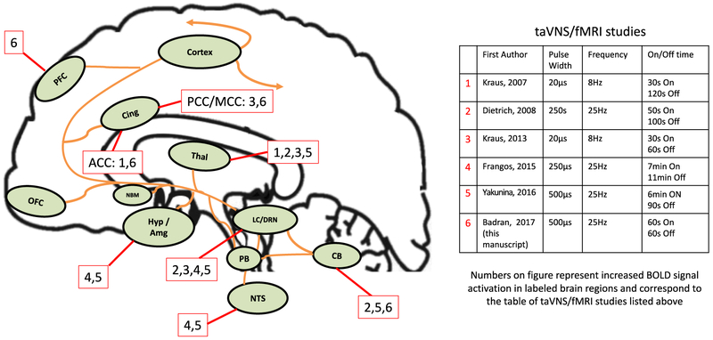
Background: Electrical stimulation of the auricular branch of the vagus nerve (ABVN) via transcutaneous auricular vagus nerve stimulation (taVNS) may influence afferent vagal networks. There have been 5 prior taVNS/fMRI studies, with inconsistent findings due to variability in stimulation targets and parameters.
Objective: We developed a taVNS/fMRI system to enable concurrent electrical stimulation and fMRI acquisition to compare the effects of taVNS in relation to control stimulation.
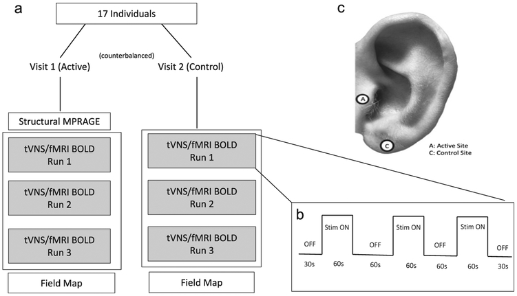
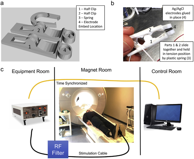
Methods: We enrolled 17 healthy adults in this single-blind, crossover taVNS/fMRI trial. Based on parameters shown to affect heart rate in healthy volunteers, participants received either left tragus (active) or earlobe (control) stimulation at 500 μs 25 HZ for 60 s (repeated 3 times over 6 min). Whole brain fMRI analysis was performed exploring the effect of: active stimulation, control stimulation, and the comparison. Region of interest analysis of the midbrain and brainstem was also conducted.
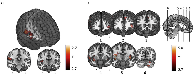
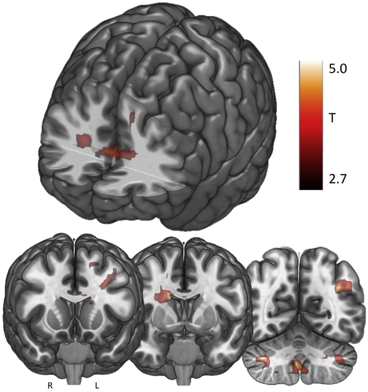
Results: Active stimulation produced significant increased BOLD signal in the contralateral postcentral gyrus, bilateral insula, frontal cortex, right operculum, and left cerebellum. Control stimulation produced BOLD signal activation in the contralateral postcentral gyrus. In the active vs. control contrast, tragus stimulation produced significantly greater BOLD increases in the right caudate, bilateral anterior cingulate, cerebellum, left prefrontal cortex, and mid-cingulate.
Conclusion: Stimulation of the tragus activates the cerebral afferents of the vagal pathway and combined with our review of the literature suggest that taVNS is a promising form of VNS. Future taVNS/fMRI studies should systematically explore various parameters and alternative stimulation targets aimed to optimize this novel form of neuromodulation.
Keywords: Anterior cingulate cortex (ACC); Ear stimulation; Transcutaneous auricular vagus nerve stimulation (taVNS); fMRI.
Copyright © 2017 Elsevier Inc. All rights reserved.
Figures
Comment in
-
Transcutaneous nerve stimulation via the tragus: are we really stimulating the vagus nerve?Brain Stimul. 2018 Jul-Aug;11(4):945-946. doi: 10.1016/j.brs.2018.03.018. Epub 2018 Apr 5. Brain Stimul. 2018. PMID: 29661599 No abstract available.
-
Tragus or cymba conchae? Investigating the anatomical foundation of transcutaneous auricular vagus nerve stimulation (taVNS).Brain Stimul. 2018 Jul-Aug;11(4):947-948. doi: 10.1016/j.brs.2018.06.003. Epub 2018 Jun 6. Brain Stimul. 2018. PMID: 29895444 Free PMC article. No abstract available.
References
-
- Ben-Menachem E, Mañon-Espaillat R, Ristanovic R, Wilder B, Stefan H, Mirza W, et al. Vagus nerve stimulation for treatment of partial seizures: 1. A controlled study of effect on seizures. Epilepsia 1994;35(3):616–26. - PubMed
-
- Handforth A, DeGiorgio C, Schachter S, Uthman B, Naritoku D, Tecoma E, et al. Vagus nerve stimulation therapy for partial-onset seizures a randomized active-control trial. Neurology 1998;51(1):48–55. - PubMed
-
- Benarroch EE, editor. The central autonomic network: functional organization, dysfunction, and perspective Mayo Clin Proc. Elsevier; 1993. - PubMed
-
- Kalia M, Sullivan JM. Brainstem projections of sensory and motor components of the vagus nerve in the rat. JComp Neurol 1982;211(3):248–64. - PubMed
-
- Van Bockstaele EJ, Peoples J, Valentino RJ. Anatomic basis for differential regulation of the rostrolateral peri–locus coeruleus region by limbic afferents. Biol Psychiatr 1999;46(10):1352–63. - PubMed
Publication types
MeSH terms
Grants and funding
LinkOut - more resources
Full Text Sources
Other Literature Sources
Medical
