. 2018 Feb 20;131(4):jcs211748.
doi: 10.1242/jcs.211748.

Characterization of a novel RP2-OSTF1 interaction and its implication for actin remodelling

Affiliations

Characterization of a novel RP2-OSTF1 interaction and its implication for actin remodelling

Rodanthi Lyraki et al. J Cell Sci. .

Abstract

Retinitis pigmentosa 2 (RP2) is the causative gene for a form of X-linked retinal degeneration. RP2 was previously shown to have GTPase-activating protein (GAP) activity towards the small GTPase ARL3 via its N-terminus, but the function of the C-terminus remains elusive. Here, we report a novel interaction between RP2 and osteoclast-stimulating factor 1 (OSTF1), an intracellular protein that indirectly enhances osteoclast formation and activity and is a negative regulator of cell motility. Moreover, this interaction is abolished by a human pathogenic mutation in RP2. We utilized a structure-based approach to pinpoint the binding interface to a strictly conserved cluster of residues on the surface of RP2 that spans both the C- and N-terminal domains of the protein, and which is structurally distinct from the ARL3-binding site. In addition, we show that RP2 is a positive regulator of cell motility in vitro, recruiting OSTF1 to the cell membrane and preventing its interaction with the migration regulator Myo1E.

Keywords: Actin; OSTF1; RP2; Retinitis pigmentosa.


Conflict of interest statement

Competing interestsThe authors declare no competing or financial interests.

Figures
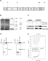
Fig. 1.
Fig. 1.
RP2 directly interacts with OSTF1. (A) Schematic showing the main protein domains of OSTF1. PR, proline-rich region; SH3, Src-homology 3 domain; ANK, ankyrin repeats; AC, acidic amino acid cluster. (B) A direct interaction between RP2 and OSTF1 was identified in a yeast two-hybrid screen and confirmed in two directions in a targeted yeast two-hybrid assay. –W –L, medium without tryptophan and leucine; –W –L –H, medium without tryptophan, leucine and histidine.  (C) Exogenous RP2 and OSTF1 co-immunoprecipitate from HEK293T lysates. Lysates from HEK293T cells previously transfected with plasmids for expression of RP2–FLAG and/or Myc–OSTF1 were subjected to anti-FLAG immunoprecipitation (IP) followed by immunoblotting with anti-Myc or anti-FLAG antibodies. (D) Bovine retinal extracts were subjected to pulldown assays using recombinant GST–RP2 or GST–OSTF1 as bait, followed by immunoblotting for endogenous OSTF1 or RP2, respectively. Ponceau staining shows the GST-tagged proteins. (E) Thermodynamic characterization of the OSTF1–RP2 interaction by ITC. 0.585 mM His-tagged OSTF1 was injected into the sample cell containing 52 μM full-length RP2.
Fig. 2.
Fig. 2.
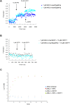
Binding of OSTF1 on RP2 does not affect the RP2–ARL3 interaction. (A,B) Complex formation between recombinant OSTF1–His, RP2 and ARL3 (A), as well as between recombinant OSTF1–His and ARL3 (B), was monitored on the basis of change in fluorescence polarization signal. The fluorescently labelled G-domain of ARL3 was used in the analysis in the following states: mantGppNHp (a non-hydrolysable GTP analog)-bound ARL3 and mantGDP-bound ARL3. (C) Binding to OSTF1 does not affect the catalytic activity of RP2. A GTPase charcoal assay was performed with catalytic concentrations of RP2 (the GAP) (0.2 μM) and ARL3 (small G protein) (20 μM) bound to 60 nM [32P-γ]GTP, in the presence or absence of 5 μM of OSTF1. GTP hydrolysis is presented as the ratio of the count at each time-point to the total count at each time-point (c.p.m. ratio) over time.
Fig. 3.
Fig. 3.
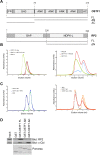
The N-terminal domain of RP2, and both the SH3 and ANK domains of OSTF1 participate in the RP2–OSTF1 interaction. (A) Diagram of the deletion constructs of OSTF1–His and RP2 that were used in the analytical gel filtration and pulldown assays. RP2 ΔN corresponds to residues 230–350, while OSTF1 ΔN corresponds to residues 75–214 and OSTF1 ΔC to residues 1–73. (B) Analytical gel filtration graphs showing complex formation or not between recombinant full-length OSTF1–His and either full-length RP2 (left) or RP2 ΔN (right). (C) Analytical gel filtration graphs showing complex formation between recombinant full-length RP2 and either OSTF1 ΔN (left) or OSTF1 ΔC (right). (D) To confirm the participation of both OSTF1 domains in the interaction with RP2, bovine retinal lysates were subjected to pulldown assays using either full-length or truncated recombinant GST–OSTF1 as bait, as indicated, followed by immunoblotting for endogenous RP2 and Cbl. Ponceau staining shows the GST-tagged protein.
Fig. 4.
Fig. 4.
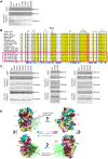
Mapping of the OSTF1 binding site of RP2 by site-directed mutagenesis. (A) The RP2–OSTF1 interaction is abolished by the R211L mutation, but not by other missense pathogenic mutations. HEK293T cell lysates previously transfected with plasmids containing WT RP2–V5 or pathogenic mutant forms of the protein were subjected to pulldown assays using recombinant GST–OSTF1 as bait. Subsequently, they were subjected to immunoblotting using an antibody against the V5 tag. Ponceau staining shows the presence of the GST-tagged protein. (B) Section of the multiple sequence alignment of RP2 protein orthologues, encompassing the critical OSTF1 interaction position (R211) which is strictly conserved among vertebrates. Non-vertebrate chordates and other phyla are shown within the pink border. An interesting strictly conserved residue in all species tested is highlighted in red (F241 in human RP2). (C) Identification of RP2 residues that are critical for the RP2–OSTF1 interaction to occur. HEK293T cell lysates previously transfected with plasmids containing WT RP2–V5 or the same protein encompassing non-destabilizing missense mutations were subjected to pulldown assays using recombinant GST–OSTF1 or GST–ARL3 Q71L as bait. The ARL3 Q71L mutant is defective in GTP hydrolysis and thus can bind RP2 stably. They were then analysed by immunoblotting using an antibody against the V5 tag. Ponceau staining shows the presence of the GST-tagged protein. (D) The ARL3-binding area on RP2, as well as the residues that we identified as important for the RP2–OSTF1 interaction (highlighted in red), belong to distinct strictly conserved clusters. The crystal structure of the RP2–ARL3 complex (PDB ID: 3BH7) is shown in four views rotated by 90° about the y-axis via ConSurf (Glaser et al., 2003; Ashkenazy et al., 2010) analysis. RP2 is shown in surface representation, where residues are colour-coded according to their conservation grade in vertebrates, with turquoise-through-maroon indicating variable to strictly conserved. The light yellow colour represents positions for which the inferred conservation level was assigned with low confidence. ARL3 GTPase is shown in green colour in a stick representation.
Fig. 5.
Fig. 5.
A docking model of the ternary OSTF–RP2–ARL3 complex compatible with experimental mutagenesis data. Shown here is a single structure of OSTF–RP2–ARL3 complex from the cluster of predicted structures with highest confidence value (cluster 1) (Fig. S4), which comprised approximately half of all docking models compatible with the experimental data, and which showed a much more strongly conserved binding surface on OSTF1 than the others.
Fig. 6.
Fig. 6.
Cells lacking RP2 expression display motility defects. (A) hTERT-RPE1 cell lines were engineered to be RP2 null but express OSTF1 at WT levels. WT and RP2-null hTERT-RPE1 cell lysates (G and I clones, two biological repeats are shown) were subjected to immunoblotting using antibodies for endogenous OSTF1, RP2 and β-actin, as a loading control. (B) WT and RP2-null hTERT-RPE1 cells (G and I clones) were subjected to a wound healing assay. Images on the left show the state of the wound in two representative time points after wound induction (0 h). The yellow colour represents the wound area, while the purple colour at the cell boundaries shows the area that has been covered by migrating cells. The graph on the right shows the area of the wound that was covered by migrating cells 10 h after wound induction (mean±s.e.m. for six technical repeats). *P=0.0103 for I versus WT, ****P<0.0001 for G versus WT (two-tailed unpaired t-tests). (C) The random migration of sparsely plated individual WT and RP2-null hTERT-RPE1 cells (G and I clones) was analysed over a 9-h period. Left panels show representative tracks followed by individual cells. The graph on the right shows the average distance covered by individual cells (n=40 for each cell line) in μm (mean±s.e.m. for four technical repeats, ****P<0.0001 for G versus WT, and for I versus WT (two-tailed t-test and two-tailed t-test with Welch's correction, respectively).
Fig. 7.
Fig. 7.
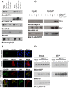
RP2 overexpression leads to OSTF1 translocation to the membrane compartment and dissociation from Myo1E. (A) HEK293T cells previously transfected with empty plasmid or a plasmid containing an RP2–V5 expression construct were subjected to detergent-based subcellular fractionation. The fractions were then analysed by immunoblotting using antibodies against the V5 tag, endogenous OSTF1, endogenous ARL3, endogenous histone H3 (marker for the nuclear fraction) and endogenous integrin β1 (marker for the membrane fraction). While RP2 overexpression led to a redistribution of OSTF1 pools to the membrane compartment, it did not affect the localization of ARL3. WCL, whole-cell lysate; CF, cytoplasmic fraction; MOF, membrane and organelle fraction; CyNF, cytoplasmic and nuclear fraction. (B) HeLa cells previously transfected with plasmids containing RP2–emGFP and/or V5–OSTF1 constructs were fixed and subjected to immunofluorescence analysis using an antibody to V5 tag in order to investigate the localization of V5–OSTF1. While V5–OSTF1 is cytosolic on its own, RP2 overexpression leads to recruitment of V5–OSTF1 to the plasma membrane. (C) HEK293T cells were transiently transfected with plasmids containing different combinations of V5–Myo1E, RP2–V5 and FLAG–OSTF1 constructs. The lysates were subjected to anti-FLAG immunoprecipitation (IP) to assess how much V5–Myo1E associated with FLAG-OSTF1 in the presence or absence of RP2–V5 overexpression, and immunoblotting using antibodies to V5 and FLAG tags. (D) Stimulation of the ERK1/2 pathway by serum stimulation slightly stabilizes the RP2–OSTF1 interaction. Cells previously transiently transfected with a vector containing an RP2–V5 construct were stimulated with 10% fetal bovine serum for the indicated time duration (in min) following overnight serum deprivation. The lysates were then subjected to anti-V5 immunoprecipitation and immunoblotting using antibodies to V5 tag, endogenous OSTF1 and endogenous phospho-ERK1/2.

References

    1. Ashkenazy H., Erez E., Martz E., Pupko T. and Ben-Tal N. (2010). ConSurf 2010: calculating evolutionary conservation in sequence and structure of proteins and nucleic acids. Nucleic Acids Res. 38, W529-W533. 10.1093/nar/gkq399 - DOI - PMC - PubMed
    1. Ashkenazy H., Abadi S., Martz E., Chay O., Mayrose I., Pupko T. and Ben-Tal N. (2016). ConSurf 2016: an improved methodology to estimate and visualize evolutionary conservation in macromolecules. Nucleic Acids Res. 44, W344-W350. 10.1093/nar/gkw408 - DOI - PMC - PubMed
    1. Chapple J. P., Hardcastle A. J., Grayson C., Spackman L. A., Willison K. R. and Cheetham M. E. (2000). Mutations in the N-terminus of the X-linked retinitis pigmentosa protein RP2 interfere with the normal targeting of the protein to the plasma membrane. Hum. Mol. Genet. 9, 1919-1926. 10.1093/hmg/9.13.1919 - DOI - PubMed
    1. Chapple J. P., Hardcastle A. J., Grayson C., Willison K. R. and Cheetham M. E. (2002). Delineation of the plasma membrane targeting domain of the X-linked retinitis pigmentosa protein RP2. Invest. Ophthalmol. Vis. Sci. 43, 2015-2020. - PubMed
    1. Chen L., Wang Y., Wells D., Toh D., Harold H., Zhou J., Digiammarino E. and Meehan E. J. (2006). Structure of the SH3 domain of human osteoclast-stimulating factor at atomic resolution. Acta Crystallogr. Sect. F Struct. Biol. Cryst. Commun. 62, 844-848. 10.1107/S1744309106030004 - DOI - PMC - PubMed

Publication types

MeSH terms

Supplementary concepts

LinkOut - more resources