. 2018 Jan 26;7(1):bio027391.
doi: 10.1242/bio.027391.

miR-9a mediates the role of Lethal giant larvae as an epithelial growth inhibitor in Drosophila

Affiliations

miR-9a mediates the role of Lethal giant larvae as an epithelial growth inhibitor in Drosophila

Scott G Daniel et al. Biol Open. .

Abstract

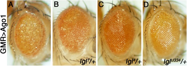
Drosophila lethal giant larvae (lgl) encodes a conserved tumor suppressor with established roles in cell polarity, asymmetric division, and proliferation control. Lgl's human orthologs, HUGL1 and HUGL2, are altered in human cancers, however, its mechanistic role as a tumor suppressor remains poorly understood. Based on a previously established connection between Lgl and Fragile X protein (FMRP), a miRNA-associated translational regulator, we hypothesized that Lgl may exert its role as a tumor suppressor by interacting with the miRNA pathway. Consistent with this model, we found that lgl is a dominant modifier of Argonaute1 overexpression in the eye neuroepithelium. Using microarray profiling we identified a core set of ten miRNAs that are altered throughout tumorigenesis in Drosophila lgl mutants. Among these are several miRNAs previously linked to human cancers including miR-9a, which we found to be downregulated in lgl neuroepithelial tissues. To determine whether miR-9a can act as an effector of Lgl in vivo, we overexpressed it in the context of lgl knock-down by RNAi and found it able to reduce the overgrowth phenotype caused by Lgl loss in epithelia. Furthermore, cross-comparisons between miRNA and mRNA profiling in lgl mutant tissues and human breast cancer cells identified thrombospondin (tsp) as a common factor altered in both fly and human breast cancer tumorigenesis models. Our work provides the first evidence of a functional connection between Lgl and the miRNA pathway, demonstrates that miR-9a mediates Lgl's role in restricting epithelial proliferation, and provides novel insights into pathways controlled by Lgl during tumor progression.

Keywords: Drosophila; Epithelial growth; miRNA.


Conflict of interest statement

Competing interestsThe authors declare no competing or financial interests.

Figures

Fig. 1.
Fig. 1.
lgl interacts genetically with Argonaute 1 (AGO1) in the eye. (A) Overexpression of AGO1 using GMR-Gal4 results in a rough eye phenotype. (B-D) Three independent alleles of lgl, namely lgl1, lgl4 and lglU334, can dominantly suppress the GMR>AGO1 phenotype. Genotypes as indicated. N=at least 10 adults were imaged per genotype.
Fig. 2.
Fig. 2.
lgl brain tissue and dysregulated microRNAs. (A-D) Cephalic complexes and ventral ganglia of third instar larvae. (A) Day 0 of control's third instar. (B-D) lgl mutants do not pupate as normal, day 0, 3, and 5 of lgl third instar are shown. (E) Graph of up- and down-regulated microRNAs from microarrays done on tissue from the brains shown in (A-D). Log of fold-change (logFC) is estimated from a linear model of the expression values as computed by the microarray analysis package, limma. The microRNAs listed here, represent ones that were dysregulated across all time-points for mutants, when compared to controls. All microRNAs shown were found to be significantly dysregulated with P<0.05, Benjamini-Hochberg multiple testing correction. Error bars show standard error of the mean.
Fig. 3.
Fig. 3.
miR-9a and lgl exhibit genetic interactions in the wing epithelium. (A) en-GAL4 control, posterior compartment P as indicated; (B) en-GAL4; UAS-lglRNAi; (C) en-GAL4; UAS-miR-9a; (D) en-GAL4; UAS-lglRNAi/UAS-miR-9a; (E) en-GAL4; miR-9aF80/+; (F) en-GAL4; UAS-lglRNAi/miR-9aF80. Insets show incomplete cross-vein phenotype. (G) Graph of posterior wing region area divided by total wing area. Genotypes as indicated. ***P<0.001, *P<0.05, n.s., not significant; student's t-test was used to determine statistical significance. For number of wings analysed, see Materials and Methods. Box and whisker plots show median, upper and lower quartiles, highest and lowest points, and outliers.
Fig. 4.
Fig. 4.
microRNA targeting strategy and dysregulated mRNAs. (A) Significantly dysregulated microRNAs were matched to mRNAs based on predictions given by the miRanda algorithm. Additionally, matches were validated by their direction of deregulation; upregulated miRNAs matched downregulated mRNAs while downregulated miRNAs matched upregulated mRNAs. (B) The matching mRNAs after applying our targeting strategy (see text). Log of fold-change (logFC) is estimated from a linear model of the expression values as computed by the microarray analysis package, limma. All mRNAs shown were found to be significantly dysregulated with P<0.05, Benjamini-Hochberg multiple testing correction. Error bars show standard error of the mean.
Fig. 5.
Fig. 5.
microRNA targeting network. MicroRNAs (center) target multiple mRNAs (outer ring); in turn, some mRNAs are targeted by a number of microRNAs (e.g. Klp98A is targeted by miR-993 and miR-275). The spectrum of maximum downregulation (−2.6 logFC) to maximum upregulation (3.3 logFC) is denoted by a standard red to green gradient. Only upregulated mRNAs are targeted by downregulated microRNAs, while downregulated mRNAs are targeted by upregulated microRNAs. Width of lines connecting microRNA to mRNA represents strength of targeting, as defined by the mirSVR score of the miRanda targeting algorithm. mirSVR score heat map as shown. Green is upregulated, and red is downregulated miRs or genes.

Similar articles

Cited by

References

    1. Abu-Safieh L., Abboud E. B., Alkuraya H., Shamseldin H., Al-Enzi S., Al-Abdi L., Hashem M., Colak D., Jarallah A., Ahmad H. et al. (2011). Mutation of IGFBP7 causes upregulation of BRAF/MEK/ERK pathway and familial retinal arterial macroaneurysms. Am. J. Hum. Genet. 89, 313-319. 10.1016/j.ajhg.2011.07.010 - DOI - PMC - PubMed
    1. Almeida M. S. and Bray S. J. (2005). Regulation of post-embryonic neuroblasts by Drosophila Grainyhead. Mech. Dev. 122, 1282-1293. 10.1016/j.mod.2005.08.004 - DOI - PubMed
    1. Ashburner M., Ball C. A., Blake J. A., Botstein D., Butler H., Cherry J. M., Davis A. P., Dolinski K., Dwight S. S., Eppig J. T. et al. (2000). Gene ontology: tool for the unification of biology. The Gene Ontology Consortium. Nat. Genet. 25, 25-29. 10.1038/75556 - DOI - PMC - PubMed
    1. Barh D., Malhotra R., Ravi B. and Sindhurani P. (2010). MicroRNA let-7: an emerging next-generation cancer therapeutic. Curr. Oncol. 17, 70-80. 10.3747/co.v17i1.356 - DOI - PMC - PubMed
    1. Batra S. K., McLendon R. E., Koo J. S., Castelino-Prabhu S., Fuchs H. E., Krischer J. P., Friedman H. S., Bigner D. D. and Bigner S. H. (1995). Prognostic implications of chromosome 17p deletions in human medulloblastomas. J. Neurooncol. 24, 39-45. 10.1007/BF01052657 - DOI - PubMed