Review
. 2018 Jan 22;19(1):318.
doi: 10.3390/ijms19010318.

Microglia and Aging: The Role of the TREM2-DAP12 and CX3CL1-CX3CR1 Axes

Affiliations
Review

Microglia and Aging: The Role of the TREM2-DAP12 and CX3CL1-CX3CR1 Axes

Carmen Mecca et al. Int J Mol Sci. .

Abstract

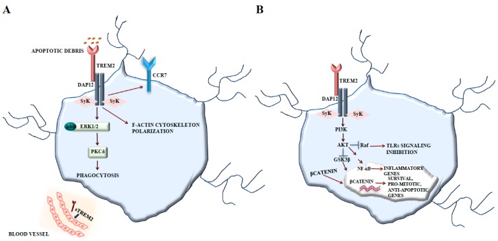
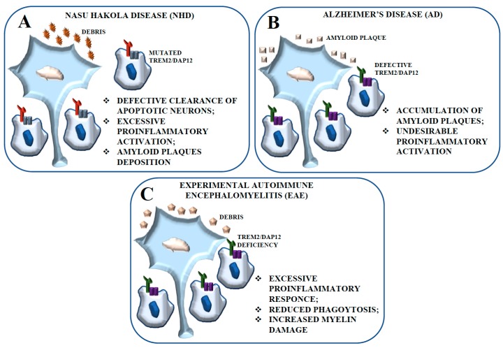
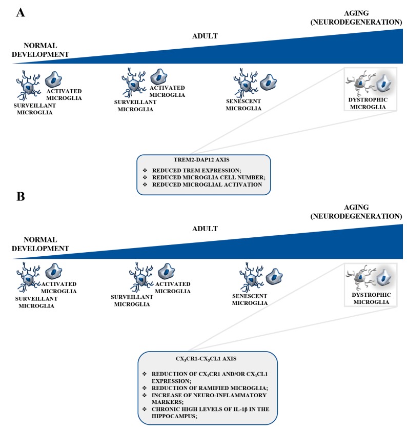
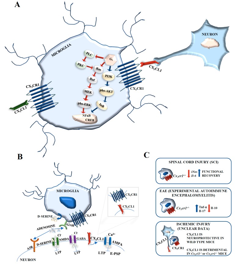
Depending on the species, microglial cells represent 5-20% of glial cells in the adult brain. As the innate immune effector of the brain, microglia are involved in several functions: regulation of inflammation, synaptic connectivity, programmed cell death, wiring and circuitry formation, phagocytosis of cell debris, and synaptic pruning and sculpting of postnatal neural circuits. Moreover, microglia contribute to some neurodevelopmental disorders such as Nasu-Hakola disease (NHD), and to aged-associated neurodegenerative diseases, such as Alzheimer's disease (AD), Parkinson's disease (PD), and others. There is evidence that human and rodent microglia may become senescent. This event determines alterations in the microglia activation status, associated with a chronic inflammation phenotype and with the loss of neuroprotective functions that lead to a greater susceptibility to the neurodegenerative diseases of aging. In the central nervous system (CNS), Triggering Receptor Expressed on Myeloid Cells 2-DNAX activation protein 12 (TREM2-DAP12) is a signaling complex expressed exclusively in microglia. As a microglial surface receptor, TREM2 interacts with DAP12 to initiate signal transduction pathways that promote microglial cell activation, phagocytosis, and microglial cell survival. Defective TREM2-DAP12 functions play a central role in the pathogenesis of several diseases. The CX3CL1 (fractalkine)-CX3CR1 signaling represents the most important communication channel between neurons and microglia. The expression of CX3CL1 in neurons and of its receptor CX3CR1 in microglia determines a specific interaction, playing fundamental roles in the regulation of the maturation and function of these cells. Here, we review the role of the TREM2-DAP12 and CX3CL1-CX3CR1 axes in aged microglia and the involvement of these pathways in physiological CNS aging and in age-associated neurodegenerative diseases.

Keywords: CX3CL1; CX3CR1; DAP12; TREM2; aged microglia; aging.


Conflict of interest statement

The authors declare no conflict of interest.

Figures

Figure 1
Figure 1
TREM2-DAP12 signaling. (A) TREM2 signals via DAP12 and stimulates the protein tyrosine kinase ERK, regulating actin polymerization and cytoskeleton organization. TREM2 upregulates the cell surface expression of the chemokine receptor CCR7 and promotes chemokine-directed migration towards CCR7 ligands. TREM2 activation also stimulates microglial phagocytosis via ERK. (B) TREM2 promotes microglial survival by activating Wnt/β-catenin signaling. TREM2 interacts with DAP12 and activates PI3K/Akt signaling, thereby inactivating GSK3β and stabilizing β-catenin that translocates into the nucleus, thereby activating pro-mitotic and anti-apoptotic genes. PI3K/Akt pathway activation by TREM2-DAP12 contributes to the regulation of NF-κB and to the inhibition of TLR signaling by blocking MAPK signaling at the RAF level.
Figure 2
Figure 2
Schematic representation of the contribution of the TREM2-DAP12 pathway to NHD, AD, and Experimental Autoimmune Encephalomyelitis (EAE). (A) The defective expression of TREM2 and DAP12 in microglial cells is considered tightly related to the inhibition of apoptotic neuron clearance observed in NHD. Impaired phagocytosis could be responsible for the excessive proinflammatory activation of microglial cells resulting in neurodegeneration. (B) The lack of function of TREM2, -DAP12, or both in AD patients leads to failing amyloid engulfment, determining an amyloid-associated dementia. These conditions would cause accumulation of amyloid-β plaques with secondary and undesirable proinflammatory microglial activation and proinflammatory activation. (C) The EAE model is an autoimmune demyelinating disorder characterized by the destruction of myelin proteins that recapitulates several aspects of the human disease multiple sclerosis (MS). The lack of function of TREM2, DAP12, or both results in an excessive inflammatory response, with decreased phagocytosis by microglial cells and increased myelin damage.
Figure 3
Figure 3
TREM2-DAP12 and CX3CL1-CX3CR1 signaling in aged microglia. (A) TREM2 expression is reduced in aged mice. A progressive decline in TREM2 expression might occur during aging in certain areas of the human brain, reducing microglial cell numbers and the microglial response to the breakdown of astrocytes, oligodendrocytes, neurons, and myelin. These events would create a fertile ground for neurodegeneration. TREM2 deficiency hinders the accumulation of microglia during aging. (B) CX3CL1 expression levels physiologically decrease in aged rodents which show a reduction of ramified microglia and a rise of neuroinflammatory markers. Reduced levels of CX3CL1 and CX3CR1 in the aged brain greatly alter the overall functionality of this pathway, with morphological and functionally impaired microglia. During aging, reduced neurogenesis occurs in the hippocampus. The pharmacological knockout or genetic deletion of CX3CR1 lead to decreased neurogenesis in the dentate gyrus of the mouse hippocampus, with an IL-1β-dependent decline of both the survival and the proliferation rate of the neuronal progenitors. The physiological decline in neuronal CX3CL1 might contribute to increased microglia activation.
Figure 4
Figure 4
CX3CL1-CX3CR1 signaling. (A) CX3CL1 induces transient phosphorylation of Akt and ERK1/2. Soluble CX3CL1 activates the transcription factor CREB and ERK1/2 and induces the translocation of NF-κB p65 subunit to the nucleus. NF-κB p65 subunit nuclear translocation is avoided by a specific inhibitor of PI3-K, suggesting that CX3CL1-CX3CR1 activates NF-κB through Akt. (B) CX3CL1, acting through CX3CR1, modulates α-amino-3-hydroxy-5-metyl-4-isoxazolepropionic acid receptor (AMPA) phosphorylation leading to increased calcium entry and inhibition of both excitatory postsynaptic potentials and long-term potentiation. CX3CL1 can increase inhibitory postsynaptic currents, possibly by enhancing neuronal responsiveness to GABA-mediated chloride entry. CX3CL1 may activate CX3CR1 on microglia with consequent adenosine release that, in turn, could activate A3R receptors on neurons. Adenosine activates A2AR on microglial cells and induces the release of d-serine. The adenosine released by microglia has also been involved in neuroprotection by activating A1R receptor subtypes in neurons. (C) CX3CL1-CX3CR1 involvement in spinal cord injury, experimental autoimmune encephalomyelitis, and ischemic injury.

References

    1. Del Rio-Hortega P. Microglia. In: Penfield W., editor. Cytology and Cellular Pathology of the Nervous System. Hoeber; New York, NY, USA: 1932. pp. 483–534.
    1. Alliot F., Godin I., Pessac B. Microglia derive from progenitors, originating from the yolk sac, and which proliferate in the brain. Brain Res. Dev. Brain Res. 1999;117:145–152. doi: 10.1016/S0165-3806(99)00113-3. - DOI - PubMed
    1. Ginhoux F., Greter M., Leboeuf M., Nandi S., See P., Gokhan S., Mehler M.F., Conway S.J., Nq L.G., Stanley E.R., et al. Fate Mapping Analysis Reveals That Adult Microglia Derive from Primitive Macrophages. Science. 2010;330:841–845. doi: 10.1126/science.1194637. - DOI - PMC - PubMed
    1. Graeber M.B., Tetzlaff W., Streit W.J., Kreutzberg G.W. Microglial cells, but not astrocytes, undergo mitosis following rat facial nerve axotomy. Neurosci. Lett. 1998;85:317–321. doi: 10.1016/0304-3940(88)90585-X. - DOI - PubMed
    1. Solomon J.N., Lewis C.A., Ajami B., Corbel S.Y., Rossi F.M., Krieger C. Origin and distribution of bone marrow-derived cells in the central nervous system in a mouse model of amyotrophic lateral sclerosis. Glia. 2006;53:744–775. doi: 10.1002/glia.20331. - DOI - PubMed

MeSH terms