Review
. 2018 Jan 22;19(1):325.
doi: 10.3390/ijms19010325.

Heat Shock Proteins and Autophagy Pathways in Neuroprotection: from Molecular Bases to Pharmacological Interventions

Affiliations
Review

Heat Shock Proteins and Autophagy Pathways in Neuroprotection: from Molecular Bases to Pharmacological Interventions

Botond Penke et al. Int J Mol Sci. .

Abstract

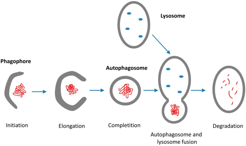
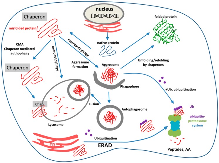
Neurodegenerative diseases (NDDs) such as Alzheimer's disease, Parkinson's disease and Huntington's disease (HD), amyotrophic lateral sclerosis, and prion diseases are all characterized by the accumulation of protein aggregates (amyloids) into inclusions and/or plaques. The ubiquitous presence of amyloids in NDDs suggests the involvement of disturbed protein homeostasis (proteostasis) in the underlying pathomechanisms. This review summarizes specific mechanisms that maintain proteostasis, including molecular chaperons, the ubiquitin-proteasome system (UPS), endoplasmic reticulum associated degradation (ERAD), and different autophagic pathways (chaperon mediated-, micro-, and macro-autophagy). The role of heat shock proteins (Hsps) in cellular quality control and degradation of pathogenic proteins is reviewed. Finally, putative therapeutic strategies for efficient removal of cytotoxic proteins from neurons and design of new therapeutic targets against the progression of NDDs are discussed.

Keywords: Hsp-inducers; autophagy; autophagy modulating drugs; endoplasmic reticulum associated degradation; heat shock proteins; neurodegenerative diseases; neuroprotection; ubiquitin-proteasome system.


Conflict of interest statement

The authors declare no conflict of interest.

Figures

Figure 1
Figure 1
Multiple roles of membranes in stress management: acting as cellular stress sensors, which can also interact with specific Hsps, and as such can serve as a novel target of various pharmacological interventions affecting both the expression and cellular localization/distribution of Hsps.
Figure 2
Figure 2
Three signal pathways of ER-stress activate UPR and lead to either cell survival or apoptosis. Under normal conditions, ER chaperon GRP78 binds all the three ER-stress sensors (PERK: protein kinase RNA like ER-kinase; IRE1α: inositol requiring enzyme 1α; ATF6: activating transcription factor 6). Under ER-stress, GRP78 dissociates from the sensors. PERK and IRE1α become phosphorylated and form oligomers, and ATF6 translocates to the Golgi. ATF6, ATF4, and XBP1 activate UPR target genes to enhance the capacity of the ER to cope with unfolded proteins. Activation of Sig-1R inhibits the three branches of UPR. (Abbreviations: eIF2α: eukaryotic translation initiation factor 2α; XBP1: X-box binding protein 1 (spliced form); TRAF2: TNF-associated factor-2; ATF4: transcriptional activator factor-4; MT: mitochondrion).
Figure 3
Figure 3
Schematic representation of the ER-associated protein degradation (ERAD) pathway: recognition, retro-translocation, and ubiquitination. Red line: protein/polypeptide chain; blue star: misfolded domain; orange circle: Cdc48 ATPase; purple dots: ubiquitin molecules.
Figure 4
Figure 4
Macroautophagy is a multistep process: initiation, elongation, completion, fusion, and lysosomal degradation. Red coil: misfolded protein; red lines: oligopeptide and amine acids; blue dots: lysosomal enzymes.
Figure 5
Figure 5
The most important pathways of intracellular protein degradation: endoplasmic reticulum associated-degradation (ERAD), the ubiquitin-proteasome system (UPS), and autophagy pathways. Purple dots: ubiquitin molecules; red coil: misfolded aggregated proteins; red lines: oligopeptides or amino acids.

References

    1. Kopito R.R. Aggresomes, inclusion bodies and protein aggregation. Trends Cell Biol. 2000;10:524–530. doi: 10.1016/S0962-8924(00)01852-3. - DOI - PubMed
    1. Balch W.E., Morimoto R.I., Dillin A., Kelly J.W. Adapting proteostasis for disease intervention. Science. 2008;319:916–919. doi: 10.1126/science.1141448. - DOI - PubMed
    1. Lim J., Yue Z.Y. Neuronal aggregates: Formation, clearance, and spreading. Dev. Cell. 2015;32:491–501. doi: 10.1016/j.devcel.2015.02.002. - DOI - PMC - PubMed
    1. Orr M.E., Oddo S. Autophagic/lysosomal dysfunction in Alzheimer’s disease. Alzheimers Res. Ther. 2013;5:53. doi: 10.1186/alzrt217. - DOI - PMC - PubMed
    1. Tarasoff-Conway J.M., Carare R.O., Osorio R.S., Glodzik L., Butler T., Fieremans E., Axel L., Rusinek H., Nicholson C., Zlokovic B.V., et al. Clearance systems in the brain—Implications for Alzheimer disease. Nat. Rev. Neurol. 2015;11:457–470. doi: 10.1038/nrneurol.2015.119. - DOI - PMC - PubMed

MeSH terms

LinkOut - more resources