. 2018 Jan 23;18(1):94.
doi: 10.1186/s12885-018-4000-y.

Testes-specific protease 50 (TSP50) promotes invasion and metastasis by inducing EMT in gastric cancer

Affiliations

Testes-specific protease 50 (TSP50) promotes invasion and metastasis by inducing EMT in gastric cancer

Qing-Hua Cao et al. BMC Cancer. .

Abstract

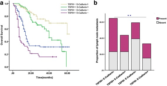
Background: TSP50 (testes-specific protease 50) has been reported to be a candidate oncogene and is overexpressed in various cancers. Our previous study demonstrated that TSP50 protein is elevated in gastric cancer, and its high expression is associated with unfavorable prognosis and lymph node metastasis. However, the role of TSP50 in gastric cancer remains elusive.

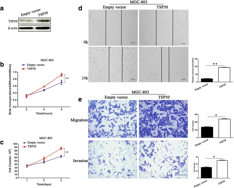
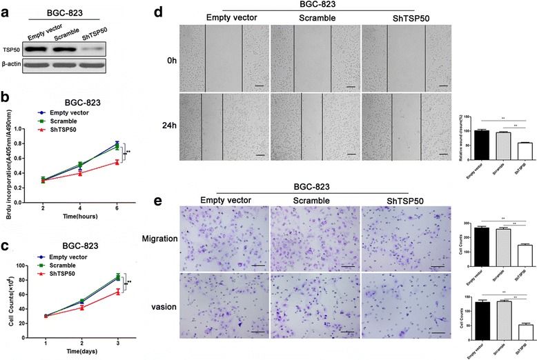
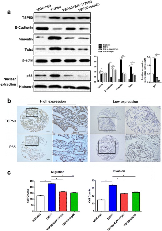
Methods: qRT-PCR, western blot were used to determine TSP50 expression in gastric cancer cell lines. Role of TSP50 in proliferation and invasion was examined by BrdU incorporation assay, cell count, wound healing and transwell assay. Immunohistochemistry and western blot were performed to identify the potential mechanisms involved.

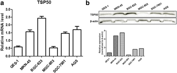
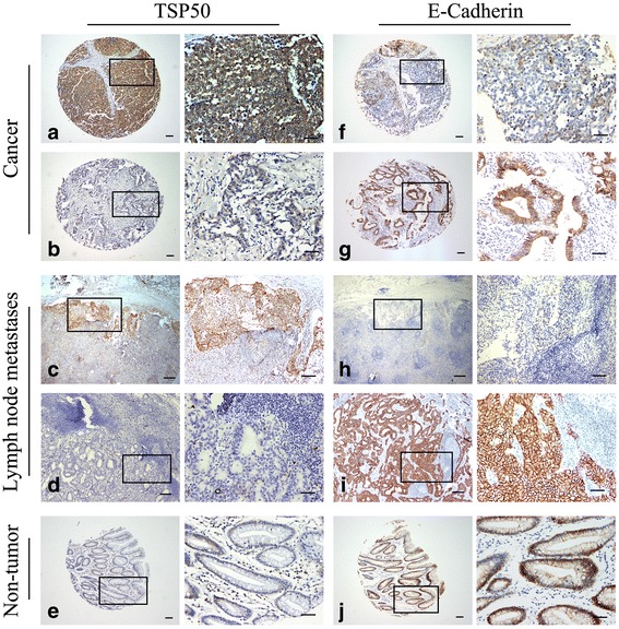
Results: TSP50 was highly expressed in most of the gastric cancer cell lines at both mRNA and protein levels. Up-regulation of TSP50 in gastric cancer cells enhanced proliferation and invasiveness, whereas down-regulation of TSP50 by its specific shRNA decreased it. A negative correlation between TSP50 and E-Cadherin was found in gastric cancer tissues, and combination of them improves the prediction for prognosis and lymph node metastasis. Mechanistic studies revealed that overexpression of TSP50 increased the expression of epithelial-to-mesenchymal transition (EMT) markers including Vimentin, and Twist, and decreased the epithelial marker E-Cadherin. NF-κB signaling pathway is involved in the regulatory effects of TSP50 on EMT, migration and invasion in gastric cancer cells.

Conclusion: TSP50 promotes the proliferation, migration and invasion of gastric cancer cells involving NF-κB dependent EMT activation. Targeting TSP50 may provide a novel therapeutic strategy for the management of gastric cancer.

Keywords: EMT; Gastric cancer; Invasion; Proliferation; TSP50.


Conflict of interest statement

Ethics approval and consent to participate

All the samples were collected with patient’s informed consent after approval from the Institute Research Medical Ethics Committee of the First Affiliated Hospital, Sun Yat-sen University. Details of tissue microarray construction were shown in previous study.

Consent for publication

Not applicable.

Competing interests

The authors declare that they have no competing interests.

Publisher’s Note

Springer Nature remains neutral with regard to jurisdictional claims in published maps and institutional affiliations.

Figures

Fig. 1
Fig. 1
Elevated expression of TSP50 in gastric cancer cell at mRNA and protein level. a Relative expression level of TSP50 was evaluated by quantitative real time PCR in gastric cancer cells and human gastric epithelial cell line GES-1. bTSP50 protein was measured by western blot analysis in gastric cancer cells and GES-1. β-actin served as an inner control. Data are expressed as the mean ± SD from three independent experiments
Fig. 2
Fig. 2
Overexpression of TSP50 promotes proliferation, migration and invasion in MGC-803 cell. a MGC-803 cell transfected with TSP50 overexpression plasmid. The efficiency of transfection was confirmed by western blot. β-actin served as an inner control. b BrdU assay showed a significant increase in BrdU uptake in MGC-803 cell transfected with TSP50 overexpression plasmid compared to that of the control group. (** P < 0.01). c Cell count experiment detected remarkable increase of cell number in overexpression-TSP50 MGC-803 cell compared to that of the control group. (** P < 0.01). d In wound closure assay, the migratory speed of MGC-803 cell transfected with TSP50 overexpression plasmid increased significantly compared to that of the vector control. (** P < 0.01). e In the transwell assay, migrated cells significantly increased after transfected with TSP50 overexpression plasmid. (* P < 0.05). In the transwell invasion assay, invasiveness was quantified by cells through Matigel and it was showed more cells in MGC-803 cell transfected with TSP50 overexpression plasmid than vector control. (* P < 0.05). All data are expressed as the mean ± SD from three independent experiments. Scale bar = 100 μm
Fig. 3
Fig. 3
Knockdown of TSP50 inhibits proliferation, migration and invasion in BGC-823 cell. a The efficiency of shTSP50 transfection was confirmed by western blot. β-actin served as an inner control. b BrdU assay showed a significant decrease in BrdU uptake in shTSP50 cell compared to that of the control groups. (** P < 0.01). c Cell count experiment detected remarkable reduction of cell number in shTSP50 cell compared to that of the control groups. (** P < 0.01). d In wound closure assay, the migratory speed of shTSP50 cell decreased significantly compared to that of the control groups. (** P < 0.01). e In the transwell assay, migrated cells significantly reduced after transfected with shTSP50. (** P < 0.01). In the transwell invasion assay, invasiveness was quantified by cells through Matigel and it was showed fewer cells in shTSP50 BGC-823 cell than control groups. (** P < 0.01). All data are expressed as the mean ± SD from three independent experiments. Scale bar = 100 μm
Fig. 4
Fig. 4
The negative correlation between TSP50 expression and E-Cadherin expression in gastric cancer tissues. a, b, c, d Representative images of high or low expression of TSP50 immunostaining in gastric cancer and lymph node metastasis lesion. f, g, h, i Immunostaining of E-Cadherin showed the contrary status in the same case compared with TSP50 immunostaining. e Low expression of TSP50 and (j) high expression of E-Cadherin in the same normal gastric mucosal tissue. In panel TSP50 and E-cadherin, the right panels displayed representative TSP50 and E-cadherin proteins expression in selected zone with enlarged view. Scale bar = 100 μm
Fig. 5
Fig. 5
Combination of TSP50 and E-Cadherin improves prognostic value for gastric cancer patients, and is closely related to lymph node metastasis. a Kaplan–Meier estimated of overall survival of gastric cancer patients. Patient groups were separated according to expression of TSP50 and E-Cadherin. High expression of TSP50 and low level of E-Cadherin group had the worst overall survival rates, whereas low TSP50 expression and high E-Cadherin expression group had the best prognosis. b High expression of TSP50 and low level of E-Cadherin group was closely related to the present status of lymph node metastasis. (** P < 0.01)
Fig. 6
Fig. 6
TSP50 induces EMT through NF-κB signaling pathway. a Expression levels of TSP50, E-Cadherin, Vimentin, Twist and nuclear p65 in MGC803 cell were determined by western blot analysis. β-actin and Histone1 were served as inner control of total protein and nuclear protein respectively. (*P < 0.05, **P < 0.01) (b)The positive association between TSP50 and p65 expression was showed in the same cohort of clinical gastric cancer tissue microarray using immunohistochemical staining. In panel high expression and low expression, the right panels displayed representative TSP50 and p65 proteins expression in selected zone with enlarged view. Scale bar = 100 μm. c Treatment with BAY117082 or transfection of shp65 reverted the effect of TSP50 on the migration and invasion in MGC803 cell partially. (*P < 0.05). All data are expressed as the mean ± SD from three independent experiments

References

    1. Chen W, Zheng R, Baade PD, Zhang S, Zeng H, Bray F, Jemal A, Yu XQ, He J. Cancer statistics in China, 2015. CA Cancer J Clin. 2016;66(2):115–32. doi: 10.3322/caac.21338. - DOI - PubMed
    1. Siegel RL, Miller KD, Jemal A. Cancer statistics, 2017. CA Cancer J Clin. 2017;67(1):7–30. doi: 10.3322/caac.21387. - DOI - PubMed
    1. Macdonald JS. Gastric cancer--new therapeutic options. N Engl J Med. 2006;355(1):76–77. doi: 10.1056/NEJMe068121. - DOI - PubMed
    1. Torre LA, Bray F, Siegel RL, Ferlay J, Lortet-Tieulent J, Jemal A. Global cancer statistics, 2012. CA Cancer J Clin. 2015;65(2):87–108. doi: 10.3322/caac.21262. - DOI - PubMed
    1. Xu HP, Yuan L, Shan J, Feng H. Localization and expression of TSP50 protein in human and rodent testes. Urology. 2004;64(4):826–832. doi: 10.1016/j.urology.2004.05.012. - DOI - PubMed

Publication types