

Let-7c Inhibits the Proliferation, Invasion, and Migration of Glioma Cells via Targeting E2F5
- PMID: 29362021
- PMCID: PMC7844676
- DOI: 10.3727/096504018X15164123839400
Let-7c Inhibits the Proliferation, Invasion, and Migration of Glioma Cells via Targeting E2F5
Abstract
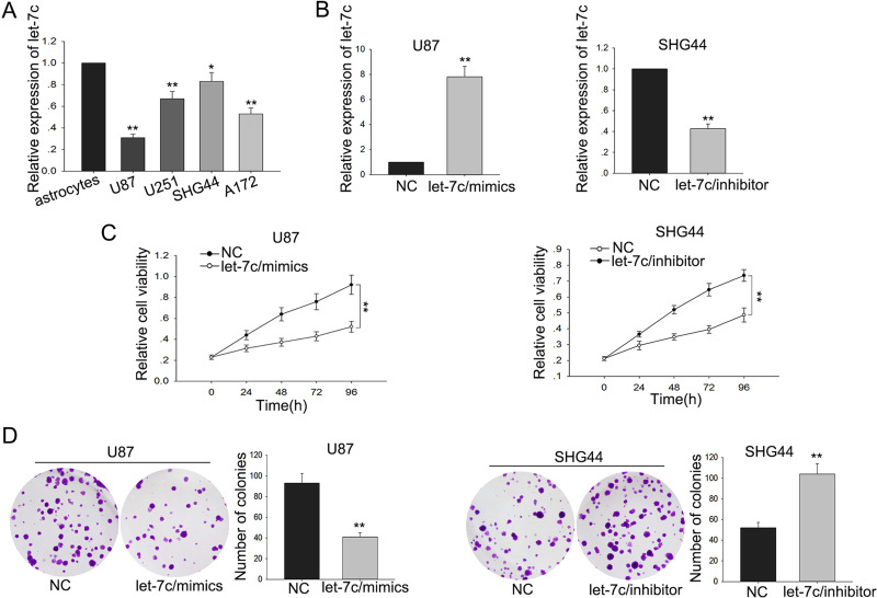
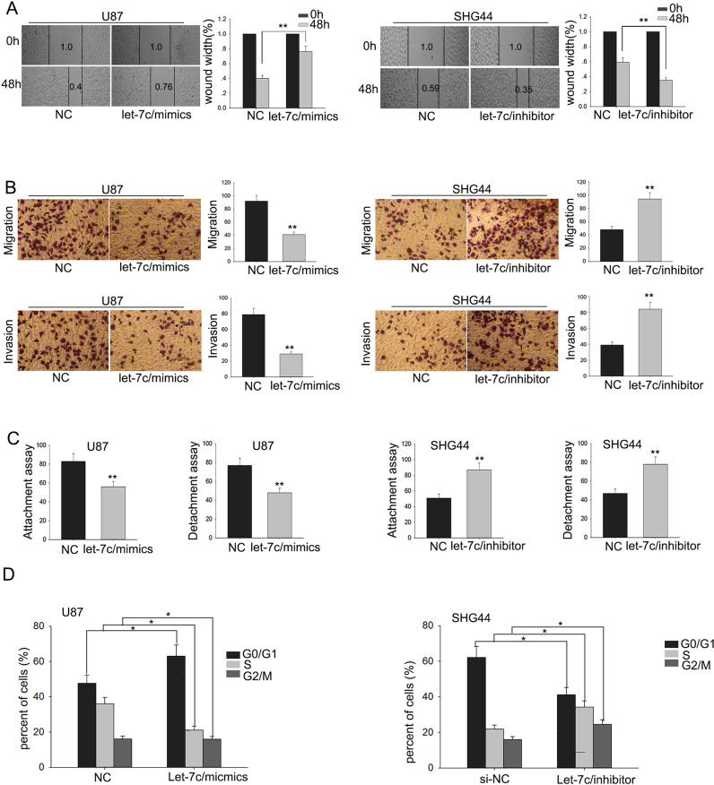
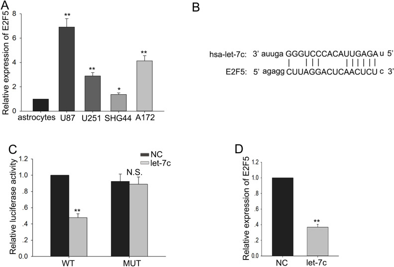
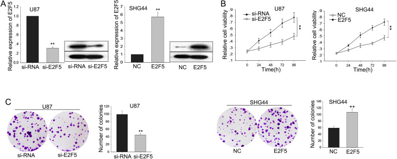
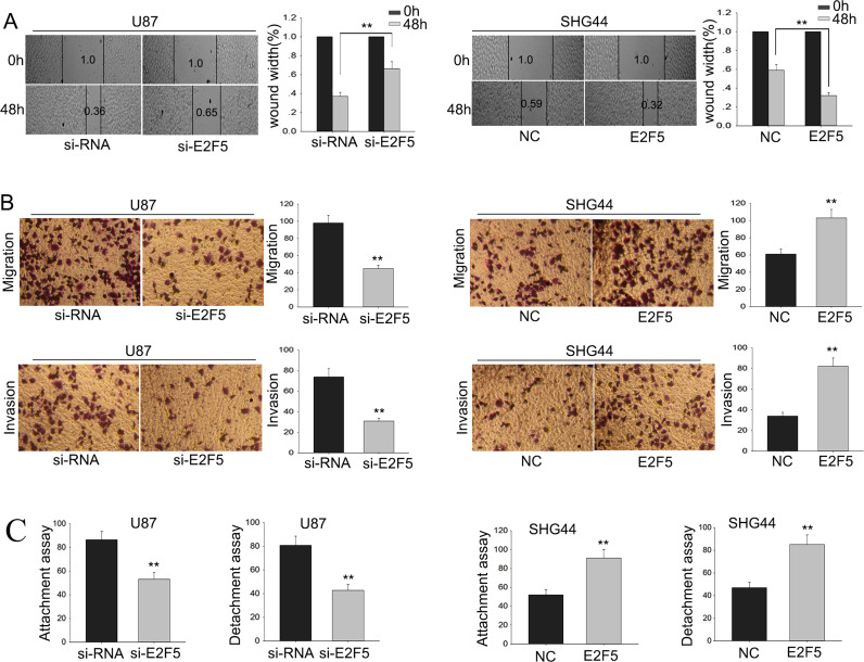
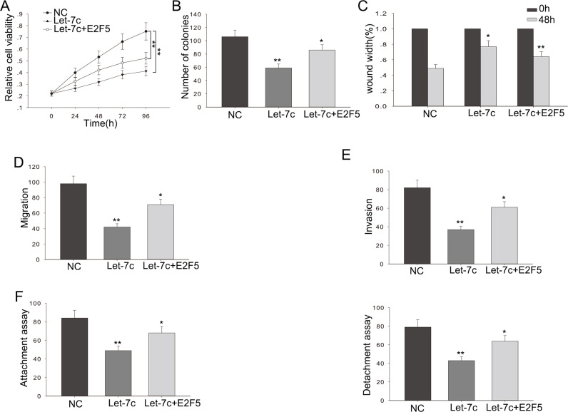
As a member of the miRNA family, let-7c has been identified as a tumor suppressor in many cancers. However, the molecular biological function of let-7c in glioma has not been elucidated. The aim of this study was to explore let-7c expression levels and evaluate its function in glioma cells. We first measured the expression of let-7c in four glioma cell lines and a normal cell line by quantitative real-time reverse transcription polymerase chain reaction (qRT-PCR), and the results showed that let-7c was downregulated in glioma cells. By applying gain-of-function and loss-of-function assays, the experiments suggested that dysregulation of let-7c could obviously affect cell proliferation, metastasis, and invasion. Based on online bioinformatics analysis and Dual-Luciferase Reporter assays, we found that E2F5 was a target gene of let-7c and contributed to the function of let-7c in glioma cells. Our investigations indicated that loss of let-7c contributed to the progression of glioma cells.
Conflict of interest statement
The authors declare no conflicts of interest.
Figures
Similar articles
-
Let-7a suppresses glioma cell proliferation and invasion through TGF-β/Smad3 signaling pathway by targeting HMGA2.Tumour Biol. 2016 Jun;37(6):8107-19. doi: 10.1007/s13277-015-4674-6. Epub 2015 Dec 29. Tumour Biol. 2016. Retraction in: Tumour Biol. 2021;43(1):351-354. doi: 10.3233/TUB-219010. PMID: 26715270 Retracted.
-
MicroRNA‑34a inhibits esophageal squamous cell carcinoma progression by targeting E2F5.J BUON. 2019 Nov-Dec;24(6):2514-2522. J BUON. 2019. PMID: 31983127
-
E2F5 promotes prostate cancer cell migration and invasion through regulation of TFPI2, MMP-2 and MMP-9.Carcinogenesis. 2020 Dec 31;41(12):1767-1780. doi: 10.1093/carcin/bgaa043. Carcinogenesis. 2020. PMID: 32386317
-
Hsa-let-7c exerts an anti-tumor function by negatively regulating ANP32E in lung adenocarcinoma.Tissue Cell. 2020 Aug;65:101372. doi: 10.1016/j.tice.2020.101372. Epub 2020 May 7. Tissue Cell. 2020. PMID: 32746998
-
Molecular and Cellular Complexity of Glioma. Focus on Tumour Microenvironment and the Use of Molecular and Imaging Biomarkers to Overcome Treatment Resistance.Int J Mol Sci. 2020 Aug 6;21(16):5631. doi: 10.3390/ijms21165631. Int J Mol Sci. 2020. PMID: 32781585 Free PMC article. Review.
Cited by
-
Exosomes of A549 Cells Induced Migration, Invasion, and EMT of BEAS-2B Cells Related to let-7c-5p and miR-181b-5p.Front Endocrinol (Lausanne). 2022 Jul 8;13:926769. doi: 10.3389/fendo.2022.926769. eCollection 2022. Front Endocrinol (Lausanne). 2022. PMID: 35898472 Free PMC article.
-
Epigenetic Drugs for Cancer and microRNAs: A Focus on Histone Deacetylase Inhibitors.Cancers (Basel). 2019 Oct 10;11(10):1530. doi: 10.3390/cancers11101530. Cancers (Basel). 2019. PMID: 31658720 Free PMC article. Review.
-
MAPK Is a Mutual Pathway Targeted by Anxiety-Related miRNAs, and E2F5 Is a Putative Target for Anxiolytic miRNAs.Biomolecules. 2023 Mar 16;13(3):544. doi: 10.3390/biom13030544. Biomolecules. 2023. PMID: 36979479 Free PMC article.
-
Inhibition of HDACs Suppresses Cell Proliferation and Cell Migration of Gastric Cancer by Regulating E2F5 Targeting BCL2.Life (Basel). 2021 Dec 17;11(12):1425. doi: 10.3390/life11121425. Life (Basel). 2021. PMID: 34947956 Free PMC article.
-
Foxf1-mediated co-regulation of miR-495 and let-7c modulates epicardial cell migration and myocardial specification.Cell Mol Life Sci. 2025 Jun 25;82(1):254. doi: 10.1007/s00018-025-05735-4. Cell Mol Life Sci. 2025. PMID: 40555850 Free PMC article.
References
-
- Furnari FB, Fenton T, Bachoo RM, Mukasa A, Stommel JM, Stegh A, Hahn WC, Ligon KL, Louis DN, Brennan C, Chin L, DePinho RA, Cavenee WK. Malignant astrocytic glioma: Genetics, biology, and paths to treatment. Genes Dev. 2007;21(21):2683–710. - PubMed
MeSH terms
Substances
LinkOut - more resources
Full Text Sources
Other Literature Sources
Research Materials