

Integration of Oncogenes via Sleeping Beauty as a Mouse Model of HPV16+ Oral Tumors and Immunologic Control
- PMID: 29362220
- PMCID: PMC6056342
- DOI: 10.1158/2326-6066.CIR-16-0358
Integration of Oncogenes via Sleeping Beauty as a Mouse Model of HPV16+ Oral Tumors and Immunologic Control
Abstract
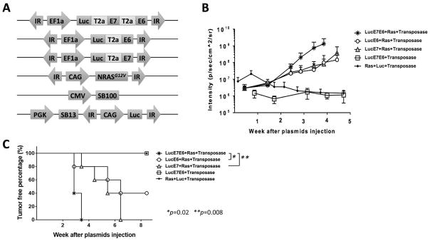
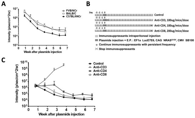
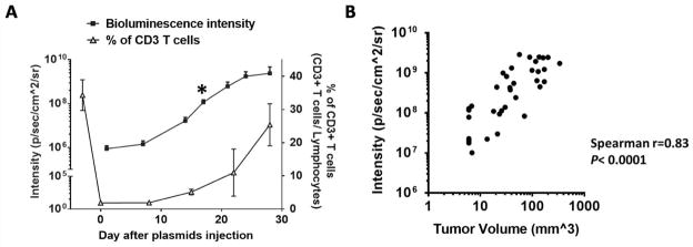
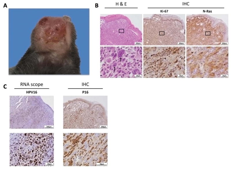
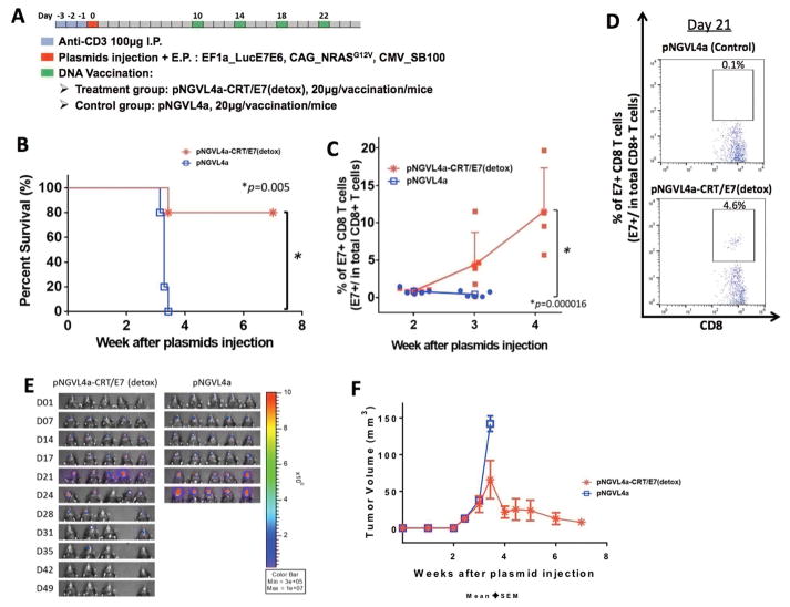
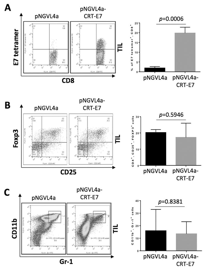
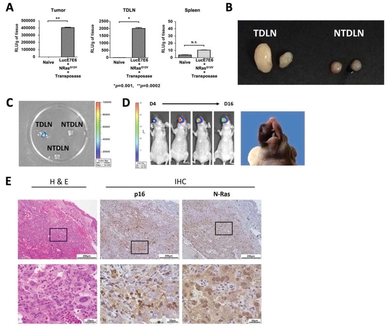
Human papillomavirus type 16 (HPV16) is the etiologic factor for cervical cancer and a subset of oropharyngeal cancers. Although several prophylactic HPV vaccines are available, no effective therapeutic strategies to control active HPV diseases exist. Tumor implantation models are traditionally used to study HPV-associated buccal tumors. However, they fail to address precancerous phases of disease progression and display tumor microenvironments distinct from those observed in patients. Previously, K14-E6/E7 transgenic mouse models have been used to generate spontaneous tumors. However, the rate of tumor formation is inconsistent, and the host often develops immune tolerance to the viral oncoproteins. We developed a preclinical, spontaneous, HPV16+ buccal tumor model using submucosal injection of oncogenic plasmids expressing HPV16-E6/E7, NRas G12V , luciferase, and sleeping beauty (SB) transposase, followed by electroporation in the buccal mucosa. We evaluated responses to immunization with a pNGVL4a-CRT/E7(detox) therapeutic HPV DNA vaccine and tumor cell migration to distant locations. Mice transfected with plasmids encoding HPV16-E6/E7, NRas G12V , luciferase, and SB transposase developed tumors within 3 weeks. We also found transient anti-CD3 administration is required to generate tumors in immunocompetent mice. Bioluminescence signals from luciferase correlated strongly with tumor growth, and tumors expressed HPV16-associated markers. We showed that pNGVL4a-CRT/E7(detox) administration resulted in antitumor immunity in tumor-bearing mice. Lastly, we demonstrated that the generated tumor could migrate to tumor-draining lymph nodes. Our model provides an efficient method to induce spontaneous HPV+ tumor formation, which can be used to identify effective therapeutic interventions, analyze tumor migration, and conduct tumor biology research. Cancer Immunol Res; 6(3); 305-19. ©2018 AACR.
©2018 American Association for Cancer Research.
Conflict of interest statement
T.-C. Wu is a co-founder of and has an equity ownership interest in Papivax LLC. He also owns Papivax Biotech Inc. stock options and is a member of Papivax Biotech Inc.’s Scientific Advisory Board. Additionally, under a licensing agreement between Papivax Biotech Inc. and the Johns Hopkins University, Dr. Wu and Dr. Hung are entitled to royalties on an invention described in this article. This arrangement has been reviewed and approved by the Johns Hopkins University in accordance with its conflict of interest policies. Yung-Nien Chang is the Chief Scientific Officer of Papivax Biotech Inc. and owns stock options in Papivax Biotech Inc. Other co-authors have declared that no conflict of interest exists.
Figures
Similar articles
-
Control of Spontaneous HPV16 E6/E7 Expressing Oral Cancer in HLA-A2 (AAD) Transgenic Mice with Therapeutic HPV DNA Vaccine.J Biomed Sci. 2021 Sep 13;28(1):63. doi: 10.1186/s12929-021-00759-x. J Biomed Sci. 2021. PMID: 34517865 Free PMC article.
-
Development of a Novel Mouse Model of Spontaneous High-Risk HPVE6/E7-Expressing Carcinoma in the Cervicovaginal Tract.Cancer Res. 2021 Sep 1;81(17):4560-4569. doi: 10.1158/0008-5472.CAN-21-0399. Epub 2021 Jul 2. Cancer Res. 2021. PMID: 34215618 Free PMC article.
-
Development of a Spontaneous HPV16 E6/E7-Expressing Head and Neck Squamous Cell Carcinoma in HLA-A2 Transgenic Mice.mBio. 2022 Feb 22;13(1):e0325221. doi: 10.1128/mbio.03252-21. Epub 2022 Jan 4. mBio. 2022. PMID: 35089069 Free PMC article.
-
Comparison of preclinical efficacy of immunotherapies against HPV-induced cancers.Expert Rev Vaccines. 2024 Jan-Dec;23(1):674-687. doi: 10.1080/14760584.2024.2374287. Epub 2024 Jul 8. Expert Rev Vaccines. 2024. PMID: 38978164 Review.
-
Inducing Immunity Where It Matters: Orthotopic HPV Tumor Models and Therapeutic Vaccinations.Front Immunol. 2020 Aug 14;11:1750. doi: 10.3389/fimmu.2020.01750. eCollection 2020. Front Immunol. 2020. PMID: 32922389 Free PMC article. Review.
Cited by
-
STAT1-Deficient HPV E6/E7-Associated Cancers Maintain Host Immunocompetency against Therapeutic Intervention.Vaccines (Basel). 2024 Apr 17;12(4):430. doi: 10.3390/vaccines12040430. Vaccines (Basel). 2024. PMID: 38675812 Free PMC article.
-
Control of Spontaneous HPV16 E6/E7 Expressing Oral Cancer in HLA-A2 (AAD) Transgenic Mice with Therapeutic HPV DNA Vaccine.J Biomed Sci. 2021 Sep 13;28(1):63. doi: 10.1186/s12929-021-00759-x. J Biomed Sci. 2021. PMID: 34517865 Free PMC article.
-
Preclinical studies of RA475, a guanidine-substituted spirocyclic candidate RPN13/ADRM1 inhibitor for treatment of ovarian cancer.PLoS One. 2024 Jul 11;19(7):e0305710. doi: 10.1371/journal.pone.0305710. eCollection 2024. PLoS One. 2024. PMID: 38990850 Free PMC article.
-
Generation of a spontaneous murine HPV + oral cancer model with site-specific oncogene insertion using CRISPR-SONIC.Cell Biosci. 2025 Jun 18;15(1):84. doi: 10.1186/s13578-025-01427-5. Cell Biosci. 2025. PMID: 40533862 Free PMC article.
-
Genetically engineered mouse models of head and neck cancers.Oncogene. 2023 Aug;42(35):2593-2609. doi: 10.1038/s41388-023-02783-7. Epub 2023 Jul 20. Oncogene. 2023. PMID: 37474617 Free PMC article. Review.
References
-
- Mehanna H, Beech T, Nicholson T, El-Hariry I, McConkey C, Paleri V, et al. Prevalence of human papillomavirus in oropharyngeal and nonoropharyngeal head and neck cancer--systematic review and meta-analysis of trends by time and region. Head & neck. 2013;35(5):747–55. doi: 10.1002/hed.22015. - DOI - PubMed
-
- Ostor AG. Natural history of cervical intraepithelial neoplasia: a critical review. International journal of gynecological pathology : official journal of the International Society of Gynecological Pathologists. 1993;12(2):186–92. - PubMed
Publication types
MeSH terms
Substances
Grants and funding
LinkOut - more resources
Full Text Sources
Other Literature Sources
Medical
Molecular Biology Databases
Research Materials
Miscellaneous
