. 2018 Apr 2;217(4):1269-1285.
doi: 10.1083/jcb.201708116. Epub 2018 Jan 23.

A prion-like domain in Hsp42 drives chaperone-facilitated aggregation of misfolded proteins

Affiliations

A prion-like domain in Hsp42 drives chaperone-facilitated aggregation of misfolded proteins

Tomas Grousl et al. J Cell Biol. .

Abstract

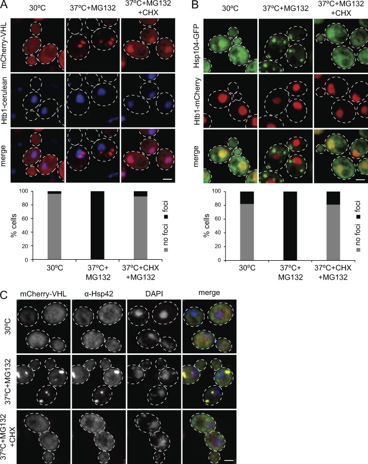
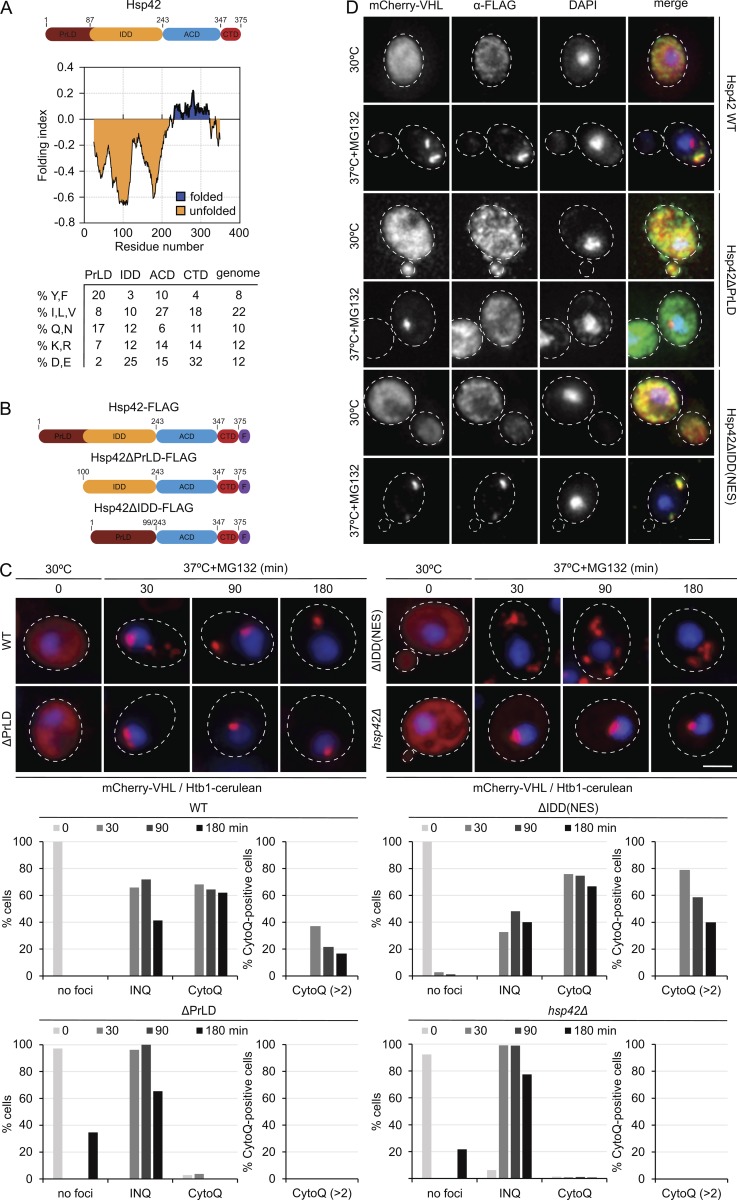
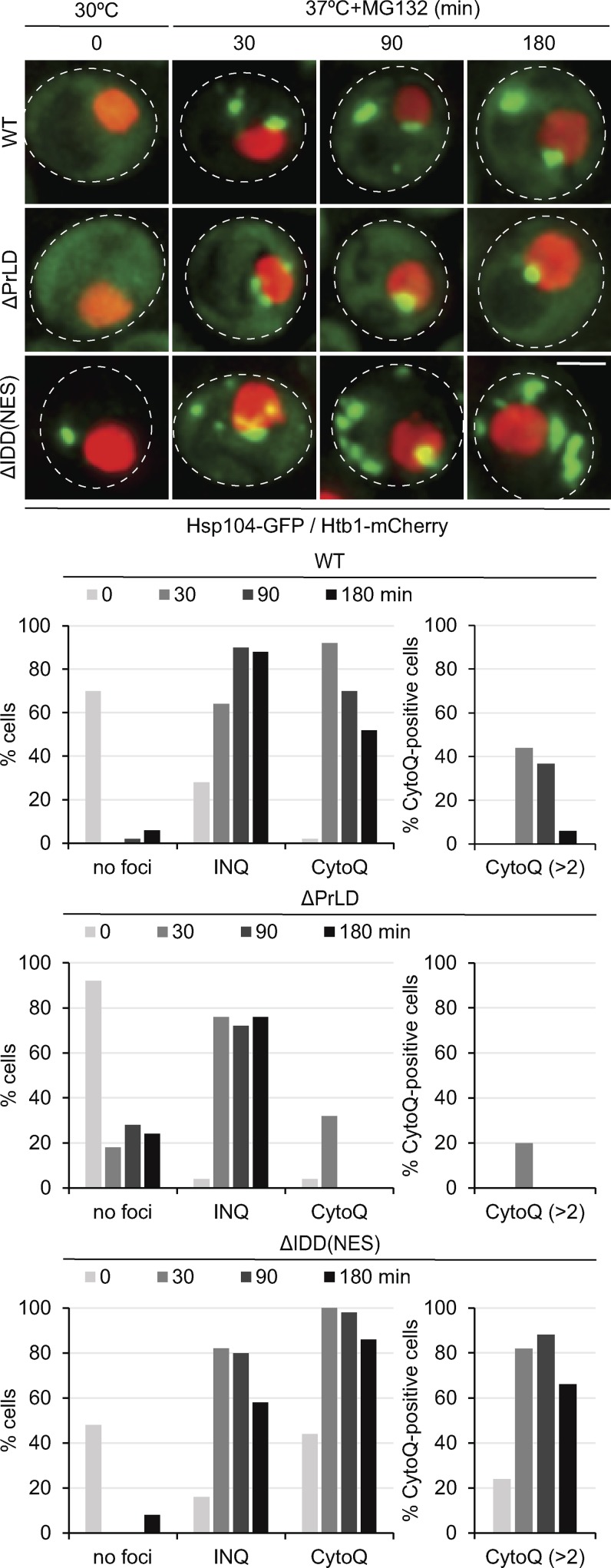
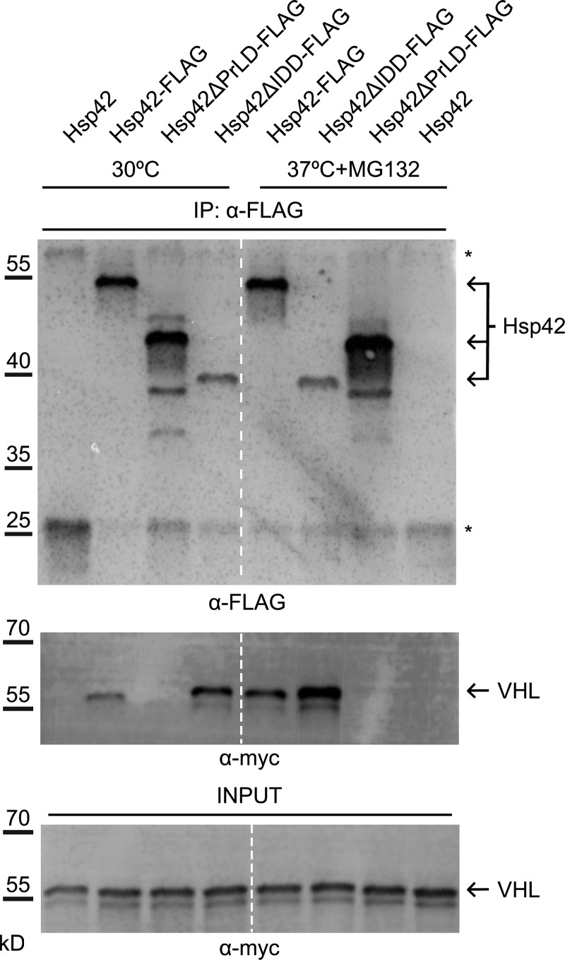
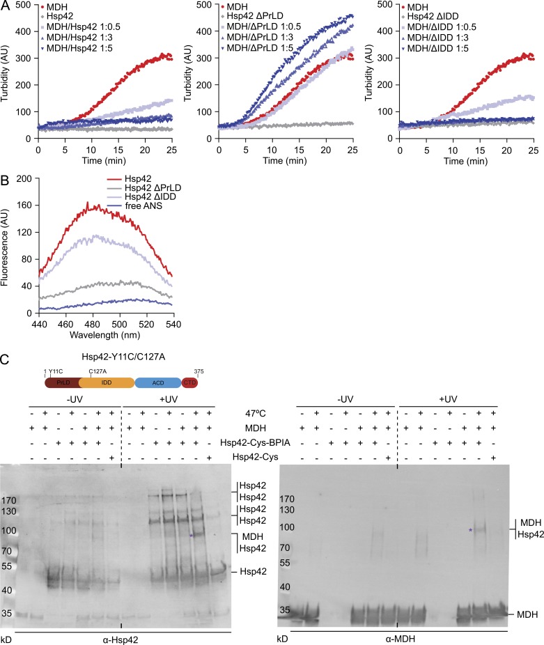
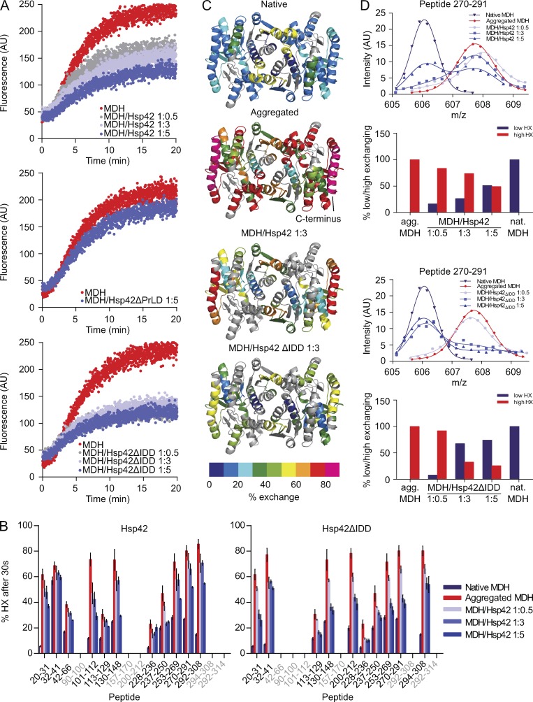
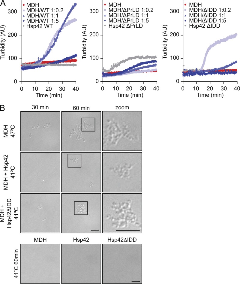
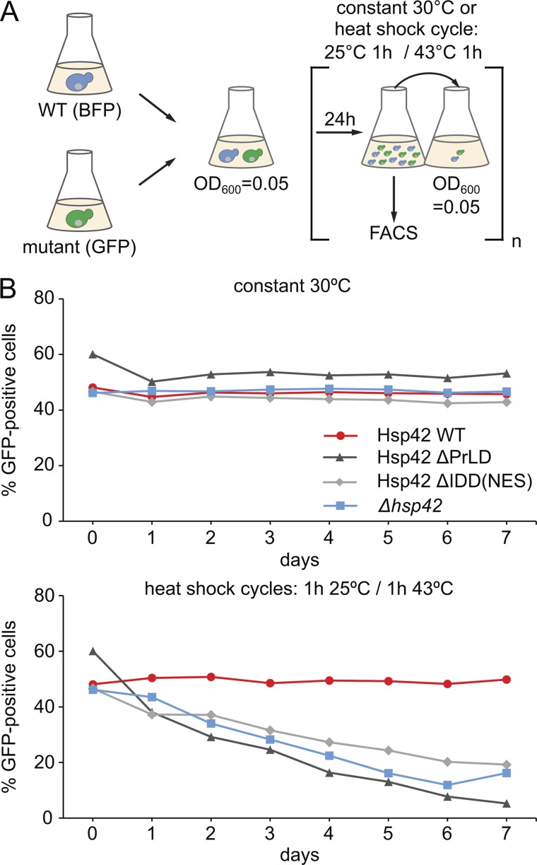
Chaperones with aggregase activity promote and organize the aggregation of misfolded proteins and their deposition at specific intracellular sites. This activity represents a novel cytoprotective strategy of protein quality control systems; however, little is known about its mechanism. In yeast, the small heat shock protein Hsp42 orchestrates the stress-induced sequestration of misfolded proteins into cytosolic aggregates (CytoQ). In this study, we show that Hsp42 harbors a prion-like domain (PrLD) and a canonical intrinsically disordered domain (IDD) that act coordinately to promote and control protein aggregation. Hsp42 PrLD is essential for CytoQ formation and is bifunctional, mediating self-association as well as binding to misfolded proteins. Hsp42 IDD confines chaperone and aggregase activity and affects CytoQ numbers and stability in vivo. Hsp42 PrLD and IDD are both crucial for cellular fitness during heat stress, demonstrating the need for sequestering misfolded proteins in a regulated manner.


Figures

Figure 1.
Figure 1.
The formation of large protein aggregates depends on translation. (A–C) Localization patterns of mCherry-VHL (A), Hsp104-GFP (B), and Hsp42 (C) in S. cerevisiae cells and their superposition with the nuclear marker Htb1-cerulean (A and B) or DNA stained by DAPI (C) upon control conditions (30°C), and proteotoxic stress in the absence or presence of CHX (37°C for 30 min + MG132 ± CHX) are shown. Maximal projections of widefield z stack images with corresponding quantifications (n > 50/sample) are presented. Bars, 2 µm.
Figure 2.
Figure 2.
The Hsp42 PrLD is essential for CytoQ formation. (A) Hsp42 domain organization. Hsp42 consists of an NTE composed of two IDDs (PrLD and IDD) followed by the ACD and a CTE. Prediction of intrinsically unfolded protein segments according to Foldindex (Prilusky et al., 2005) and the relative abundance of specific amino acids within each Hsp42 domain are shown. The average amino acid abundance of the yeast proteome is included as reference. (B) Schematic diagrams of Hsp42 WT and deletion constructs. F, FLAG tag. (C) mCherry-VHL localization patterns in S. cerevisiae hsp42Δ cells expressing Hsp42 WT or the indicated Hsp42 deletion mutants and their superposition with the nuclear marker Htb1-cerulean upon control conditions (30°C) and proteotoxic stress (37°C + MG132) are shown. The number of cells showing CytoQ (mCherry-VHL foci distant from the Htb1-cerulean signal) or INQ (mCherry-VHL foci adjacent or overlapping with the Htb1-cerulean signal) inclusions was quantified (n > 50/sample). (D) Immunofluorescence images of Hsp42 WT and deletion mutants and their superposition with mCherry-VHL and DAPI upon control conditions (30°C) and proteotoxic stress (37°C for 30 min + MG132) are shown. Maximal projections of widefield z stack images are presented. Bars, 2 µm.
Figure 3.
Figure 3.
Distinct functions of Hsp42 PrLD and IDD in CytoQ formation. Localization patterns of Hsp104-GFP and their superposition with the nuclear marker Htb1-mCherry in S. cerevisiae cells expressing Hsp42 WT or deletion mutants upon control conditions (30°C) and proteotoxic stress (37°C + MG132). The number of cells showing CytoQ (mCherry-VHL foci distant from the Htb1-cerulean signal) or INQ (mCherry-VHL foci adjacent or overlapping with the Htb1-cerulean signal) inclusions was quantified at the indicated time points (n > 50 cells). Maximal projections of widefield z stack images are presented. Bar, 2 µm.
Figure 4.
Figure 4.
Hsp42 PrLD is essential for mCherry–VHL interaction. Coimmunoprecipitation of Hsp42 WT and deletion mutants (all FLAG-tagged) and mCherry-myc-VHL. Cells were grown at control conditions (30°C) or stressed (37°C for 45 min + MG132) before extract preparation. Hsp42 proteins were precipitated using anti-FLAG agarose, and the amount of bound Hsp42 and mCherry-myc-VHL was quantified by immunoblot analysis using anti-FLAG and anti-myc antibodies. Fractions were analyzed for the content of FLAG-tagged proteins and mCherry–VHL interaction (anti-myc). The sizes of Hsp42 proteins are indicated (arrows). Asterisks indicate coeluting mouse IgG heavy and light chains. VHL input controls are provided. The dashed line indicates intervening lanes have been spliced out. IP, immunoprecipitation.
Figure 5.
Figure 5.
PrLD is the major Hsp42 substrate interaction site. (A) MDH was denatured for 30 min at 47°C in the absence or presence of Hsp42 WT and deletion mutants at indicated ratios. All control Hsp42 proteins were heated alone. The formation of MDH aggregates was followed by turbidity measurements. (B) Overall surface hydrophobicity of Hsp42 WT and deletion mutants probed by ANS fluorescence. Fluorescence of free ANS is provided as reference. (C) Cross-linking of BPIA-labeled Hsp42-Y11C/C127A (Hsp42-Cys) to unfolded MDH. MDH and Hsp42 alone or MDH/Hsp42-Cys (unlabeled and BPIA-labeled) mixtures (1:5 ratio) were incubated at 30°C or 47°C. Samples were afterward exposed to UV (+/− UV), and cross-link products were analyzed by immunoblot analysis using MDH and Hsp42-specific antibodies. Asterisks indicate Hsp42-MDH cross-link products. A schematic diagram of Hsp42-Y11C/C127A is shown at top.
Figure 6.
Figure 6.
Hsp42ΔIDD exhibits superior chaperone activity. (A) Coaggregation of MDH-YFP (FRET donor) and MDH labeled with 7-diethylcoumarin-3-carboxylic acid (FRET acceptor) was monitored by FRET (increased acceptor fluorescence) at 47°C in the absence or presence of Hsp42 WT and deletion mutants. (B) Relative proton/deuteron exchange in native or aggregated MDH and MDH coaggregated with Hsp42 WT and Hsp42ΔIDD (at indicated MDH/sHsp ratios) after 30 s incubation in D2O. Aggregated MDH and MDH–sHsp complexes were formed by incubation for 30 min at 47°C. Error bars denote SD for each point based on three repetitions. All data were corrected for deuteron losses caused by back exchange using a 100% deuterated control. Grey regions could not be detected. (C) HX heat maps of native, aggregated, and Hsp42-complexed states (threefold excess of Hsp42) of the MDH dimer structure (PDB ID: 1MLD) are shown. Peptic peptides are colored according to their exchange behavior (% exchange). Grey regions could not be detected. (D) Bimodal distribution of isotope peaks of indicated MDH peptides derived from MDH–Hsp42 (WT or ΔIDD) complexes. Top: intensity versus m/z diagrams for the indicated peptic MDH fragment after 30 s HX at 30°C. Bottom: fractions of native-like (low HX) and aggregate-like (high HX) populations.
Figure 7.
Figure 7.
Aggregase function of Hsp42 deletion mutants. (A) MDH was denatured for 60 min at 41°C in the absence or presence of Hsp42 WT and deletion mutants at the indicated ratios. As a control, Hsp42 proteins were heated alone. The formation of MDH aggregates was followed by turbidity measurements. (B) Complexes of MDH with Hsp42 WT or Hsp42ΔIDD formed at 41°C at equimolar ratios were analyzed by light microscopy. MDH aggregated at 47°C served as reference. As control, MDH and Hsp42 proteins were heated alone. Widefield images are presented. Bars, 5 µm.
Figure 8.
Figure 8.
PrLD and IDD are crucial for cytoprotective Hsp42 activity during heat stress. (A) Schematic diagram of the growth competition assay. S. cerevisiae WT cells expressing BFP and indicated hsp42 mutant cells expressing GFP were mixed 1:1. Mixtures were either grown at 30°C or were subjected to heat shock cycles, switching repetitively between 25°C and 43°C. Each day, the proportion of WT and mutant cells was determined by FACS, and cell mixtures were diluted to OD600 0.05. (B) Fractions of Hsp42 WT and Hsp42 deletion mutants or hsp42Δ deletion strain in the mixed cultures upon constant growth at 30°C or repetitive heat stress are plotted. The representative result of one out of three biological replicates is shown.

Comment in

Similar articles

Cited by

References

    1. Alberti S., Halfmann R., King O., Kapila A., and Lindquist S.. 2009. A systematic survey identifies prions and illuminates sequence features of prionogenic proteins. Cell. 137:146–158. 10.1016/j.cell.2009.02.044 - DOI - PMC - PubMed
    1. Balchin D., Hayer-Hartl M., and Hartl F.U.. 2016. In vivo aspects of protein folding and quality control. Science. 353:aac4354 10.1126/science.aac4354 - DOI - PubMed
    1. Basha E., Friedrich K.L., and Vierling E.. 2006. The N-terminal arm of small heat shock proteins is important for both chaperone activity and substrate specificity. J. Biol. Chem. 281:39943–39952. 10.1074/jbc.M607677200 - DOI - PubMed
    1. Chen B., Retzlaff M., Roos T., and Frydman J.. 2011. Cellular strategies of protein quality control. Cold Spring Harb. Perspect. Biol. 3:a004374 10.1101/cshperspect.a004374 - DOI - PMC - PubMed
    1. Cherkasov V., Grousl T., Theer P., Vainshtein Y., Glässer C., Mongis C., Kramer G., Stoecklin G., Knop M., Mogk A., and Bukau B.. 2015. Systemic control of protein synthesis through sequestration of translation and ribosome biogenesis factors during severe heat stress. FEBS Lett. 589:3654–3664. 10.1016/j.febslet.2015.10.010 - DOI - PubMed

Publication types

MeSH terms

Substances

Associated data

LinkOut - more resources