

The Ancient Link between G-Protein-Coupled Receptors and C-Terminal Phospholipid Kinase Domains
- PMID: 29362235
- PMCID: PMC5784254
- DOI: 10.1128/mBio.02119-17
The Ancient Link between G-Protein-Coupled Receptors and C-Terminal Phospholipid Kinase Domains
Abstract
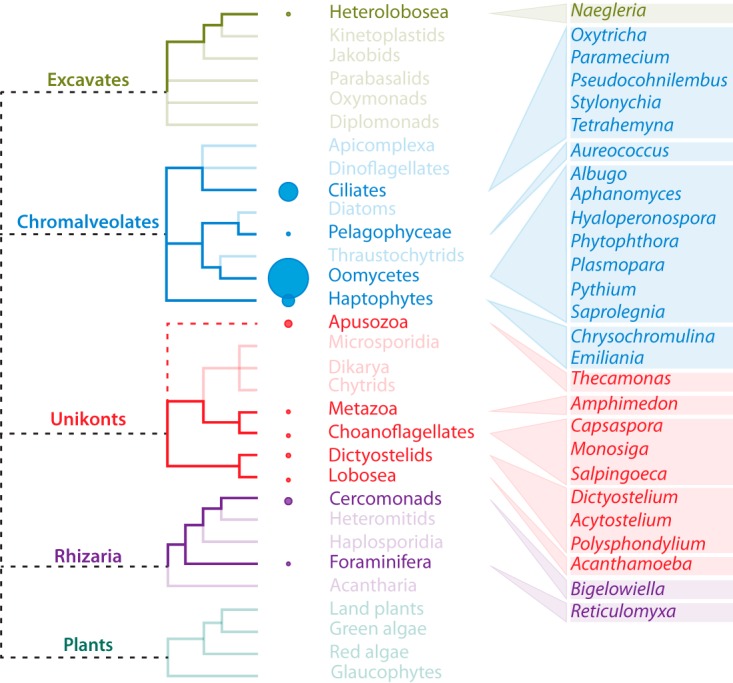
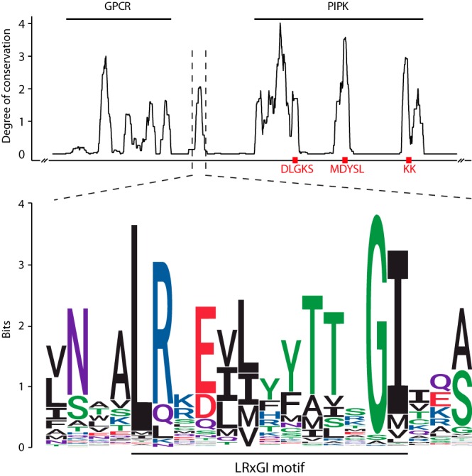
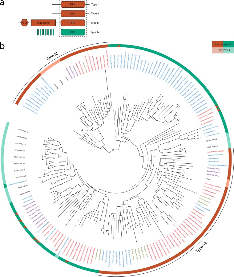
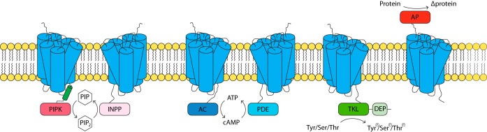
Sensing external signals and transducing these into intracellular responses requires a molecular signaling system that is crucial for every living organism. Two important eukaryotic signal transduction pathways that are often interlinked are G-protein signaling and phospholipid signaling. Heterotrimeric G-protein subunits activated by G-protein-coupled receptors (GPCRs) are typical stimulators of phospholipid signaling enzymes such as phosphatidylinositol phosphate kinases (PIPKs) or phospholipase C (PLC). However, a direct connection between the two pathways likely exists in oomycetes and slime molds, as they possess a unique class of GPCRs that have a PIPK as an accessory domain. In principle, these so-called GPCR-PIPKs have the capacity of perceiving an external signal (via the GPCR domain) that, via PIPK, directly activates downstream phospholipid signaling. Here we reveal the sporadic occurrence of GPCR-PIPKs in all eukaryotic supergroups, except for plants. Notably, all species having GPCR-PIPKs are unicellular microorganisms that favor aquatic environments. Phylogenetic analysis revealed that GPCR-PIPKs are likely ancestral to eukaryotes and significantly expanded in the last common ancestor of oomycetes. In addition to GPCR-PIPKs, we identified five hitherto-unknown classes of GPCRs with accessory domains, four of which are universal players in signal transduction. Similarly to GPCR-PIPKs, this enables a direct coupling between extracellular sensing and downstream signaling. Overall, our findings point to an ancestral signaling system in eukaryotes where GPCR-mediated sensing is directly linked to downstream responses.IMPORTANCE G-protein-coupled receptors (GPCRs) are central sensors that activate eukaryotic signaling and are the primary targets of human drugs. In this report, we provide evidence for the widespread though limited presence of a novel class of GPCRs in a variety of unicellular eukaryotes. These include free-living organisms and organisms that are pathogenic for plants, animals, and humans. The novel GPCRs have a C-terminal phospholipid kinase domain, pointing to a direct link between sensing external signals via GPCRs and downstream intracellular phospholipid signaling. Genes encoding these receptors were likely present in the last common eukaryotic ancestor and were lost during the evolution of higher eukaryotes. We further describe five other types of GPCRs with a catalytic accessory domain, the so-called GPCR-bigrams, four of which may potentially have a role in signaling. These findings shed new light onto signal transduction in microorganisms and provide evidence for alternative eukaryotic signaling pathways.
Keywords: G-protein-coupled receptors; Phytophthora; cell signaling; oomycetes; phospholipid-mediated signaling.
Copyright © 2018 van den Hoogen et al.
Figures
Similar articles
-
Novel phosphatidylinositol phosphate kinases with a G-protein coupled receptor signature are shared by Dictyostelium and Phytophthora.Trends Microbiol. 2006 Sep;14(9):378-82. doi: 10.1016/j.tim.2006.07.006. Epub 2006 Jul 31. Trends Microbiol. 2006. PMID: 16876997
-
Genomewide analysis of phospholipid signaling genes in Phytophthora spp.: novelties and a missing link.Mol Plant Microbe Interact. 2006 Dec;19(12):1337-47. doi: 10.1094/MPMI-19-1337. Mol Plant Microbe Interact. 2006. PMID: 17153918
-
Identification of G protein-coupled receptor signaling pathway proteins in marine diatoms using comparative genomics.BMC Genomics. 2013 Jul 24;14:503. doi: 10.1186/1471-2164-14-503. BMC Genomics. 2013. PMID: 23883327 Free PMC article.
-
Sensing and transduction of nutritional and chemical signals in filamentous fungi: Impact on cell development and secondary metabolites biosynthesis.Biotechnol Adv. 2019 Nov 1;37(6):107392. doi: 10.1016/j.biotechadv.2019.04.014. Epub 2019 Apr 26. Biotechnol Adv. 2019. PMID: 31034961 Review.
-
Superfamily of G-protein coupled receptors (GPCRs)--extraordinary and outstanding success of evolution.Postepy Hig Med Dosw (Online). 2014 Oct 31;68:1225-37. doi: 10.5604/17322693.1127326. Postepy Hig Med Dosw (Online). 2014. PMID: 25380205 Review.
Cited by
-
Exchanges at the Plant-Oomycete Interface That Influence Disease.Plant Physiol. 2019 Apr;179(4):1198-1211. doi: 10.1104/pp.18.00979. Epub 2018 Dec 11. Plant Physiol. 2019. PMID: 30538168 Free PMC article. Review.
-
GPCR Modulation in Breast Cancer.Int J Mol Sci. 2018 Dec 2;19(12):3840. doi: 10.3390/ijms19123840. Int J Mol Sci. 2018. PMID: 30513833 Free PMC article. Review.
-
GPCR-bigrams: Enigmatic signaling components in oomycetes.PLoS Pathog. 2018 Jul 5;14(7):e1007064. doi: 10.1371/journal.ppat.1007064. eCollection 2018 Jul. PLoS Pathog. 2018. PMID: 29975773 Free PMC article. No abstract available.
-
Mining oomycete proteomes for metalloproteases leads to identification of candidate virulence factors in Phytophthora infestans.Mol Plant Pathol. 2021 May;22(5):551-563. doi: 10.1111/mpp.13043. Epub 2021 Mar 3. Mol Plant Pathol. 2021. PMID: 33657266 Free PMC article.
-
Allosteric Regulation of G-Protein-Coupled Receptors: From Diversity of Molecular Mechanisms to Multiple Allosteric Sites and Their Ligands.Int J Mol Sci. 2023 Mar 24;24(7):6187. doi: 10.3390/ijms24076187. Int J Mol Sci. 2023. PMID: 37047169 Free PMC article. Review.
References
Publication types
MeSH terms
Substances
LinkOut - more resources
Full Text Sources
Other Literature Sources
